

Vascular occlusion (VO) after filler means filler is blocking a facial artery, from inside or by pressing on it, and the skin shows it first: pallor that outlasts the needle, then a violet net-like pattern across the artery’s territory within minutes. Treatment starts on those signs: stop injecting, give hyaluronidase at once and scan to locate the block while it works. Starting within 6 hours improved outcomes in Alhusain’s 2026 meta-analysis of 231 patients, and the Dutch national referral center counted one event per 6,558 treatments (Schelke 2020). This guide covers the skin stages, bruise against occlusion on the lips, what Color, Power and PW Doppler show during an event, the hyaluronidase doses published for guided and unguided treatment, the steps when vision changes, and the probe to keep charged in the treatment room.


Коротка відповідь
Treat blanching, a violet net-like pattern along one artery or a capillary refill over 3 seconds against the other side as a vascular occlusion: start hyaluronidase at once and scan to find the blocked artery. For that scan I recommend the Suresult L20Pro at $3,720. In Schelke’s 2023 series of 39 events that a mean 1,519 IU of flooding had not resolved, hyaluronidase placed beside the blocked trunk under Doppler restored flow in under a minute at a first dose averaging 95 IU, and all 39 healed without scars. The L20Pro scans at 16/20 MHz, above the 15 MHz floor of the 2025 Delphi consensus, reaches from the dorsal nasal artery 2.5 mm under the skin to the facial artery 8 mm deep beside the ala, and runs Color, Power and PW Doppler with Needle Enhance in the free Suresult app, with three months of FaceAtlas AI included.
Короткий список
- Every facial territory, nose to temple, in one probe: Suresult L20Pro, $3,720.
- Lips, nose and glabella at 24 MHz, with a 10 mm preset for the shallowest arteries: Suresult L24Pro, $4,185.
- Face plus MSK, vascular and nerve work across the body: Suresult L14Pro, $3,441.
- The 192-element siblings in the same bands: Suresult L20, L24 and L14, from $2,139.
This guide comes from two independent physician device reviewers with deep ultrasound knowledge. I built the clinical sections from consensus guidelines (CMAC 2021, the ACE Group 2020, the 2025 Delphi panel on ultrasound in aesthetic injectables, the WFUMB 2025 position paper and the 2026 UK vision-loss consensus) and from published case series, each cited where its numbers appear. For the equipment table I read the maker pages and stores for the Suresult L20Pro, Clarius L20 HD3, Butterfly iQ3, GE Vscan Air SL and Philips Lumify L12-4 on October 26 and 27, 2026, and judged each probe on what an event scan needs: frequency against the depth of the facial arteries, the Doppler modes in the app, and what those modes cost to keep.
The probe to keep in the treatment room for a vascular event
An event scan asks three things of a probe: enough frequency to resolve arteries 2.5 to 8 mm under the skin, Color and PW Doppler that read slow flow, and a charged battery and a paired tablet the moment a patient blanches. The 2025 Delphi panel of 15 experts from 11 countries set a linear probe of at least 15 MHz as the minimum, accepted handheld devices, and made ultrasound-guided injection mandatory in complication management. Prices checked October 26 and 27, 2026.
| Пристрій | Найкраще для | Частота та глибина | Doppler in the app | Ціна | Software fee for Doppler |
|---|---|---|---|---|---|
 Suresult L20Pro | Every facial territory in one probe, from the dorsal nasal artery 2.5 mm deep to the facial artery 8 mm deep beside the ala | 16/20 MHz; 10, 20, 30 and 40 mm depth presets; 256 elements | Color, Power and PW Doppler, Needle Enhance and DICOM export in the free Suresult app for iOS, Android and Windows | $3,720 | Free Suresult app with every Doppler mode; FaceAtlas AI included for three months, renewable at $50 monthly or $500 yearly |
 Clarius L20 HD3 | Facial imaging up to 20 MHz, with T-Mode AI Aesthetics labels in the Essentials membership | 8–20 MHz, imaging to 4 cm | Color and Power Doppler with the scanner; PW Doppler and Needle Enhance with Essentials | $5,395 at the Clarius store | Essentials, which adds PW Doppler: $695 a year or $1,785 for three years |
 Butterfly iQ3 | Whole-body point-of-care scanning from a single probe | 1–12 MHz, imaging to 30 cm | Color Doppler with Core; Power Doppler, PW Doppler and Needle Viz with Advanced | $3,899 at the Butterfly store, plus a Core or Advanced membership | Core membership $299 yearly; Advanced $420 yearly, or $1,500 covering five years |
| GE Vscan Air SL | Linear and sector arrays in one probe, for vascular and cardiac checks | Linear 3–12 MHz to 8 cm; sector 1.6–3.7 MHz to 24 cm | Color and PW Doppler in the Vscan Air app | $4,999 at the GE HealthCare store | Color and PW in the core app; optional Innovations subscription for cardiac and bladder tools, $500 a year or $1,299 for three years |
| Philips Lumify L12-4 | Practices already running the Lumify app on other Philips transducers | 4–12 MHz, imaging to 12 cm | Steerable Color and PW Doppler in the Lumify app | $10,307.95 outright at PartsSource; by quote through Philips | Color and PW included with an outright purchase |
Frequency decides which arteries show. The Delphi minimum is a 15 MHz linear probe, with a footnote that 12 MHz serves in some cases, and Velthuis’s 2021 Doppler guide reports that 12 MHz does not always show the small infraorbital and supraorbital arteries. The six centers in Sigrist’s 2026 series scanned every event at 18 to 24 MHz. In Desyatnikova and Schelke’s 2022 handheld series, 18 patients with filler occlusions recovered completely after 50 to 300 U per event (median 100), placed with a Philips Lumify at 12 MHz, a Clarius L20 at 20 MHz or a GE Vscan Air at 12 MHz. The WFUMB 2025 position paper makes Color Doppler a requirement in every aesthetic study and calls PW Doppler essential to rule out an occlusion. The frequency bands behind these choices are set out in the guide to high-frequency ultrasound machines.
Compare carefully what keeping PW Doppler costs, because PW is the mode that measures the flow. Clarius puts PW in its Essentials membership and Butterfly in its Advanced membership; GE, Philips and Suresult include it in the app. Five-year totals for each probe, software included, are in the посібник із вартості УЗД в естетичній медицині.
The Suresult L20Pro covers that range from 16 to 20 MHz with presets from 10 to 40 mm, so the dorsal nasal arteries at 2.5 mm and the facial artery at 8 mm beside the ala sit in one field. Its PW gate’s sample volume, steer and angle are set on screen, and the app reads PS, ED, PI and RI on the trace, the values published event series compare with the other side of the face. Every Suresult L-series probe is 510(k)-cleared under K211321, rated IP67 and drop-tested.
Signs, symptoms and stages of vascular occlusion after filler
The first signs of vascular occlusion after filler follow the timeline the ACE Group sets out (King 2020): blanching at once, livedo within minutes to tens of minutes, a capillary refill over 3 seconds within minutes, and a blue-gray cast over tens of minutes to hours. The CMAC guideline (Murray 2021) groups the skin changes into five stages, with coagulation able to come before the pustules, with them or hidden under them. CMAC treats pallor and livedo as stages that still reverse: neither one makes necrosis inevitable.
| Stage | What the skin shows | When it appears |
|---|---|---|
| Stage 1: pallor | White skin that stays white after the needle leaves; adrenaline in the local anesthetic masks it | Instantly; lasts seconds or persists |
| Stage 2: livedo reticularis | A violet, net-like pattern across the territory of the blocked artery | Within minutes; lasts 24–36 hours |
| Dusky blue-gray | A gray hue as the tissue starts to infarct, easy to mistake for a bruise | Tens of minutes to hours |
| Stage 3: pustules | Pustules and blisters over the ischemic area | Around day 3 (72 hours) |
| Stage 4: coagulation | Blackening, firm tissue | Over several days |
| Stage 5: slough or eschar | 5a: moist cream, yellow or green slough; 5b: dry black eschar | Days |
| Repair | Granulation, then remodeling | Granulation 2–4 weeks; remodeling from day 21 |
Capillary refill is the bedside test. Press the area for 5 seconds with a fingertip or a small flat object, release, and time the color’s return on both sides. CMAC calls under 2 seconds normal; the ACE Group puts normal at 2 seconds for men under 65, 3 seconds for women under 65 and 4 seconds in elderly patients, and reads anything over 3 seconds as a sign of vascular compromise. A delayed refill points to the artery; a brisk refill on bluish skin points to the venous side, which presents later with a dull pain or none and dark discoloration.
Pain is the least reliable sign. Lidocaine in the syringe masks it, early pain is not always present, and pain away from the injection point is a warning sign of its own (King 2020). Adrenaline mixed with lidocaine masks the blanching, so CMAC leaves it out and allows 30 to 45 minutes to assess a patient after injecting the nose, glabella or forehead. Sigrist’s 100 referrals presented with livedo in 46, blanching in 38, pain in 25, pustules in 20 and ecchymosis in 16.
Bruise or vascular occlusion: how to tell them apart
A bruise is blood that has leaked into the skin; an occlusion is skin that has lost its blood supply. Both turn purple, and the ACE Group notes that the dusky phase of an occlusion mimics a bruise, but a bruise does not blanch. Blanching, and a pattern that follows one artery’s path, belong to the occlusion. Two checks separate them at the chair, blanching under pressure and capillary refill timed against the other side, and Doppler settles the rest.
| Check | Bruise | Vascular occlusion |
|---|---|---|
| Що це таке | Blood leaked into the skin (King 2020) | Blocked arterial flow: filler inside the artery or pressing on it (Murray 2021; Kroumpouzos 2023) |
| Pressure test | Does not blanch (King 2020) | Pale at once, then a net-like pattern along the blocked artery's path, reaching skin away from the injection point (King 2020; Murray 2021) |
| Capillary refill | A hematoma limits the test, yet refill can still be timed against the other side (Rasheed 2026) | Over 3 seconds against the other side (King 2020; Rasheed 2026) |
| When it shows | Within a few hours of the injection (Figueiredo 2024) | At once in most events; compression signs the next day (Schelke 2023; Figueiredo 2024) |
| Допплер | High-frequency ultrasound cleared 15 of 57 suspected occlusions (Yao 2026) | Absent or reduced flow, silent perforators, compensatory flow around a silent area (Sigrist 2026) |
| How often after filler | Day-3 bruising in 48.3% of 60 landmark-guided lower-face patients, 23.3% with Doppler mapping first (Li 2026) | 1 in 6,558 treatments (Schelke 2020) |
Two signs carried the diagnosis in Yao’s 2026 series of 57 suspected occlusions after hyaluronic acid injections: a change in skin color (odds ratio 30.4) and a delayed capillary refill (odds ratio 185.9). Together they predicted the occlusions that high-frequency ultrasound confirmed with an area under the curve of 0.944. Ultrasound confirmed 42 of the 57 and cleared the other 15.
A hematoma can hide an occlusion. In Rasheed and Itani’s 2026 case, a large bruise after nasolabial fold and upper-lip filler covered the livedo and the color change, yet capillary refill still ran over 3 seconds in places against the other side. Color Doppler and microflow imaging found the angular and superior labial arteries blocked and the perforators silent, after a blind 24,000 IU of hyaluronidase had failed. Guided injections of 1,560 IU into the deposits, in two cycles, restored flow, and the skin healed within a month.
What Color, Power and PW Doppler show in a vascular occlusion
On Doppler, a filler vascular occlusion shows as absent flow in the perforating arteries (42 of 100 patients) or in a named artery such as the angular or a labial artery (35 of 100), with compensatory flow around the silent area in 26, a string sign in 18, flow diversion in 16 and a peak systolic velocity raised above the other side or the adjacent segment in 16 (Sigrist 2026). Those rates come from the largest Doppler series of filler events, from six centers in Brazil, Chile, Colombia, the Netherlands and the United States. Every one of the 100 patients was scanned with an 18 to 24 MHz linear probe in Color, Power and spectral Doppler, with the velocity scale set near 3 cm/s, abundant gel, minimal probe pressure and the healthy side alongside for comparison. Twelve of the 100 had a normal scan, which the authors attribute to flow in small vessels below the 2 cm/s a scanner detects, or to flow that earlier hyaluronidase had already restored. One patient can show several patterns.
| Pattern | Що показує УЗД | Patients of 100 |
|---|---|---|
| Absent perforator flow | No color signal in the small perforating arteries, even at optimized settings | 42 |
| Absent flow in a named artery | No signal in an axial artery such as the angular or a labial artery | 35 |
| Compensatory flow | Increased signal around a silent area | 26 |
| String sign | A thin line of color through a narrowed vessel | 18 |
| Flow diversion | Flow detouring through branches around the silent segment | 16 |
| Raised peak systolic velocity | PSV above the other side or the adjacent segment | 16 |
| Normal scan | Color and spectral signal preserved | 12 |
| Tardus–parvus | A damped, delayed upstroke with a resistance index under 0.4 | 9 |
| Staccato | High resistance: a short systolic spike with almost no diastolic flow | 8 |
Two patterns travel together: absent perforator flow and compensatory flow around it (phi 0.37). Pattern rates showed no significant difference between the 79 patients who had hyaluronidase before the scan and the 21 who had not. Among 117 arterial involvements, the superior labial artery led (18%), followed by the angular artery (11%) and the inferior labial, supratrochlear and dorsal nasal arteries (9% each). Hyaluronic acid caused 98 of the 100 events and calcium hydroxylapatite 2.
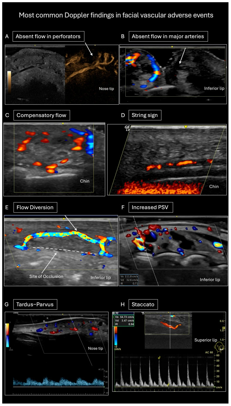
Other series add what B-mode shows at the block. In all 39 of Schelke’s 2023 cases, an anechoic or hypoechoic oval of hyaluronic acid sat beside or around the arterial trunk, the trunk itself was hypervascular, and the symptomatic skin above it was hypovascular and held no filler. Azizi’s 2025 cases showed the blocked artery as a noncompressible tube without flow, and the WFUMB position paper describes a serrated arterial wall with a hypoechoic lumen and an attenuated PW signal just beyond the block. Most filler obstructions are partial, the WFUMB panel notes, so a thin or slowed signal counts as much as an empty vessel. Velthuis’s 2021 Doppler guide adds the hemodynamics: the vessel before the block widens with turbulent red and blue flow, and collaterals dilate around it in a pattern the team calls a Medusa head.
Settings that catch slow flow
Facial arteries under the skin run slowly: the WFUMB panel puts hypodermal peak systolic velocities at 15 cm/s or less, against a usual detection floor of 2 cm/s. Set the Color scale low, near the 3 cm/s Sigrist’s centers used, lower the pulse repetition frequency and the wall filter until the pulsatile signal stands out, switch to Power Doppler for the faintest flow, and use the PW gate to confirm an arterial waveform and read PSV and RI on both sides. Use abundant gel and keep the probe light: pressure collapses these vessels, and Velthuis’s team scans the nose and the supraorbital area with no pressure at all. Veins compress under the probe; a blocked artery stays a noncompressible tube.
Vascular occlusion treatment protocol: the first hour with a probe in the room
Vascular occlusion treatment starts on the clinical signs, without waiting for imaging: stop injecting, start hyaluronidase, then scan to locate the block, target it under guidance and rescan until flow returns. In Sigrist’s centers hyaluronidase went in promptly once the signs were recognized, and the Doppler scan followed, often on the same day as treatment. The same team writes that occlusions of the perforators and the microvasculature are hard to treat on clinical signs alone, and that patients benefit from Doppler’s localization of the abnormal flow. Keep the probe charged and paired before the first syringe, so the scan starts within minutes. The ACE protocol adds aspirin 300 mg at once, then 75 mg daily.
- Stop injecting and look at the whole face. Check capillary refill on both sides and along the artery’s course, and video the area in good light before treatment, as CMAC directs.
- Start hyaluronidase on the clinical signs. Treatment goes ahead of the scan: CMAC infiltrates 1,500 units along the artery’s course and across the whole ischemic area, uses more than one vial when the area is large, and treats to effect (Murray 2021).
- Scan as you treat. Scan the injected area first, then the full face: the Delphi panel makes the injected-area scan mandatory and recommends the full-face scan. Compare each artery with the other side in Color, Power and PW.
- Locate the block. Follow the arterial track from the skin sign back to the trunk. Livedo maps onto the perforators of the superficial fat compartments (Schelke 2024, 125 patients), and the filler sits as an anechoic oval at the base of the trunk (Schelke 2023).
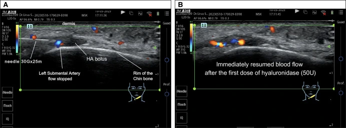
- Target the block under guidance. Bring the needle in plane, under a sterile probe cover with sterile gel, into the perivascular space or into the deposit at the base of the trunk. Schelke’s first doses averaged 95 IU; Urso’s team started at 30 IU and stepped up by 35 to 50 IU to a maximum of 150 IU.
- Rescan at once. Flow returned in under 60 seconds in Schelke’s 39 patients. Where it stays silent, inject again: in Azizi’s nasal case a second 150 U at 15 minutes brought rebound flow in the dorsal nasal artery, with a resistance index comparable to the other side.
- Record it. Save clips and the PSV and RI on both sides before and after treatment, and name the equipment in the report: the Delphi panel makes recording mandatory and WFUMB asks for the equipment in every report.
- Follow up in person. Rescan within 24 hours and again at about a week; Urso’s team rescanned at day 1 and day 20. CMAC reviews every resolved occlusion the next day, and the ACE Group gives each patient an out-of-hours number and assesses any suspected event in person.

The first hour on one page
The protocol above as a one-page sheet for the treatment room: the signs in order, the eight treatment steps, vision-loss transfer, Doppler settings for slow arteries and the emergency kit checklist. Treatment-room protocol from CMAC 2021, the ACE Group 2020, the 2025 Delphi consensus, WFUMB 2025 and the 2026 UK vision-loss consensus.
Hyaluronidase dose for vascular occlusion: guided and unguided protocols
Hyaluronidase for vascular occlusion follows one of two dosing schools. Unguided protocols flood the ischemic territory and repeat hourly until the skin recovers: CMAC infiltrates a 1,500-unit vial per round, more than one for a large area, and the high-dose pulsed protocol gives 500 IU for a small area and 1,500 IU for a larger one, as Schelke 2023 describes it. Guided protocols place a fraction of that into the deposit or beside the blocked trunk and stop when Doppler shows flow: Schelke’s first doses averaged 95 IU, and Urso’s ran from 30 IU up to 150 IU. Hyaluronidase crosses the arterial wall: in DeLorenzi’s 2014 cadaver work it broke down hyaluronic acid sealed inside intact facial-artery segments from the outside, at 4 and 24 hours, while saline controls stayed unchanged. CMAC and the ACE Group both treat around the artery rather than inside it, and Boey’s 2025 meta-analysis found no significant difference in resolution between intra-arterial and into-deposit placement (p = 0.36).
| Protocol | Dose | Repeat | Endpoint |
|---|---|---|---|
| High-dose pulsed (DeLorenzi 2017) | Scaled to the ischemic area: 500 IU for a small area and 1,500 IU for a larger one, as Schelke 2023 describes it | За годину | Capillary refill, skin color and pain resolved |
| CMAC (Murray 2021) | 1,500 units in 1 mL, infiltrated along the artery and across the ischemic area; more than one vial for a large area, treated to effect | At least hourly while capillary refill stays over 3 seconds | Capillary refill back to 3 seconds or less |
| Consensus (Cohen 2015) | At least 200 U at once | Again if no improvement within 60 minutes | Clinical improvement; best outcomes when treated within 24 hours |
| Ultrasound-guided, targeted (Schelke 2023) | Mean 95 IU (25–400) into the perivascular space at the trunk; total 122.5 IU | Second dose at 24 hours in 6 of 39, mean 51 IU | Reperfusion on Doppler in under 60 seconds; 39 of 39 healed without scars |
| Ultrasound-guided, minimal (Urso 2024) | 30 IU into the deposit, then 35–50 IU steps to a maximum of 150 IU | Top-up the next day in 2 of 5 | Flow on ultrasound; the 4 patients seen on days 1 to 3 healed on 75 to 150 IU each |
| Handheld, guided (Desyatnikova 2022) | 0.5 mL aliquots of 75 U; 50–300 U per event, median 100 | One session for 13 of 19 events, two for 6 | Complete recovery in all 18 patients |
| Review of 83 guided cases (Azizi 2025) | 30–150 IU in 67 of the 83; the authors' center starts near 350 U | Again on the rescan: 150 U more at 15 minutes in their nasal case | Flow on Doppler |
Guided treatment holds up across studies. Boey’s 2025 meta-analysis of four studies and 55 patients put complete resolution after ultrasound-guided hyaluronidase at 94.6% (95% CI 80.6–98.7), against about 77.8% for hyaluronidase in general. Two or four split doses beat one large dose in Lee’s 2020 rabbit model: four 125 IU injections 15 minutes apart kept 88.26% of the flap alive and two of 250 IU kept 81.49%, against 74.83% for a single 500 IU. Murray’s 2026 diffusion analysis explains the gap in units: cross-linked filler slows the enzyme’s spread, so placing it inside the deposit at several points does the work that volume does in a flood. CMAC’s own guideline puts guided doses at 35 to 60 units on average, up to 150. Hyaluronidase dissolves the filler, and the probe makes each unit count: it finds the deposit beside the trunk, guides the needle into it in plane and shows flow return on the rescan. Planned dissolving outside an emergency follows its own routine, set out in the guide to розчинення філера під контролем УЗД.
Radiesse (CaHA), Sculptra (PLLA) and other non-HA fillers
Occlusions also follow calcium hydroxylapatite (Radiesse) and poly-L-lactic acid (Sculptra) fillers, and hyaluronidase still has a role after them. The 2020 CaHA consensus (van Loghem) gives 600 U of hyaluronidase for every 0.1 mL of CaHA injected, checks reperfusion every 30 to 60 minutes, and repeats every 2 hours for 3 to 4 cycles when flow does not return within the hour. Kadouch’s team resolved two CaHA events and one PLLA event with hyaluronidase injected into the lesion under ultrasound, without necrosis or scarring. On ultrasound, CaHA shows as hyperechoic deposits with posterior shadowing, and PMMA as bright dots with comet-tail artifacts (Tao 2022). Sodium thiosulfate dissolved CaHA injected into porcine skin samples (Robinson 2018) but left intra-arterial CaHA intact in human cadaver arteries at 24 hours (Yankova 2021), and a 2024 review calls it too slow for an emergency (Wollina 2024).
Vision loss and blindness after filler: the first minutes
Vision loss after filler most often begins immediately or within 10 minutes of the injection (Sansome 2026), and 62.7% of the cases in Doyon’s 2024 literature review presented with no perception of light. An ophthalmic artery occlusion after filler takes vision with ptosis and ophthalmoplegia; a central retinal artery occlusion takes vision alone (Goodman 2020). CMAC cites work showing retinal infarction within 12 to 15 minutes of a complete block. The nose, forehead and glabella account for 40.6%, 27.7% and 19.0% of the 365 cases published from 2018 to 2023, and hyaluronic acid for 79.6% (Doyon 2024).
- Stop and transfer. Send the patient to the nearest emergency department without delay. The 2026 UK consensus asks every injector to keep the department’s details printed, to go with the patient when possible, and to consider phoning ahead on the way.
- For hyaluronic acid filler, give hyaluronidase without delaying transfer. The UK consensus allows it where the filler went in or near the supraorbital notch or the supratrochlear vessels, from a clinician competent in the technique and only when it does not significantly delay transfer; it gives retrobulbar and peribulbar injection no place. Goodman’s consensus injects 1,500 units in 2 mL of 1% lidocaine at the filler site and over any skin showing ischemia. Walker’s consensus sends four 1,500 IU vials with the patient.
- Use the minutes before the ambulance. While transfer is arranged, and as long as they add no delay: ocular massage in 5-second presses with 10 seconds off for up to 5 minutes, the patient supine at 15 to 45°; rebreathing into a paper bag for 10 minutes in every 30; one drop of timolol 0.5% in the affected eye; and 300 mg of aspirin unless contraindicated (Sansome 2026).
- Write down the times. Walker’s record form logs every intervention, the time of onset, the filler type and amount, the site, needle or cannula, the signs, the treatment after vision loss, the time the ambulance arrived and the first hospital interventions.
The hospital confirms the block. In Huang’s 2019 series of 10 patients who lost vision after cosmetic facial injections (7 autologous fat, 3 hyaluronic acid), color Doppler flow imaging of the orbit showed absent or sharply reduced retrobulbar flow in all six ophthalmic artery occlusions, and absent or reduced central retinal artery flow in the three central retinal artery occlusions. Partial vision at onset predicted improvement (P < .001): of 318 cases with recorded outcomes in Doyon's review, 6.0% recovered fully and 25.8% partly.
Vascular occlusion after lip filler
The early signs of vascular occlusion in the lips are the skin signs read against the other side of the lip: blanching at once, a violet net-like pattern along the path of the blocked labial artery, and capillary refill over 3 seconds (King 2020). A bruise beside them does not blanch. Lips sit in CMAC’s moderate risk grade, yet they carry a large share of the events: 31 of 162 hyaluronic acid occlusions by needle in Alam’s 2021 survey (19.1%), second only to the nasolabial folds, and the labial arteries in 17 of 44 Dutch referrals. They also recover well. Of the 34 lip occlusions in Alam’s survey, 33 left no sequelae, and all 17 Dutch labial cases recovered fully after Doppler-guided hyaluronidase.
The labial arteries run deeper than the lip line suggests. In Cotofana’s 2020 ultrasound study of 41 volunteers, the superior labial artery lay 5.6 mm deep and the inferior labial artery 5.2 mm, in the submucosal plane in 58.5% of measurements, intramuscular in 36.2% and subcutaneous in 5.3%. A 2026 meta-analysis puts the pooled averages at 4.59 to 4.90 mm (Trzeciak 2026). Both sit inside the 10 mm preset of the L20Pro and L24Pro. Prevention comes before the needle: map the labial arteries before you inject, following each one in Color or Power Doppler from the commissure to the midline.
A lip occlusion from compression can surface the next day. Figueiredo’s 2024 patient arrived 24 hours after 1 mL of hyaluronic acid with lidocaine in both lips, with pain and ecchymosis over the right upper lip, the nose and the oral vestibule. Color Doppler on a 7 to 16 MHz linear probe showed reduced flow in the superior labial artery against the other side, and five hyaluronidase applications totaling 5 mL over 3 hours restored it before discharge. The authors place bruises within a few hours of the injection and compression signs on the following day. Kroumpouzos’s 2023 review of lip and perioral complications puts ultrasound to the next question: it separates an embolus inside the artery from a filler deposit pressing on it. The lip also appears among vision-loss sites: 1 of 226 in the American Academy of Ophthalmology’s 2025 report, and one orbital infarction after hyaluronic acid lip filler in Diala’s 2026 case.
Vascular occlusion risk by facial site
Risk follows the arteries. CMAC grades the glabella, nose and forehead very high; the temples, nasolabial folds, tear troughs, periorbital area and medial cheek high; the lips, perioral area and anterior cheek moderate; and the jawline, marionette area, lateral cheek, submalar area, preauricular area and chin low (Murray 2021, adapted from the Goodman consensus). Each row pairs a site with the arteries measured there and the events counted in published series. Mapping these arteries before the first injection is the subject of the guide to ultrasound-guided filler injections.
| Зона | CMAC grade | Arteries and measured depth | Events in published series |
|---|---|---|---|
| Glabella | Very high | Supratrochlear artery 3.34 ± 0.6 mm deep at rest, supraorbital artery 3.54 ± 0.8 mm (Cotofana 2020, 41 volunteers) | 18 of 162 needle occlusions with hyaluronic acid (11.1%) and the highest mean severity of any common site; 7 of 19 glabellar occlusions left a scar (Alam 2021) |
| Nose | Very high | Dorsal nasal arteries 2.52 and 2.66 mm under the skin, left and right (Ortiz Middleton 2025, 50 women); the tip and ala depend on an end artery (King 2020) | 18 of 162 (11.1%) (Alam 2021); in nonsurgical rhinoplasty, occlusion in 0.35%, necrosis in 0.08% and vision loss in 0.09% of 8,604 patients (DeVictor 2021) |
| Лоб | Very high | Supratrochlear, supraorbital and central forehead branches; the frontal branch of the superficial temporal artery 2.7 mm under the skin at the lateral canthal line (Phumyoo 2020) | 8 of the 10 vascular obstructions in 290,307 injection sites (Tamura 2025); 27.7% of vision-loss cases (Doyon 2024) |
| Носогубна складка | High | Facial artery 6.27 mm deep at the mandibular border and 8.04 mm beside the nasal ala, pooled from ten studies (Pourani 2025); angular artery 3.6 ± 1.7 mm under the skin at the pyriform space (Franceschelli 2024, 332 hemifaces) | 39 of 162 (24.1%), the most occluded site (Alam 2021); all 13 patients with an early complication after nasolabial fold injection had vascular compromise (Nishikawa 2023) |
| Tear trough and medial cheek | High | Infraorbital artery and its branches (Barber-García 2026) | Cheek 21 of 162 (13.0%) (Alam 2021); necrosis in infraorbital territory after 48 symptom-free hours (Chauhan 2019); infraorbital compression with gum ischemia, resolved with 300 IU, the last 100 IU guided by ultrasound (Barber-García 2026) |
| Temple | High | Superficial temporal artery and its frontal branch | 21 of 226 vision-loss injection sites (9%) (Foster 2025); superficial temporal artery in 3 of 44 Dutch referrals (Schelke 2020); 1 of 162 needle occlusions (Alam 2021) |
| Lips | Moderate | Superior labial artery 5.6 mm and inferior labial artery 5.2 mm deep, submucosal in 58.5% of measurements (Cotofana 2020, 41 volunteers) | 31 of 162 (19.1%), with 33 of 34 lip occlusions free of sequelae (Alam 2021); labial arteries in 17 of 44 Dutch referrals, all recovered (Schelke 2020) |
| Chin, jawline and marionette area | Низький | Submental and mental arteries | Chin 3, jawline 3 and marionette area 4 of 162 (Alam 2021); submental artery in 7 of 44 Dutch referrals, 2 of them after calcium hydroxylapatite (Schelke 2020) |
The nose leads the most recent vision-loss review, with 40.6% of 365 cases (Doyon 2024). Nose filler necrosis occurred in 0.08% of 8,604 nonsurgical rhinoplasty patients, and vision loss in 0.09% (DeVictor 2021). Its tip and ala depend on an end artery with little collateral flow (King 2020), and Park’s 2011 referral series names the glabella and the nasal ala as the two danger zones for necrosis. A prior rhinoplasty adds to the risk: in Rivkin’s 2,488 nonsurgical rhinoplasty procedures it raised the odds of an adverse event (odds ratio 1.51), and all seven patients in Robati’s 2018 necrosis series had had a cosmetic rhinoplasty more than three years earlier. The dorsal nasal arteries run 2.5 to 2.7 mm under the skin, inside the 10 mm preset of the L20Pro and L24Pro, and Velthuis’s team scans the nose with no probe pressure at all.
Delayed vascular occlusion: hours to days after filler
Delayed vascular occlusion after filler surfaces hours to days after the injection, once the patient has gone home: published cases began at 6, 15 and 36 hours (Bravo 2015), after 48 symptom-free hours (Chauhan 2019) and at 2 days (Cassiano 2020). It is the smaller group: 38 of the 39 events in Schelke’s 2023 series began immediately, and CMAC places most ischemic changes instantly or within a few hours. For the later ones CMAC lists three mechanisms: an embolus lodging in a narrower arteriole downstream, hyaluronic acid drawing in water until the bolus blocks or compresses the vessel, and platelets gathering on a partial bolus. Hirsch’s 2007 case shows the second: slow swelling after a Juvéderm injection compressed the vessel, and the impending necrosis that surfaced late was managed with hyaluronidase. The ACE Group adds an embolus in skin with poor collateral supply, where the signs surface over the following hours, and filler that sits at a bifurcation and dislodges later into a terminal branch.
| Case report | Site and artery | Onset | Treatment and outcome |
|---|---|---|---|
| Bravo 2015, three patients | Left zygomatic area (zygomaticofacial artery); right nasolabial fold twice (angular artery) | 36, 6 and 15 hours; no signs during the injection or on the day | Hyaluronidase in all three; complete healing in the first, five days after treatment |
| Chauhan 2019 | Cheek, infraorbital-artery territory | 48 symptom-free hours, then pulsating pain | Three hourly pulses of 500 units; healed by day 15 |
| Cassiano 2020 | Лоб | 2 days: pain, erythema and edema | Treated within 24 hours of onset; healed with a discreet scar |
| Figueiredo 2024 | Upper and lower lips (superior labial artery, compression) | Seen at 24 hours | 5 mL of hyaluronidase in five applications over 3 hours; flow confirmed on Doppler before discharge |
| Azizi 2025 | Nose (dorsal nasal artery) | Seen 2 days after the injection, with pustules | 150 U under ultrasound, 150 U more at 15 minutes; rebound flow in the dorsal nasal artery |
Late events reach the eye too: in Taylor’s 2020 anatomical study, blindness was delayed by up to 2 weeks in some cases, and Walker’s consensus sorts vision loss into immediate (minutes), delayed (1 to 24 hours) and late (days to weeks) onset. Kroumpouzos’s 2023 review times perioral complications as immediate within 24 hours, early from 24 hours to 4 weeks, and late after 4 weeks. The ACE Group gives every filler patient an out-of-hours number and sees any suspected event in person, never by photo or message. Guided treatment still helps late: in the Dutch referral series, one infraorbital patient received Doppler-guided hyaluronidase 8 months after the injection and recovered fully.
Skin necrosis after filler: the treatment window by day
Skin necrosis after filler announces itself around day 3, when pustules and blisters rise over the ischemic area (Murray 2021; Farmer 2021). Hong’s team found that after 3 days a crust forms over a red base with a fibrous rim, and advised starting treatment no later than day 3 to head off necrosis. Earlier is better at every step. In a rabbit-ear model, 750 IU of hyaluronidase given 4 hours after the filler shrank the necrotic area, while the same dose at 24 hours made no difference (Kim 2011). DeLorenzi reports no partial or complete skin loss under the high-dose pulsed protocol when it started within 2 days of onset, and Alhusain’s 2026 meta-analysis of 21 studies and 231 patients found that treatment within 6 hours improved outcomes, with skin necrosis resolving in 78.4% overall.
Once the skin breaks down, wound care leads. CMAC makes it the priority after 48 hours, or once blisters form at about 72 hours (Murray 2021). Granulation lasts 2 to 4 weeks with the size and depth of the wound bed, and remodeling begins as soon as 21 days after the injury (Farmer 2021). Rescans across that period show whether flow has returned to the territory: Urso’s team rescanned at day 1 and day 20, and Azizi’s at 1 hour, 24 hours and 1 week.
How common vascular occlusion is after filler
Vascular occlusion occurs after 1 in 6,558 filler treatments by the Dutch national referral center’s count (Schelke 2020, range 1 in 5,300 to 1 in 8,000), and after 1 in 6,410 needle syringes against 1 in 40,882 cannula syringes in Alam’s 2021 survey of 370 US dermatologists. Each study counts a different denominator: treatments, syringes, injection sites or patients. The Dutch center counted every occlusion referred over 25 months against a national estimate of 138,496 filler treatments a year, and Alam’s team asked its dermatologists about 1.7 million syringes injected over the preceding ten years.
| Дослідження | Setting | Events | Rate |
|---|---|---|---|
| Schelke 2020 | Netherlands national referral center, January 2018 to January 2020 | 44 referred occlusions in 25 months, against 138,496 treatments a year | 1 in 6,558 treatments (range 1 in 5,300 to 1 in 8,000); all recovered after Doppler-guided hyaluronidase |
| Alam 2021 | 370 US board-certified dermatologists, preceding 10 years, 1.7 million syringes | 176 occlusions with needles, 13 with cannulas | 1 per 6,410 needle syringes and 1 per 40,882 cannula syringes; 85% left no long-term sequelae |
| Tamura 2025 | Clinic database in Japan, October 2020 to April 2024, 209,083 patients | 10 vascular obstructions in 290,307 hyaluronic acid injection sites, 8 of them on the forehead | 1 per 29,031 injection sites |
| Nishikawa 2023 | Nationwide multicenter study in Japan | 18 cases of vascular compromise in 41,775 cases (58,533 injection sites) | 0.043% of cases |
| DeVictor 2021 | Meta-analysis of nonsurgical rhinoplasty | 30 occlusions, 7 necroses and 8 vision losses in 8,604 patients | 0.35%, 0.08% and 0.09% |
| Miranda Cerna 2026 | Systematic review of lip augmentation techniques, 16 studies | No occlusions | 0 of 3,692 patients |
Occlusions concentrate where the arteries are: in Alam’s survey the nasolabial folds and lips led the count, and the glabella carried the highest mean severity. Needles produced occlusions about six times as often per syringe as cannulas, and 106 of the 370 dermatologists (28.6%) reported at least one occlusion. The guide to choosing a cannula or a needle by area covers the gauge for each site and how the tip shows on the scan. CMAC asks every injector to recognize the clinical presentation and to have clear protocols and referral pathways in place before the first event.
The vascular occlusion emergency kit
A vascular occlusion emergency kit starts with hyaluronidase: the 10-Point Plan (Heydenrych 2021) keeps at least five 1,500 IU vials of hyaluronidase in the clinic, the Goodman consensus asks for at least 7,500 units on site, and CMAC reconstitutes at 1,500 units per mL. Store the enzyme below 25 °C and reconstitute it with a local anesthetic without adrenaline (Murray 2021, hyaluronidase guideline).
| Пункт | What to stock | Guideline |
|---|---|---|
| Hyaluronidase | At least five 1,500 IU vials, 7,500 units in total, reconstituted at 1,500 units per mL; four vials travel with a patient who loses vision | Heydenrych 2021; Goodman 2020; Murray 2021; Walker 2021 |
| Пам’ять | Below 25 °C, with expiry dates tracked by reminder | Murray 2021 (hyaluronidase guideline); Heydenrych 2021 |
| Aspirin | 300 mg, chewable | Sansome 2026; Heydenrych 2021 |
| Eye drops | Timolol 0.5%; apraclonidine; bimatoprost | Sansome 2026; Heydenrych 2021 |
| Paper bag | For rebreathing, 10 minutes in every 30 | Sansome 2026 |
| Vision check | A Snellen chart app and a light source | Heydenrych 2021 |
| Anaphylaxis and resuscitation | Adrenaline, an airway, breathing and circulation kit, antihistamines, IV steroids and vasopressors | Murray 2021; King 2020; Heydenrych 2021 |
| Paperwork | Flowcharts, the local emergency department's details printed, a vision-loss record form | Murray 2021; Sansome 2026; Walker 2021 |
| Ультразвук | A linear probe of at least 15 MHz with Color, Power and PW Doppler, charged and paired, with sterile covers and sterile gel | Velthuis 2025 (Delphi); Chammas 2025 (WFUMB) |
Suresult’s sterile hydrogel gel pads, Ø 35 × 4 mm, put a 4 mm stand-off between the probe face and the skin, so the probe reads the superficial arteries without pressing them shut: EFSUMB’s dermatologic ultrasound statement calls for gel pads to avoid compressing the superficial vascular plexus (Alfageme 2021). The pad stays on through the scan and the guided injection, and the needle passes through it. Each patient gets a fresh sterile pad. The pads are latex-free and tested for biocompatibility to ISO 10993, fit other brands’ linear probes as well as Suresult’s and have a 5-year shelf life, long enough to wait in the kit between events. A pack of 50 costs $140, shipping and tax included.

Recording the event: clips, refill video and the report
A complete record serves the next scan as much as the file. CMAC asks for a video in good light with capillary refill checked along the artery’s track, the whole ischemic area marked, contemporaneous notes with images, and a review the next day even after resolution. The Delphi panel makes ultrasound recording mandatory in complication management, and WFUMB asks every report to name the equipment.
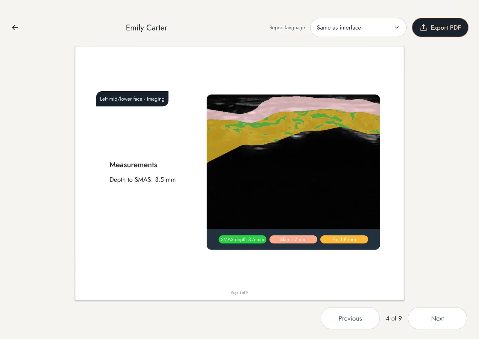
The Suresult app keeps cine loops of 100, 200, 500 or 1,000 frames and exports JPEG, PNG, MP4 and DICOM, so the before-and-after clips and the PSV and RI values on both sides go into the record and the referral letter. FaceAtlas AI, included for three months with each Pro probe, files every visit in the patient’s record with one-click comparison to the last visit, 5-second clips and a Facial Ultrasound Assessment PDF shared by email, Drive or print.
Suresult probes for a vascular event scan
The three Pro probes share a 256-element linear array 25 mm wide, a 4,400 mAh battery rated for 4 hours of scanning, a 145 g housing and the full Doppler set: Color, Power and PW with steer, gain, PRF, wall filter, gate volume and angle on screen, PS, ED, PI and RI on the trace, and automatic vessel flow measurement. Each includes three months of FaceAtlas AI. Their frequency bands and depth presets decide which arteries fill the screen; the L20 vs L24 aesthetic ultrasound guide compares them site by site.
Suresult L20Pro
16/20 MHz, 10–40 mm. The whole face in one probe: the dorsal nasal arteries at 2.5 mm and the angular artery at 3.6 mm in the 10 mm preset, the facial artery 8 mm deep beside the ala, and the deep temporal layers in the 30 and 40 mm presets. Needle Enhance makes the needle stand out on its way to the perivascular space. My pick for an injector who keeps one probe ready for every event.
256 elements · 4 hours of scanning · 145 g · Color, Power and PW Doppler
$3,720
Suresult L24Pro
18/24 MHz, 10–25 mm. The highest frequency in the family, for the shallowest arteries: the dorsal nasal arteries at 2.5 mm, the supratrochlear artery at 3.3 mm and the labial arteries at about 5 mm all sit in the 10 mm preset. Sigrist’s six centers scanned their 100 events at 18 to 24 MHz, the L24Pro’s band. My pick for a practice built on lips, noses and glabellar lines.
256 elements · 4 hours of scanning · 145 g · Color, Power and PW Doppler
$4,185
Suresult L14Pro
10/14 MHz, 20–80 mm. For clinics that treat the face and the body: facial vessels in the face, plus the MSK, nerve, vascular and carotid presets for the rest of the patient list. Its 14 MHz sits above the 12 MHz the Delphi footnote accepts in some cases, and the 72-hour nasal referral in the first-hour clip was scanned at 14 MHz, this probe’s band. My pick when one probe also covers body work.
256 elements · 4 hours of scanning · 145 g · Color, Power and PW Doppler
$3,441
All six Suresult probes in this guide pair over Wi-Fi with the free Suresult app for iOS, Android and Windows, which runs B-mode, M-mode, Color, Power and PW Doppler.
L20, L24 and L14: the 192-element siblings
The L20, L24 and L14 carry the Pro bands and depth presets with 192 elements, 2 hours of scanning per charge and the same Color, Power and PW Doppler in the app, priced at $3,000 for the L20, $3,441 for the L24 and $2,139 for the L14. FaceAtlas AI adds to any of them with a monthly license at $50, a yearly one at $500, or the lifetime license at $999, on sale until 11:59 PM Pacific Time, December 31, 2026.
Suresult L20
16/20 МГц, 10–40 мм, 192 елементи, Needle Enhance
$3,000
Suresult L24
18/24 МГц, 10–25 мм, 192 елементи, Needle Enhance
$3,441
Suresult L14
10/14 MHz, 20–80 mm, 192 elements
$2,139
З чого почати
For one probe that covers every facial artery, I recommend the L20Pro at $3,720. For a practice centered on lips, noses and glabellar lines, I choose the L24Pro at $4,185, and for a clinic that scans the body as well as the face, the L14Pro at $3,441. Each Pro ships within 24 hours with a 30-day trial and a two-year warranty that extends to three or five years, and the price at checkout already covers customs and duties: No Hidden Fees.
Vascular occlusion questions
What are the signs of vascular occlusion after filler?
The signs of vascular occlusion after filler are skin that stays white after the needle leaves, a violet net-like pattern (livedo reticularis) across one artery's territory within minutes, a capillary refill over 3 seconds against the other side, and a dusky blue-gray cast over tens of minutes to hours (King 2020; Murray 2021). Among Sigrist's 100 referrals, livedo was present in 46, blanching in 38, pain in 25, pustules in 20 and ecchymosis in 16. In Yao's 2026 series of 57 suspected occlusions, a change in skin color and a delayed capillary refill together predicted the occlusions ultrasound confirmed, with an area under the curve of 0.944.
How do I tell a bruise from a vascular occlusion after lip filler?
A bruise after lip filler does not blanch under pressure, because it is blood that has leaked into the skin; an occlusion turns pale, then shows a violet net-like pattern along the blocked artery, with capillary refill over 3 seconds against the other side (King 2020). Figueiredo's team places bruises a few hours after the injection and compression signs on the following day (Figueiredo 2024). When a hematoma hides the skin signs, Color Doppler settles it: reduced or absent labial flow against the other side means an occlusion.
What are the stages of vascular occlusion?
CMAC describes five skin stages: pallor at once; livedo reticularis within minutes, lasting 24 to 36 hours; pustules around day 3; coagulation over the following days; and slough or eschar. Between livedo and pustules the skin turns gray as the tissue starts to infarct, which the ACE Group times at tens of minutes to hours. Pallor and livedo still reverse with treatment (Murray 2021).
How soon after filler can a vascular occlusion appear?
Most appear at once: 38 of 39 events in Schelke's 2023 series began immediately, and CMAC places ischemic changes instantly or within a few hours. Published delayed cases began at 6, 15 and 36 hours (Bravo 2015), after 48 symptom-free hours (Chauhan 2019) and at 2 days (Cassiano 2020), and in Taylor's 2020 study blindness was delayed by up to 2 weeks in some cases. Give every patient an out-of-hours number and review any suspected event in person (King 2020).
How common is vascular occlusion after filler?
Vascular occlusion occurs after 1 in 6,558 filler treatments by the Dutch national referral center's count (Schelke 2020). In Alam's 2021 survey of 370 US dermatologists covering 1.7 million syringes, needles produced 1 occlusion per 6,410 syringes and cannulas 1 per 40,882, and 106 of the 370 reported at least one occlusion. Clinic databases in Japan counted 10 obstructions in 290,307 injection sites (Tamura 2025) and 18 cases of vascular compromise in 41,775 cases (Nishikawa 2023).
How common is vascular occlusion with lip filler?
Lips accounted for 31 of 162 needle occlusions with hyaluronic acid in Alam's 2021 survey of 370 US dermatologists (19.1%), second only to the nasolabial folds, and the labial arteries for 17 of 44 referrals at the Dutch national referral center, where the overall rate was 1 in 6,558 treatments (Schelke 2020). A 2026 systematic review of 16 lip augmentation studies recorded no occlusions in 3,692 patients (Miranda Cerna 2026). Lip occlusions recover well: 33 of 34 in Alam's survey left no sequelae, and all 17 Dutch labial cases recovered fully after Doppler-guided hyaluronidase.
Can a vascular occlusion from filler be reversed?
Yes. Doppler-guided hyaluronidase resolved all 44 occlusions referred to the Dutch national referral center (Schelke 2020), all 39 patients in Schelke's 2023 guided series healed without scars, and Boey's 2025 meta-analysis put complete resolution after ultrasound-guided hyaluronidase at 94.6%. Starting within 6 hours improved outcomes in Alhusain's 2026 meta-analysis of 231 patients, and CMAC treats pallor and livedo as stages that still reverse (Murray 2021). In Alam's survey 85% of occlusions left no long-term sequelae; the glabella carried the highest severity, with 7 of 19 glabellar occlusions leaving a scar (Alam 2021).
Is a vascular occlusion always painful?
No. Lidocaine in the filler masks pain, and CMAC notes that pain is not always present in the early stages. Pain away from the injection point is a warning sign of its own (King 2020). Among Sigrist's 100 referrals, pain was present in 25, against livedo in 46 and blanching in 38, so check the skin and the capillary refill whatever the patient feels.
Can filler cause blindness?
Yes. Hyaluronic acid filler accounted for 79.6% of the 365 vision-loss cases published from 2018 to 2023, and injections in the nose, forehead and glabella for 40.6%, 27.7% and 19.0% (Doyon 2024); in nonsurgical rhinoplasty, vision loss followed 0.09% of 8,604 patients (DeVictor 2021). Lip filler causes it in rare cases: the lip was 1 of 226 injection sites in the American Academy of Ophthalmology's 2025 report, 1 of the 34 lip occlusions in Alam's survey caused an ocular injury or blindness, and Diala's team described orbital infarction after hyaluronic acid lip filler in 2026. Partial vision at onset predicted improvement: of 318 cases with recorded outcomes, 6.0% recovered fully and 25.8% partly (Doyon 2024).
What should a vascular occlusion emergency kit contain?
Hyaluronidase first: at least five 1,500 IU vials, 7,500 units in total, stored below 25 °C. Add aspirin 300 mg, timolol 0.5% eye drops, a paper bag for rebreathing, a Snellen chart app and a light source, adrenaline and resuscitation equipment, printed emergency department details and flowcharts (Heydenrych 2021; Sansome 2026; Murray 2021). My addition is a linear probe of at least 15 MHz with Color and PW Doppler, charged and paired, with sterile covers and sterile gel.
What should I do first if a patient's vision changes after filler?
Stop injecting and transfer the patient to the nearest emergency department without delay, with its details printed in advance; go with the patient when possible and consider phoning ahead on the way (Sansome 2026). For hyaluronic acid filler, a clinician competent in the technique can inject hyaluronidase where the filler was placed or near the supraorbital notch or supratrochlear vessels, as long as it does not significantly delay transfer (Sansome 2026); Goodman's consensus uses 1,500 units in 2 mL of 1% lidocaine at the filler site and over any skin showing ischemia (Goodman 2020). The UK panel recommends ocular massage, rebreathing into a paper bag, one drop of timolol 0.5% and 300 mg of aspirin while transfer is arranged, as long as they add no significant delay.
How much hyaluronidase does a vascular occlusion need?
Unguided protocols give 200 to 1,500 units or more per round, and guided protocols a fraction of that. The Cohen consensus gives at least 200 U at once, the high-dose pulsed protocol gives 500 IU for a small area and 1,500 IU for a larger one, hourly, and CMAC infiltrates a 1,500-unit vial along the artery and across the territory, adds vials for a large area and treats to effect, repeating at least hourly while capillary refill stays over 3 seconds. Guided protocols place the enzyme beside the blocked trunk or into the deposit: Schelke's first doses averaged 95 IU, Urso's started at 30 IU with a 150 IU ceiling, and Desyatnikova's handheld series used 50 to 300 U per event. In Schelke's 39 patients, whom a mean 1,519 IU of flooding had not resolved, a guided mean of 122.5 IU restored flow in every case.
Джерела
- Murray G, Convery C, Walker L, et al. Guideline for the management of hyaluronic acid filler-induced vascular occlusion. J Clin Aesthet Dermatol. 2021;14(5):E61–E69. — October 27, 2026
- King M, Walker L, Convery C, et al. Management of a vascular occlusion associated with cosmetic injections. J Clin Aesthet Dermatol. 2020;13(1):E53–E58. — October 27, 2026
- Velthuis PJ, Alfageme F, Cartier H, et al. The use of ultrasound imaging in aesthetic injectables: a modified Delphi consensus. J Plast Reconstr Aesthet Surg. 2025;104:33–37. — October 27, 2026
- Chammas MC, Sigrist R, Alfageme F, et al. WFUMB position paper: consensus on best practice in aesthetic dermatologic ultrasound. Ultrasound Med Biol. 2025;51(11):2173–2193. — October 27, 2026
- Sansome S, Clague M, Convery C, et al. Consensus guidelines for the management of tissue filler-induced vision loss in the United Kingdom. Aesthet Surg J. 2026;46(5):563–568. — October 27, 2026
- Goodman GJ, Magnusson MR, Callan P, et al. A consensus on minimizing the risk of hyaluronic acid embolic visual loss and suggestions for immediate bedside management. Aesthet Surg J. 2020;40(9):1009–1021. — October 27, 2026
- Walker L, Convery C, Davies E, et al. Consensus opinion for the management of soft tissue filler induced vision loss. J Clin Aesthet Dermatol. 2021;14(12):E84–E94. — October 27, 2026
- Cohen JL, Biesman BS, Dayan SH, et al. Treatment of hyaluronic acid filler-induced impending necrosis with hyaluronidase: consensus recommendations. Aesthet Surg J. 2015;35(7):844–849. — October 27, 2026
- Murray G, Convery C, Walker L, et al. Guideline for the safe use of hyaluronidase in aesthetic medicine, including modified high-dose protocol. J Clin Aesthet Dermatol. 2021;14(8):E69–E75. — October 27, 2026
- Farmer A, Murray G, Croasdell B, et al. Facial vascular events and tissue ischemia: a guide to understanding and optimizing wound care. J Clin Aesthet Dermatol. 2021;14(12 Suppl 1):S39–S48. — October 27, 2026
- Heydenrych I, De Boulle K, Kapoor KM, et al. The 10-point plan 2021: updated concepts for improved procedural safety during facial filler treatments. Clin Cosmet Investig Dermatol. 2021;14:779–814. — October 27, 2026
- van Loghem J, Funt D, Pavicic T, et al. Managing intravascular complications following treatment with calcium hydroxylapatite: an expert consensus. J Cosmet Dermatol. 2020;19(11):2845–2858. — October 27, 2026
- Sigrist RMS, Gonzalez C, Schelke L, et al. Doppler ultrasound findings in filler-related facial vascular adverse events: an international multicenter study. Diagnostics (Basel). 2026;16(11):1587. — October 27, 2026
- Schelke LW, Velthuis PJ, Decates T, et al. Ultrasound-guided targeted vs regional flooding: a comparative study for improving the clinical outcome in soft tissue filler vascular adverse event management. Aesthet Surg J. 2023;43(1):86–96. — October 27, 2026
- Velthuis PJ, Jansen O, Schelke LW, et al. A guide to Doppler ultrasound analysis of the face in cosmetic medicine. Part 2: vascular mapping. Aesthet Surg J. 2021;41(11):NP1633–NP1644. — October 27, 2026
- Desyatnikova S, Schelke L. Treatment of filler-related vascular occlusion using handheld portable ultrasound device. J Cosmet Dermatol. 2022;21(7):3166–3168. — October 27, 2026
- Azizi N, Tootoonchi N, Khorasanizadeh F, et al. Ultrasound-guided hyaluronidase injections for the management of filler-induced arterial ischemia: a pictorial case series and systematic review of literature. Aesthet Surg J Open Forum. 2025;7:ojaf125. — October 27, 2026
- Urso SU, Molinari P, Fundarò S, Mosti G. Use of minimal amounts of hyaluronidase in the ultrasound-guided treatment of acute vascular occlusion by hyaluronic acid: a preliminary report. Aesthet Surg J Open Forum. 2024;6:ojae025. — October 27, 2026
- Schelke LW, Velthuis PJ, Mojallal A, et al. Reticulated livedoid skin patterns after soft-tissue filler-related vascular adverse events. J Am Acad Dermatol. 2024;91(1):37–42. — October 27, 2026
- Yao X, Tang W, Mao Q. Clinical features associated with HFUS-defined vascular occlusion after hyaluronic acid facial injections: a retrospective exploratory study. J Cosmet Laser Ther. 2026:1–7. — October 27, 2026
- Rasheed S, Itani I. Atypical hyaluronic acid-induced vascular occlusion masked by a hematoma: ultrasound-guided diagnosis and management. Cureus. 2026;18(5):e108472. — October 27, 2026
- Li X, Xie W. Pre-injection Doppler mapping versus landmark guidance for lower facial hyaluronic acid injection in a prospective safety study. J Vis Exp. 2026;(233). — October 27, 2026
- Boey JJE, Boey JJJ, Chen Z, et al. Superior outcomes with ultrasound-guided hyaluronidase for impending filler-induced facial skin necrosis: a systematic review and pilot meta-analysis. Aesthetic Plast Surg. 2025;49(13):3519–3525. — October 27, 2026
- Lee W, Oh W, Oh SM, et al. Comparative effectiveness of different interventions of perivascular hyaluronidase. Plast Reconstr Surg. 2020;145(4):957–964. — October 27, 2026
- Murray G, Khoshnaw S, Velthuis P. Dissolving hyaluronic acid filler: the diffusion barrier to hyaluronidase efficacy. J Cosmet Dermatol. 2026;25(4):e70818. — October 27, 2026
- DeLorenzi C. Transarterial degradation of hyaluronic acid filler by hyaluronidase. Dermatol Surg. 2014;40(8):832–841. — October 27, 2026
- DeLorenzi C. New high dose pulsed hyaluronidase protocol for hyaluronic acid filler vascular adverse events. Aesthet Surg J. 2017;37(7):814–825. — October 27, 2026
- Kadouch J, Schelke L, Groh O, et al. Intralesional hyaluronidase injection to relieve non-hyaluronic acid filler-induced vascular adverse events. Int J Dermatol. 2024;63(9):1252–1255. — October 27, 2026
- Tao Y, Wei C, Su Y, Hu B, Sun D. Emerging high-frequency ultrasound imaging in medical cosmetology. Front Physiol. 2022;13:885922. — October 27, 2026
- Robinson DM. In vitro analysis of the degradation of calcium hydroxylapatite dermal filler: a proof-of-concept study. Dermatol Surg. 2018;44(Suppl 1):S5–S9. — October 27, 2026
- Yankova M, Pavicic T, Frank K, et al. Intraarterial degradation of calcium hydroxylapatite using sodium thiosulfate: an in vitro and cadaveric study. Aesthet Surg J. 2021;41(5):NP226–NP236. — October 27, 2026
- Wollina U, Goldman A. Spontaneous and induced degradation of dermal fillers: a review. J Cutan Aesthet Surg. 2024;17(4):273–281. — October 27, 2026
- Huang P, Liu A, Ren H, Xue K. Color Doppler flow imaging of retrobulbar ocular blood flow changes in retinal artery occlusions caused by cosmetic facial filler injections. Ophthalmic Plast Reconstr Surg. 2019;35(3):227–231. — October 27, 2026
- Schelke L, Decates T, Kadouch J, et al. Incidence of vascular obstruction after filler injections. Aesthet Surg J. 2020;40(8):NP457–NP460. — October 27, 2026
- Alam M, Kakar R, Dover JS, et al. Rates of vascular occlusion associated with using needles vs cannulas for filler injection. JAMA Dermatol. 2021;157(2):174–180. — October 27, 2026
- Tamura T, Tamura T, Okumura K, et al. Serious complications of hyaluronic acid fillers: a retrospective study of 290,307 cases. Ann Plast Surg. 2025;94(6):630–633. — October 27, 2026
- Nishikawa A, Aikawa Y, Kono T. Current status of early complications caused by hyaluronic acid fillers: insights from a descriptive, observational study of 41,775 cases. Aesthet Surg J. 2023;43(8):893–904. — October 27, 2026
- DeVictor S, Ong AA, Sherris DA. Complications secondary to nonsurgical rhinoplasty: a systematic review and meta-analysis. Otolaryngol Head Neck Surg. 2021;165(5):611–616. — October 27, 2026
- Miranda Cerna VR. Hyaluronic acid injection techniques for lip augmentation: comparison of linear, retrograde, and microdeposit approaches. A systematic review. Aesthet Surg J Open Forum. 2026;8:ojag059. — October 27, 2026
- Rivkin A. Nonsurgical rhinoplasty using injectable fillers: a safety review of 2488 procedures. Facial Plast Surg Aesthet Med. 2021;23(1):6–11. — October 27, 2026
- Robati RM, Moeineddin F, Almasi-Nasrabadi M. The risk of skin necrosis following hyaluronic acid filler injection in patients with a history of cosmetic rhinoplasty. Aesthet Surg J. 2018;38(8):883–888. — October 27, 2026
- Park TH, Seo SW, Kim JK, Chang CH. Clinical experience with hyaluronic acid-filler complications. J Plast Reconstr Aesthet Surg. 2011;64(7):892–896. — October 27, 2026
- Cotofana S, Alfertshofer M, Schenck TL, et al. Anatomy of the superior and inferior labial arteries revised: an ultrasound investigation and implication for lip volumization. Aesthet Surg J. 2020;40(12):1327–1335. — October 27, 2026
- Trzeciak M, Lipski M, Gładysz T, et al. Anatomical depth of labial arteries in dermal filler procedures: a meta-analysis with clinical implications. Aesthet Surg J. 2026;46(5):478–485. — October 27, 2026
- Cotofana S, Alfertshofer M, Frank K, et al. Relationship between vertical glabellar lines and the supratrochlear and supraorbital arteries. Aesthet Surg J. 2020;40(12):1341–1348. — October 27, 2026
- Ortiz Middleton E, Aguilar A. Anatomical mapping of nasal arteries in Chilean patients using Doppler ultrasound: clinical application for injectables. Aesthet Surg J Open Forum. 2025;7:ojaf016. — October 27, 2026
- Phumyoo T, Jiirasutat N, Jitaree B, et al. Localization and topography of the arteries on the middle forehead region for eluding complications following forehead augmentation: conventional cadaveric dissection and ultrasonography investigation. J Craniofac Surg. 2020;31(7):2029–2035. — October 27, 2026
- Franceschelli A, Cazzulani M, Urso SU, et al. In vivo ultrasound study of the angular artery anatomy: practical indications for the treatment of the deep pyriform space. Aesthet Surg J Open Forum. 2024;6:ojae113. — October 27, 2026
- Pourani MR, Ebrahimzade M, Goudarzi E, et al. Evaluation of facial artery course variations, diameters, and depth using Doppler ultrasonography: a systematic review and meta-analysis. J Cosmet Dermatol. 2025;24(9):e70431. — October 27, 2026
- Barber-García C, Nevado-Sánchez E, Núñez-Rodríguez S, et al. Ultrasound-guided low-dose hyaluronidase for infraorbital artery occlusion with secondary gingival ischemia after hyaluronic acid filler injection: a case report. Diagnostics (Basel). 2026;16(13):1973. — October 27, 2026
- Foster J, Aakalu VK, Freitag SK, et al. Vision-threatening complications of soft tissue fillers: a report by the American Academy of Ophthalmology. Ophthalmology. 2025;132(8):935–944. — October 27, 2026
- Doyon VC, Liu C, Fitzgerald R, et al. Update on blindness from filler: review of prognostic factors, management approaches, and a century of published cases. Aesthet Surg J. 2024;44(10):1091–1104. — October 27, 2026
- Diala FGI, Biesman BS, Pargament JM, et al. Orbital infarction syndrome following hyaluronic acid lip filler injection. Ophthalmic Plast Reconstr Surg. 2026;42(4):e143–e146. — October 27, 2026
- Souza Felix Bravo B, Klotz De Almeida Balassiano L, Roos Mariano Da Rocha C, et al. Delayed-type necrosis after soft-tissue augmentation with hyaluronic acid. J Clin Aesthet Dermatol. 2015;8(12):42–47. — October 27, 2026
- Hirsch RJ, Lupo M, Cohen JL, Duffy D. Delayed presentation of impending necrosis following soft tissue augmentation with hyaluronic acid and successful management with hyaluronidase. J Drugs Dermatol. 2007;6(3):325–328. — October 27, 2026
- Chauhan A, Singh S. Management of delayed skin necrosis following hyaluronic acid filler injection using pulsed hyaluronidase. J Cutan Aesthet Surg. 2019;12(3):183–186. — October 27, 2026
- Cassiano D, Miyuki Iida T, Lúcia Recio A, et al. Delayed skin necrosis following hyaluronic acid filler injection: a case report. J Cosmet Dermatol. 2020;19(3):582–584. — October 27, 2026
- Figueiredo HP, Coimbra F, de Carvalho Rocha T, et al. Ultrasonography in the management of lip complications caused by hyaluronic acid. Imaging Sci Dent. 2024;54(3):296–302. — October 27, 2026
- Kroumpouzos G, Harris S, Bhargava S, Wortsman X. Complications of fillers in the lips and perioral area: prevention, assessment, and management focusing on ultrasound guidance. J Plast Reconstr Aesthet Surg. 2023;84:656–669. — October 27, 2026
- Taylor GI, Shoukath S, Gascoigne A, et al. The functional anatomy of the ophthalmic angiosome and its implications in blindness as a complication of cosmetic facial filler procedures. Plast Reconstr Surg. 2020;146(4):745. — October 27, 2026
- Hong JY, Seok J, Ahn GR, et al. Impending skin necrosis after dermal filler injection: a "golden time" for first-aid intervention. Dermatol Ther. 2017;30(2):e12440. — October 27, 2026
- Kim DW, Yoon ES, Ji YH, et al. Vascular complications of hyaluronic acid fillers and the role of hyaluronidase in management. J Plast Reconstr Aesthet Surg. 2011;64(12):1590–1595. — October 27, 2026
- Alhusain AM, Alsaif M, Alkhathami AM, et al. Hyaluronidase protocol in management of hyaluronic acid filler-related vascular complications: a systematic review and meta-analysis. Aesthetic Plast Surg. 2026;50(6):2300–2317. — October 27, 2026
- Alfageme F, Wortsman X, Catalano O, et al. European Federation of Societies for Ultrasound in Medicine and Biology (EFSUMB) position statement on dermatologic ultrasound. Ultraschall Med. 2021;42(1):39–47. — October 27, 2026
- Clarius L20 HD3. — October 27, 2026
- Butterfly Network pricing. — October 27, 2026
- Butterfly iQ3 specifications. — October 27, 2026
- GE HealthCare Vscan Air SL. — October 27, 2026
- Vscan Air SL specifications. — October 27, 2026
- Philips Lumify L12-4 linear array transducer. — October 27, 2026
- PartsSource, Philips L12-4 transducer. — October 27, 2026
- Suresult L20Pro. — October 27, 2026
- Suresult L24Pro. — October 27, 2026
- Suresult L14Pro. — October 27, 2026
- Suresult L20. — October 27, 2026
- Suresult L24. — October 27, 2026
- Suresult L14. — October 27, 2026
- FaceAtlas AI monthly license. — October 27, 2026
- FaceAtlas AI yearly license. — October 27, 2026
- FaceAtlas AI lifetime license. — October 27, 2026
- Suresult aesthetic ultrasound gel pads. — October 27, 2026

