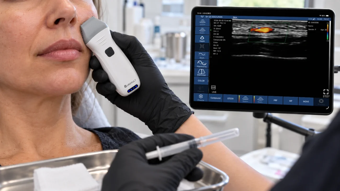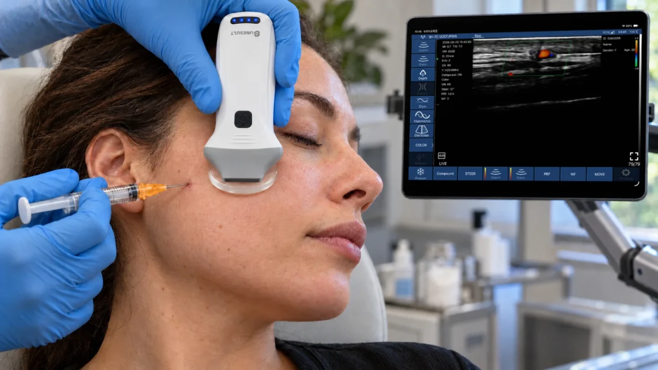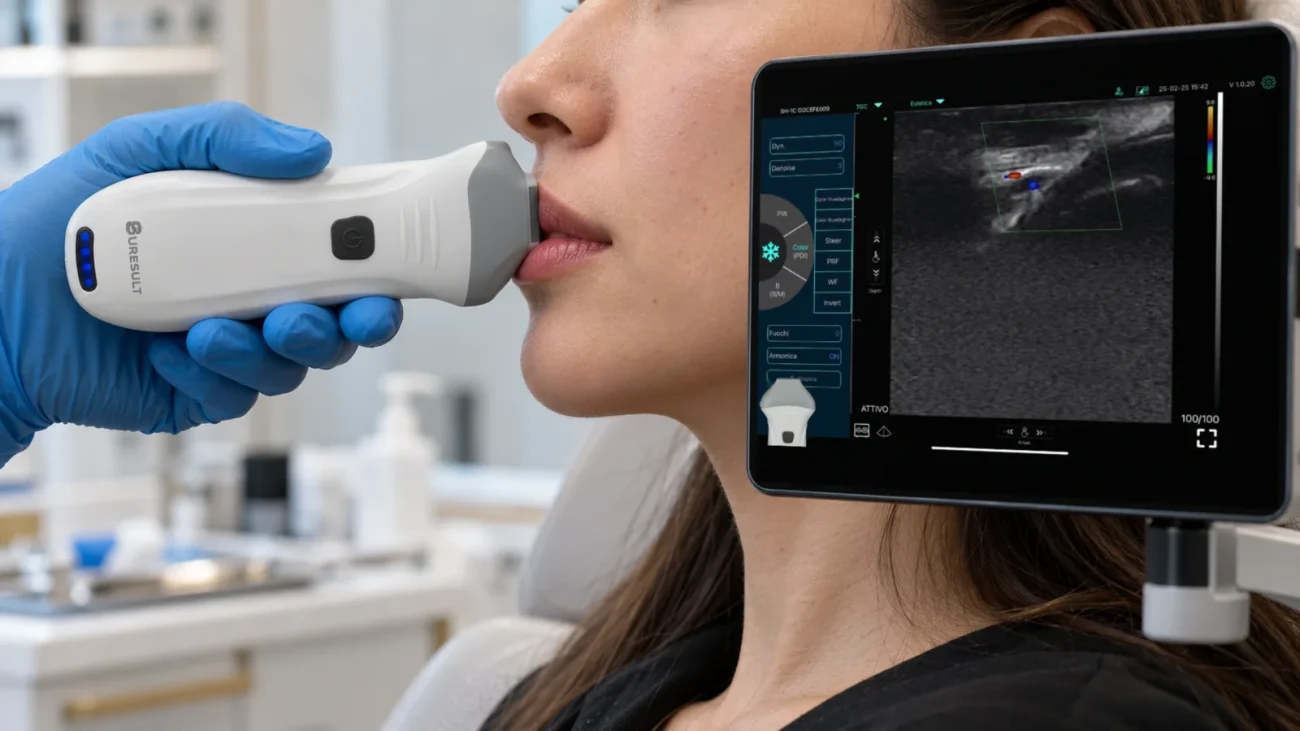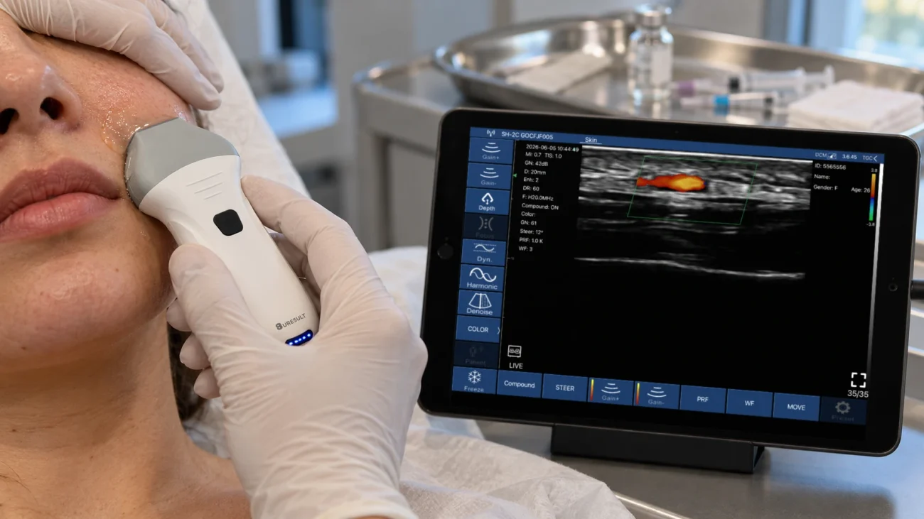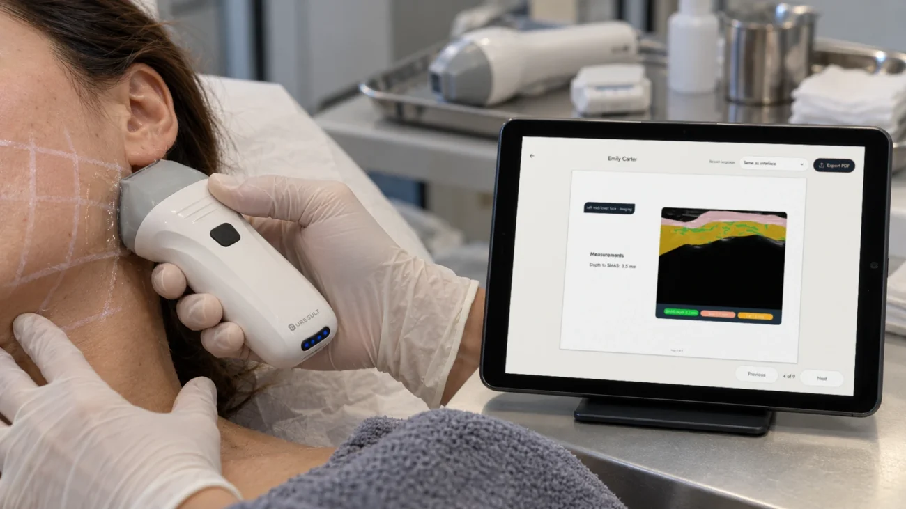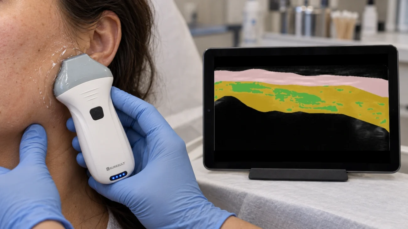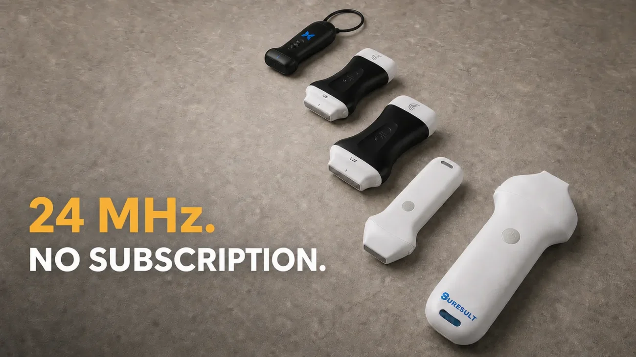
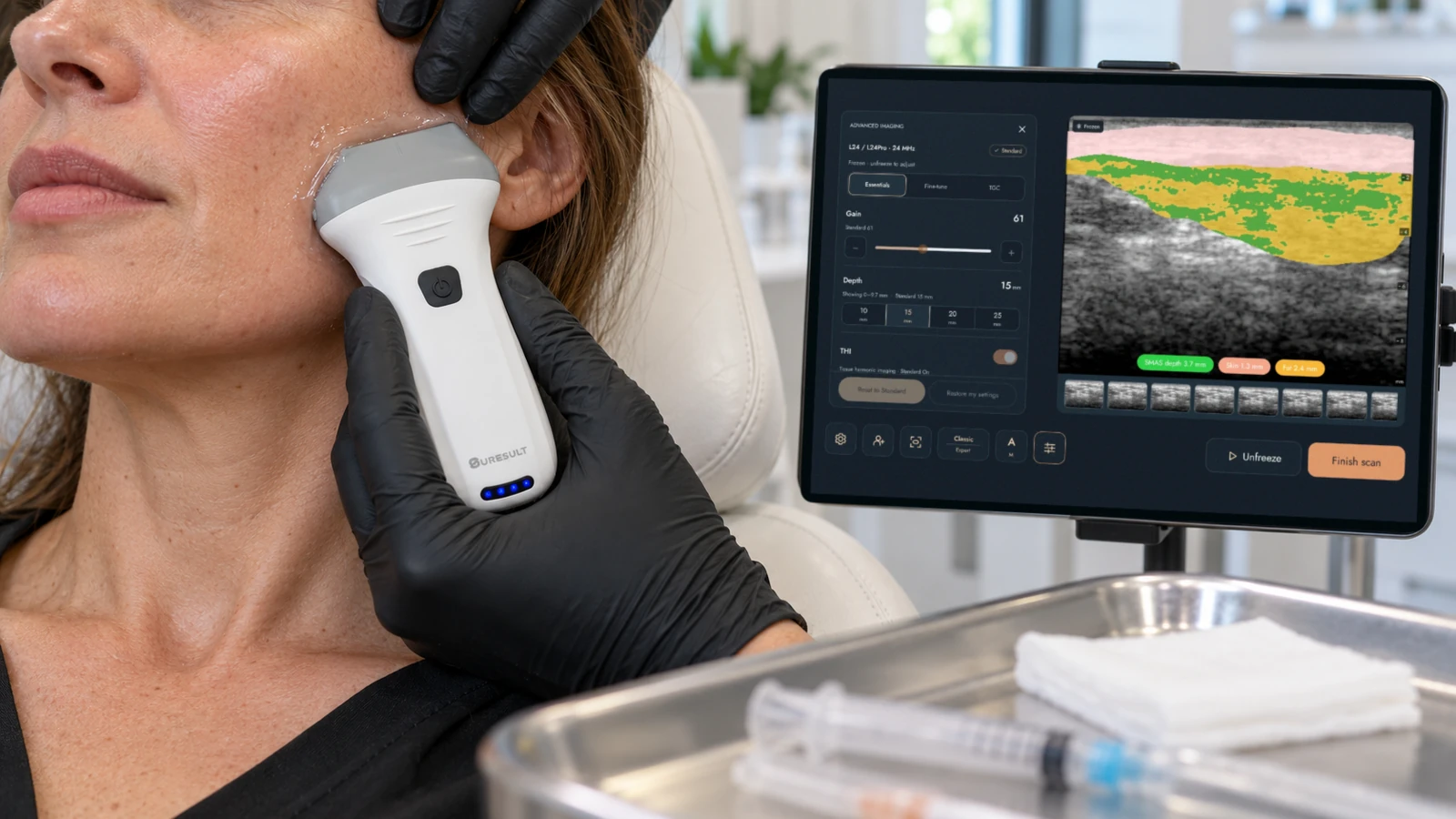
For biostimulator work I pick the Suresult L24Pro at $4,005 for a fixed visit order: map the vessels, confirm the plane as the cannula fans, and measure the dermis at the same marked point before the first session and at every follow-up. Its 18/24 MHz band resolves facial dermis measured at 0.58 to 1.30 mm in healthy adults, and its Color, Power and PW Doppler come in the price for the vessel map before the first pass. Sculptra, Radiesse, HArmonyCa and Ellansé build collagen over weeks, and the collagen response to PLLA cannot be immediately reversed the way HA filler is dissolved (Ouyang 2025), so the map and the plane come first, and the dermis shows the result at follow-up: +14.9% after two sessions of 1:4 Radiesse in the neck (Trindade de Almeida 2023), and from 1.80 to 2.70 mm after Sculptra in the upper arm (Bravo 2026). Below are the Sculptra injection technique, a diluted Radiesse chart, the Ellansé variants with Ellansé vs Sculptra, the vessels by site, cannula passes, normal dermal values and six scanners ranked.


Reikwijdte van de beoordeling: Working as an independent device reviewer with deep ultrasound knowledge, I tested every claim on this page against the Sculptra, Radiesse, Radiesse (+) and HArmonyCa labels and the Ellansé IFU, the dilution and technique consensus papers and the ultrasound studies of each plane, then scored 13 instruments from six makers on seven criteria, from top frequency to five-year cost: the Suresult L24Pro, L20Pro and L14Pro with the L24, L20 and L14, the Clarius L20 HD3 with its L15 HD3 sibling, the GE Vscan Air SL, the Butterfly iQ3, the Cortex DermaScan C with the DermaLab Combo, and the tpm DUB SkinScanner. Maker pages and US stores were read on October 27 to 31, 2026 and rechecked on November 16, 2026 (Butterfly’s plan prices on November 6); FDA databases, ClinicalTrials.gov and PubMed were read on October 30 and 31, and the FDA records again on November 16.
Quick answer: which ultrasound should you buy for biostimulator injections?
The Suresult L24Pro ($4,005) is the best ultrasound for biostimulator injections in my six-maker comparison, at 90.3/100. Its 18/24 MHz band and 10 to 25 mm presets hold the facial dermis, the subcutaneous cannula plane and the zygoma’s bone line in one window, deep enough for the deepest facial-artery reading in the mapping studies, 15.0 mm at the mandible (Ten 2021). Color, Power and PW Doppler and Needle Enhance ship with the probe, with no subscription for imaging. FaceAtlas AI, 3 months included, reads skin thickness, fat thickness and SMAS depth in millimeters, recognizes PLLA, CaHA and PCL among seven materials, and sets each visit beside the last one for the follow-up. Five years with the lifetime FaceAtlas AI license, on sale until 11:59 PM PT, December 31, 2026, total $5,004.
The Clarius L20 HD3 ranks second at 64.8: 8–20 MHz to 4 cm, Color and Power Doppler with the scanner, Auto Aesthetic Calipers and a six-zone skin protocol, with PW Doppler, Needle Enhance and DICOM in the Essentials membership.
Biostimulator ultrasound compared: six scanners side by side
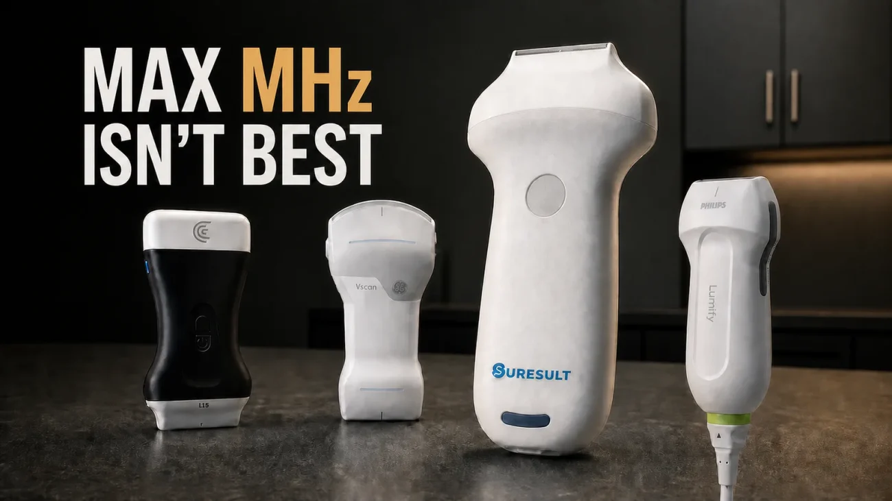
The six scanners split into two kinds: four handheld linears with Doppler that map vessels and follow a cannula, the Suresult L24Pro, Clarius L20 HD3, GE Vscan Air SL and Butterfly iQ3, and two skin consoles that measure the dermis at 20 to 50 MHz, the Cortex DermaScan C (with the DermaLab Combo in its card) and the tpm DUB SkinScanner.
| Scanner | Maximale frequentie · diepte | Doppler | Dermal-thickness and follow-up software | Functies voor naald en canule | Records | Kosten over vijf jaar en abonnement | Het beste voor |
|---|---|---|---|---|---|---|---|
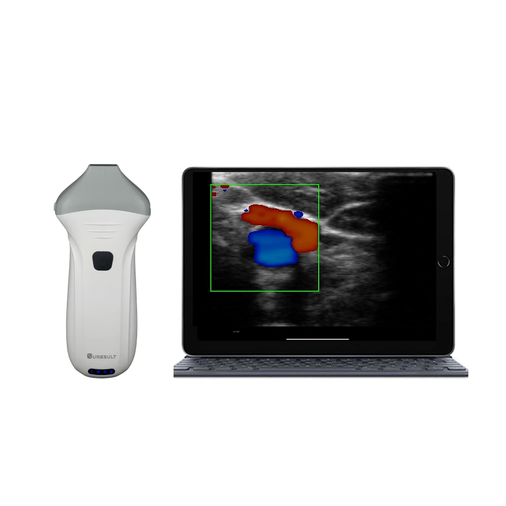 Suresult L24Pro · No. 1 · 90.3/100 |
18/24 MHz · presets at 10, 15, 20 and 25 mm · 256 elements | Color, Power and PW in the price | FaceAtlas AI, 3 months included: skin, fat and SMAS depth in mm; PLLA, CaHA and PCL among 7 materials; this visit beside the last | Needle Enhance; in-plane en out-of-plane geleidelijnen | 1,000-frame cine ≈ 165 s; DICOM export; FaceAtlas AI PDF report | $5,004 ($4,005 plus the one-time FaceAtlas AI lifetime license; imaging with no subscription) | Dermis, vessel map and the face planes to 25 mm on one probe |
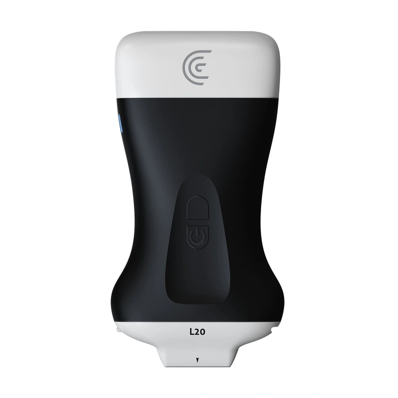 Clarius L20 HD3 · No. 2 · 64.8/100 |
8–20 MHz · images to 4 cm · 25 mm field of view | Color and Power in the price; PW with Essentials | Auto Aesthetic Calipers for skin, fat and tissue thickness; echogenicity histograms; a six-zone protocol at 1, 3 and 6 months | Needle Enhance met Essentials; centrale geleidelijn | 30 s cine; storage and DICOM with Essentials | $7,895 ($5,395 scanner plus Essentials at $2,500 one-time) | A published biostimulation protocol at 20 MHz |
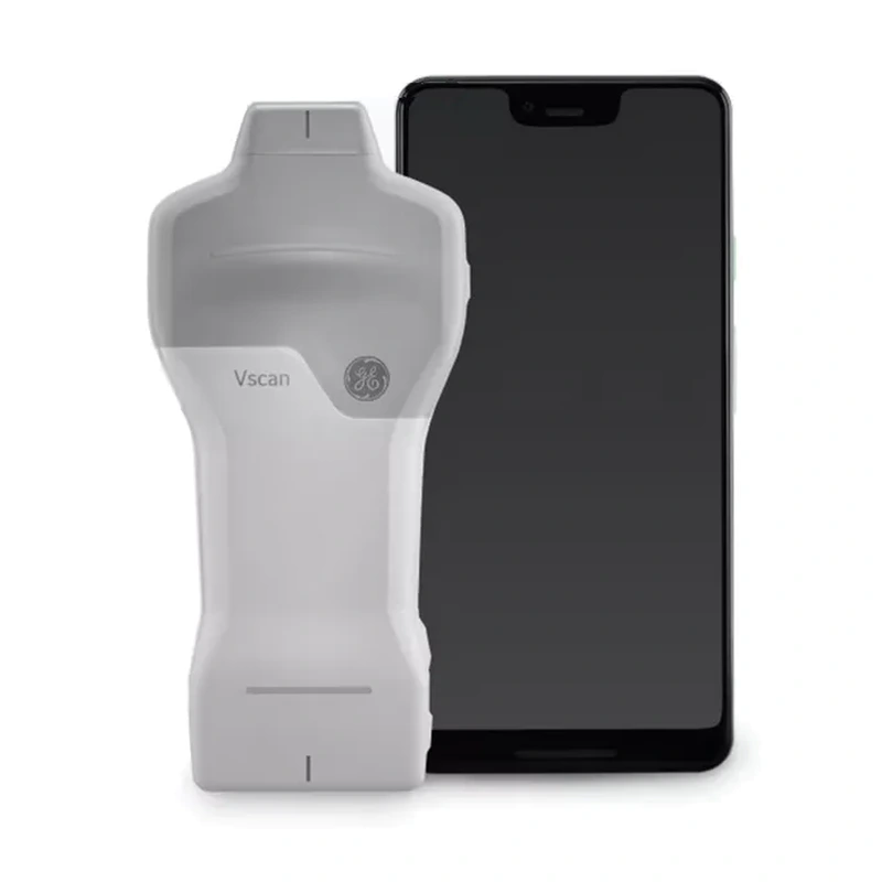 GE Vscan Air SL · No. 3 · 43.3/100 |
Linear 3–12 MHz · to 8 cm | Color and PW in the price | Caption AI, AutoEF and a bladder volume tool in the optional Innovations subscription | Middellijnmarkering | Up to 500 exams on the mobile device; DICOM | $4,999, every scored tool included without a required subscription | Color and PW Doppler at the lowest five-year rival cost |
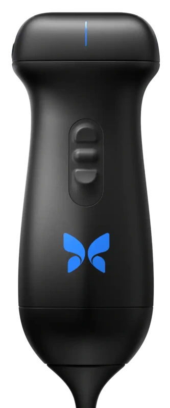 Butterfly iQ3 · No. 4 · 42.1/100 |
1–12 MHz · Vascular: Superficial preset at 1–4 cm · nerve imaging to 8 cm | Kleurendoppler, powerdoppler en PW-doppler met Advanced | Up to 4 linear calipers; Auto Bladder Volume and Auto B-Lines | Needle Viz and Biplane with Advanced; midline marker | Unlimited cloud storage with Advanced; Basic DICOM for purchase | $5,399 ($3,899 plus five years of Advanced, $1,500 one-time) | One whole-body probe with a preset for small facial vessels |
 Cortex DermaScan C · No. 5 · 39.8/100 |
20–50 MHz · 23 mm at 20 MHz, 7 mm at 50 MHz | A- and B-mode skin imaging | Average, maximum and minimum skin thickness by edge detection; old and new scans on one split screen | Built for skin measurement at 6–8 frames per second | Cine of the last 100 images, about 15 s; JPEG and TIFF files | Op offertebasis through Cortex and cyberDERM; measurement software included without a required plan | 50 MHz dermal measurement, FDA-cleared under K983945 |
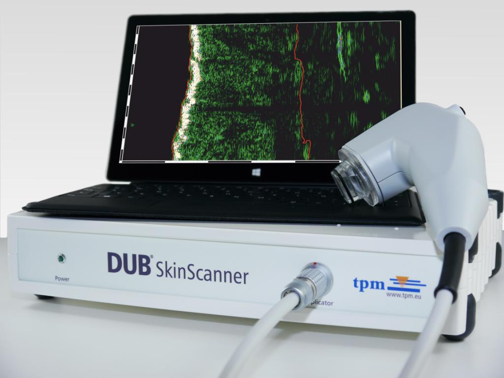 tpm DUB SkinScanner · No. 6 · 29.0/100 |
22 or 33 MHz · 6–8 mm · 48 µm axial resolution at 33 MHz | B-scan, HF mode, A-scan and Sum-A | Automatic skin thickness and density; length, area, width and depth tools | Built for skin measurement across a 12.8 mm scan width | ScanLoop playback; 7 color scales | Op offertebasis through tpm; measurement software included without a required plan | Automatic skin-thickness readings at 33 MHz |
Each five-year figure adds the software every scored tool needs to the instrument; Cortex and tpm sell by quote.
Beste keuze per toepassing: snel overzicht
- Best overall for biostimulator work: Suresult L24Pro, 90.3/100. 24 MHz for the dermis, three Doppler modes in the price for the vessel map, and FaceAtlas AI readings compared visit to visit.
- Best for a published biostimulation protocol at 20 MHz: Clarius L20 HD3, 64.8/100. A six-zone skin protocol rescanned at 1, 3 and 6 months.
- Best for Color and PW Doppler at the lowest five-year rival cost: GE Vscan Air SL, 43.3/100. Both modes in the $4,999 price, checked November 16, 2026, on a 3–12 MHz linear that images to 8 cm.
- Best whole-body probe with a facial-vessel preset: Butterfly iQ3, 42.1/100. A Vascular: Superficial preset built for small, slow-flow facial vessels.
- Best for dermal measurement at 50 MHz: Cortex DermaScan C, 39.8/100. A 20–50 MHz skin system, FDA-cleared under K983945, that reopens old scans beside new ones.
- Best for automatic skin-thickness readings at 33 MHz: tpm DUB SkinScanner, 29.0/100. Automatic thickness and density readings at 48 µm axial resolution.
Waar u elk echoapparaat kunt kopen
- Suresult L24Pro: $4,005 with every Doppler mode and Needle Enhance; FaceAtlas AI runs free for 3 months from the day you press Start, then costs $500 per year, $50 per month or $999 once for lifetime use, sold until 11:59 PM PT, December 31, 2026. Order the Suresult L24Pro linear probe at $4,005
- Clarius L20 HD3: $5,395 from the Clarius online store, checked November 16, 2026; pick the Essentials tier at checkout. Kopen bij Clarius
- GE Vscan Air SL: $4,999 at GE HealthCare’s US online shop, checked November 16, 2026, with remote training and support and a 3-year warranty. Kopen bij GE HealthCare
- Butterfly iQ3: $3,899, checked November 6, 2026, with a 3-year warranty, plus the Advanced membership for Color, Power and PW Doppler and the needle tools. See Butterfly iQ3 pricing
- Cortex DermaScan C: by quote from Cortex Technology in Aalborg and its North American distributor cyberDERM inc., with a 1-year warranty. See the DermaScan C at Cortex
- tpm DUB SkinScanner: by quote from tpm taberna pro medicum GmbH in Lüneburg. See the DUB SkinScanner at tpm
Why map the face before a biostimulator?
Map first because a biostimulator has to land in the right plane the first time. Hyaluronidase dissolves HA filler, yet the stimulation PLLA sets off on collagen cannot be immediately reversed (Ouyang 2025), and in HArmonyCa hyaluronidase dissolves the HA component, while excess superficial CaHA particles are diluted with saline (Philipp-Dormston 2025; Cavallini 2026); the HA step is covered in echogeleid oplossen van fillers. Young’s 2026 review counts at least 11 published cases of vision-impairing vascular occlusion after CaHA, most from the nasal dorsum, and 2 confirmed after PLLA, one of them from the temple; the event itself is covered in vasculaire occlusie na filler.
The 2025 Delphi panel ties scanning to the same anatomy: it holds imaging before injection to be obligatory at the temple, the forehead, the glabella, the nose and the pyriform fossa, and advises it at the zygomatic arch, the chin and the pre-auricular region, three sites that biostimulator protocols treat, on a linear of 15 MHz or more with a hockey-stick footprint favored (Velthuis 2025).
The vessels to map at each biostimulator site
Map the artery that runs in or beside each product’s plane: the superficial temporal, zygomatico-orbital and deep temporal arteries at the temple, the transverse facial artery over the zygoma, the facial artery and vein along the jawline, and the submental and mental arteries at the chin. The facial artery-depth table, site by site, is in the guide to ultrasound-guided filler injections.
| Locatie | Vessel or structure to map | Depth and layer | Plane each filler uses there | Bron |
|---|---|---|---|---|
| Temple, superficial | Superficial temporal artery branches; sentinel and middle temporal veins | In the superficial temporal fascia in all 34 temple patients, with a branch in the subcutaneous plane in 1; frontal branch 2.7 mm deep on the forehead at the lateral canthal line | Sculptra boluses in the temporal fascia; PLLA subcutaneous and interfascial fans; Ellansé interfascial fan | Teixeira 2025; Phumyoo 2020; Sigrist 2024; Sculptra US label; Chang 2026; de Melo 2017 |
| Temple, interfascial | Zygomatico-orbital artery | 5.61 mm deep, 2.52 mm across (n = 50); most trunks within 20 mm above the arch; deep branches pierce the deep temporal fascia’s superficial layer | Cannula between the two fasciae, confirmed with a test bolus | Choi 2018; Liu 2021; Desyatnikova 2022 |
| Temple, deep | Anterior and posterior deep temporal arteries | 1.3 to 2.1 mm above the bone, between the temporalis and the periosteum | Radiesse undiluted on bone; Ellansé preperiosteal bolus at low pressure | Bae 2023; Zhou 2023; Lorenc 2018 (composite volumization); de Melo 2017 |
| Zygoma and lateral cheek | Transverse facial artery; zygomaticofacial foramen | On 96% of 200 sides, 1.0 mm across, in an 8.8 mm band starting 17.0 mm below the arch | Radiesse arch boluses; HArmonyCa superficial subcutaneous fan; Sculptra subcutaneous or supraperiosteal | Koziej 2019; Vasconcelos-Berg 2024; Hernandez 2024; Cavallini 2026; Munoz-Lora 2026 |
| Jawline and mandible | Facial artery and vein | Artery 6.27 mm deep at the mandibular border (pooled, 402 arteries) and 2.5 to 15.0 mm at its mandibular origin across 168 hemifaces, in front of the masseter; vein 7.8 to 8.2 mm deep, 3.4 to 4.7 mm behind it | Radiesse (+) subdermal or at six supraperiosteal points; HArmonyCa subcutaneous boluses and threads; Sculptra subdermal along the border | Pourani 2025; Ten 2021; Vasconcelos-Berg 2025; Pistoia 2023; Wu BQ 2025; Bravo 2023; Yen 2026 |
| Kin | Submental artery, paramedian and sometimes crossing the midline; mental, inferior labial or labiomental and midline perforating arteries | Submental artery under the mentalis, over the bone; ascending mental artery 0.7 mm across at the mental protuberance | Radiesse and Ellansé on bone | Vasconcelos-Berg 2025; Tansatit 2019; Lorenc 2018 (physiochemical review); de Melo 2017 |
| Hals | The layer above the platysma, with the thyroid deeper | Skin to superficial fascia 3.71 mm at the lateral neck (2.0–5.0) in 150 adults | Radiesse 1:2 to 1:4 subdermal above the platysma; Sculptra at 8 or 17 mL per vial | Cotofana 2020 (superficial fascia study); de Almeida 2019; Trindade de Almeida 2023; Haddad 2025 |
| Décolleté | The treatment zone of about 100 cm² and the breast tissue at its lower edge | Chest dermis 1.96 mm at 50 MHz | Radiesse 1:2 subdermal by 22 G 50 mm cannula, above the breast tissue; Sculptra at 8 or 17 mL per vial | Radiesse label (S162); Wang Y 2025; Haddad 2025 |
| Buttock, thigh, abdomen and upper arm | The superficial fat over the grid and the superficial fascia that splits the gluteal subcutis | Superficial fascia 20.52 mm deep at the buttock (6.1–38.4) | Sculptra subdermal to high subcutaneous fans, 70% deeper in the upper buttock; Radiesse 1:1 to 1:4 by cannula fans | Cotofana 2020 (superficial fascia study); Ducati 2025; Haddad 2025; de Almeida 2019 |
At the temple, three fascial layers carry three different arteries, so the plane decides which artery the map has to find. Sigrist’s 2024 best-practice paper maps the superficial temporal, deep temporal and zygomaticotemporal arteries and the sentinel and middle temporal veins before the first pass, then scans after each cannula pass and keeps the tip 1 to 2 mm from the vessel. Fixed points miss the variation: Lee’s 2022 scans found arteries at the classic point, 1 cm above and 1 cm lateral to the brow tail, in 9 of 30 patients.


Over the zygoma, find the transverse facial artery before a supraperiosteal bolus: Vasconcelos-Berg’s 2024 midface guide scans each needle point for it and for the zygomaticofacial foramen, and runs cannula work in the deep subcutis, clear of the artery. Along the jawline the facial artery runs deep, in front of the masseter, with its vein behind it (Vasconcelos-Berg 2025). The subdermal plane and subcutaneous fat of the jawline held no major nerve or vessel in 72 cadavers (Suwanchinda 2018), the layer where the Radiesse (+) label and Yen’s 2026 Sculptra protocol place product.
At the chin the lower-face guide maps the submental, mental and labial arteries under the paired mentalis bellies before a bolus (Vasconcelos-Berg 2025); Radiesse and Ellansé go on bone there, so the map runs to the periosteum. On the neck the plane sits just under the dermis, above the platysma; on the décolleté the label places diluted Radiesse subdermally, in a zone that ends at the top of the intermammary cleft. On the buttock the superficial fascia sat 6.1 to 38.4 mm deep (Cotofana 2020, superficial fascia study), so measure the plane depth on every patient.
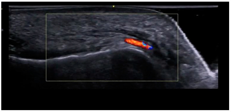
Doppler settings for small facial vessels
Set the color scale near 3 cm/s, lower the PRF and wall filter to match, and scan through plenty of gel with almost no pressure (Sigrist 2026): with detection starting at 2 cm/s, ultrasound picks up only one or two vessels in the dermis (WFUMB 2025).
| Stap of instelling | What to set | Bron |
|---|---|---|
| Koppeling | Copious gel and light pressure, so the vessel stays open | WFUMB 2025; Bustamante 2025 |
| Sweep order | Two perpendicular sweeps: gray scale, then color, then PW | WFUMB 2025 |
| Kleurvenster | Scan across the vessel, set scale and gain, steer the box along it and mark the points on the skin | Sigrist 2024 |
| Snelheidsschaal | About 3 cm/s, with PRF and wall filter matched | Sigrist 2026 |
| PRF at a lower Doppler frequency | 0.5–0.8 kHz with 5.0 MHz Doppler on a 4–16 MHz linear; inter-examiner ICC 0.891–0.967 | Bustamante 2025 |
| Wandfilter | Low; a high filter hides slow flow in small vessels | Pistoia 2023 |
| Modi | Color and spectral Doppler required; Power Doppler recommended | Velthuis 2025 |
| Arterie of vene | The vein compresses under light pressure; the artery keeps pulsing | Vasconcelos-Berg 2025 |
| Expected flow | Hypodermal arteries at 15 cm/s peak systolic velocity or less; detection from 2 cm/s | WFUMB 2025 |
Every Suresult probe carries PRF, wall filter, steer, color gain and move-and-zoom for Color and Power Doppler, and gain, steer, sample volume and angle for PW, in the price. The probe rests on a Suresult aesthetic ultrasound gel pad (Ø35 × 4 mm, $134 for 50) instead of the skin, so superficial facial arteries keep their flow on color Doppler, and the needle passes straight through the hydrogel with the image live. For a sterile field during guided passes, see steriele koppeling voor echogeleide injecties.
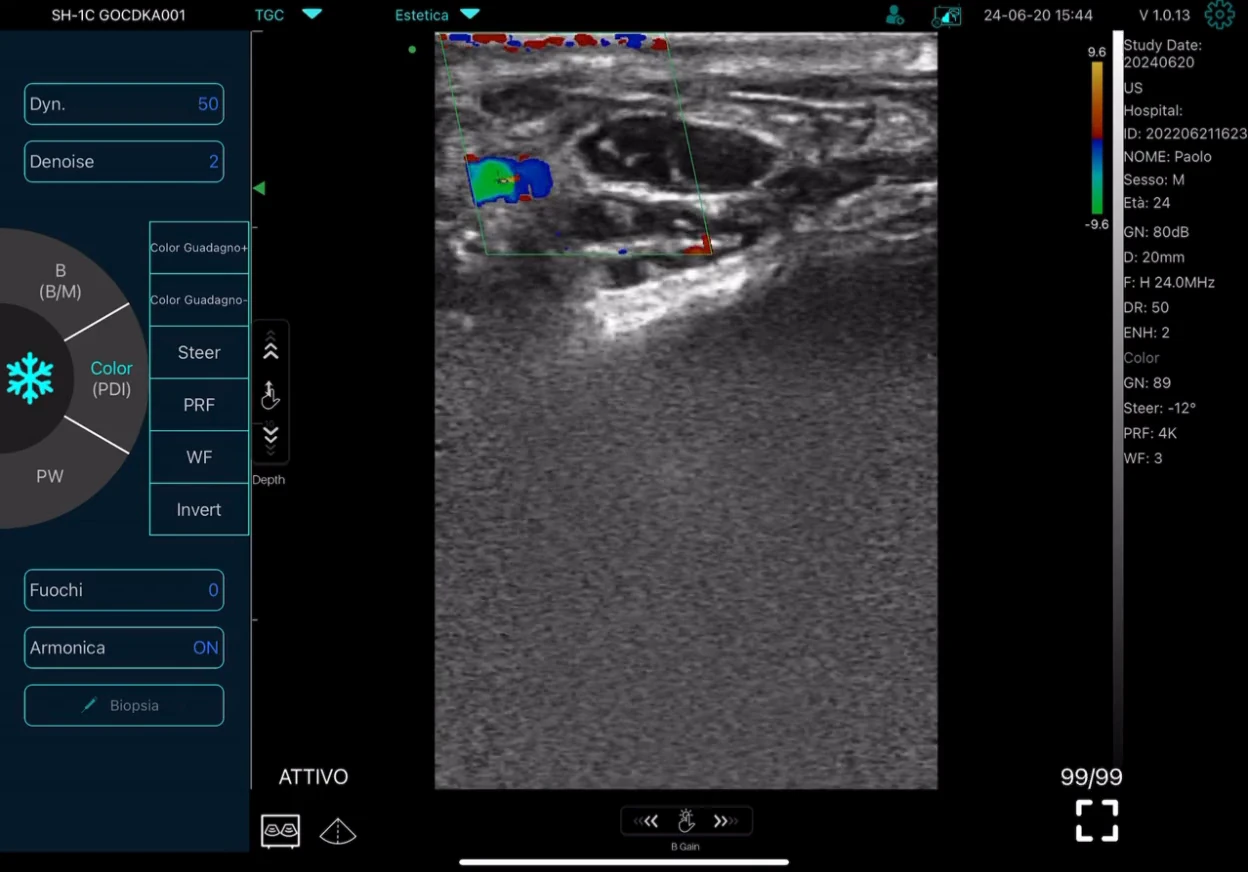
Sculptra injection technique: reconstitution, planes, injection points and the scan
The Sculptra injection technique starts with the reconstitution volume, because the volume decides the needle and the plane: on the US label, 5 mL of sterile water hydrated for at least 2 hours goes through a 26 G needle into the deep dermis, and 8 mL mixed for about a minute goes through a 25 G needle into the subdermal plane, subcutaneous or supraperiosteal. Each vial holds 150 mg of PLLA microparticles (Haddad 2025), irregular flakes of 2 to 150 µm (McCarthy 2024). Sculptra holds FDA premarket approval P030050 for facial lipoatrophy in HIV (2004), for nasolabial fold contour deficiencies and other facial wrinkles (2009) and for fine lines and wrinkles of the cheek (2023).
| Total volume per vial | How it is prepared | Needle, plane and site | Bron |
|---|---|---|---|
| 5 mL | Sterile water; stands 2 hours or more to hydrate, with no shaking; usable for up to 72 hours | 26 G needle into the deep dermis; face | Sculptra US label |
| 8 mL, plus 1 mL of 2% lidocaine as an option | 5 mL, shaken about 1 minute, then 3 mL more; injected straight away | 25 G needle into the subdermal plane, subcutaneous or supraperiosteal; face, as in the cheek pivotal trial | Sculptra US label; Baumann 2020; Fabi 2024 |
| 8 to 12 mL | Higher dilution, with fewer vials per session | All product subcutaneous, none in the dermis; face | Lam 2006 |
| 8 or 17 mL, with lidocaine | Body consensus volumes, chosen by site | 25–26 G needle or 22–25 G cannula; 8 or 17 mL on the neck and chest, 17 mL on the arms, abdomen and buttock | Haddad 2025 |
Both label routes mix the same 150 mg vial; the 8 mL route skips the 2-hour wait because its suspension kept the same properties at 0, 2, 24 and 72 hours after 1 minute of shaking (Baumann 2020), and the cheek-wrinkle pivotal trial ran it with 1 mL of 2% lidocaine for up to 4 monthly sessions, reaching 71.6% responders at month 12 against 26.1% of untreated controls (P < 0.0001; Fabi 2024).
Following the label, mark the plan with a water-soluble pencil, enter bevel-up at about 30 to 40 degrees and advance to depth; the resistance changes as the needle passes from the dermis into the subcutis. For the grid, lower the needle to 10 to 20 degrees, advance it parallel to the skin and leave a thin retrograde trail on withdrawal, stopping before the bevel shows; start at the base of the fold, then cross the first lines at right angles. The label’s other three techniques are a bolus with the needle held still in the subdermal region, a linear thread laid on slow withdrawal, and a fan of threads from one entry point. Keep every injection to 0.1 to 0.2 mL, 0.5 to 1 cm apart. Agitate the syringe between passes, massage in circles after every 3 to 4 injections and for at least 2 minutes at the end with a moisturizer, and send the patient home on 5 minutes of massage, 5 times a day, for 5 days. Under-correct at every session: treat, wait, assess.
| Locatie | Plane and technique | Tool and preparation | Protocol and result | Bron |
|---|---|---|---|---|
| Cheek, US label | Subdermal plane, subcutaneous or supraperiosteal: linear threads, boluses, fans or cross-hatching, 0.1–0.2 mL each, 0.5–1 cm apart | 25 G needle with the 8 mL preparation, plus 1 mL of 2% lidocaine as an option | One vial per cheek per session, up to 9 mL; up to 4 monthly sessions in the pivotal trial | Sculptra US label; Fabi 2024 |
| Nasolabial fold, US label | Deep dermal grid with the 5 mL preparation; subdermal threads, boluses or fans with the 8 mL | 26 G needle with 5 mL; 25 G with 8 mL plus 1 mL lidocaine | Up to 2.5 mL per fold per session with 5 mL, or 4.5 mL with 8 mL | Sculptra US label |
| Slaap | Label (lipoatrophy instructions): small boluses in the temporal fascia. Trial: retrograde subcutaneous and interfascial fans | Trial: 23 G entry needle at the hairline, level with the lateral canthus; 25–27 G blunt cannula | 174 patients randomized 2:1: 96.5% gained at least one temple-hollowing grade at month 6, controls 0% (a PLLA other than Sculptra) | Sculptra US label; Chang 2026 |
| Lateral zygoma | Both planes on each hemiface, swapped at day 60: subcutaneous left and supraperiosteal right at day 0 | Sculptra: 5 mL water shaken 1 minute, then 3 mL water and 2 mL lidocaine (10 mL); 27 G needle on bone | 20 women, 10 on Sculptra and 10 on Elleva; both planes ran the same course to day 120 | Munoz-Lora 2026 |
| Midface | Supraperiosteal needle boluses plus cannula fans at the deep dermal–subcutaneous junction | Needle and cannula | Up to 3 sessions; 6 patients (a PLLA other than Sculptra) | AL-Attab 2026 |
| Lower face and jawline | Cannula across the lateral lower cheek, the mandibular retaining zone and the prejowl sulcus; subdermal along the mandibular border | Canule | Weeks 0 and 6, plus week 12 when needed; 80 patients | Yen 2026 |
| Upper arms | Subcutaneous fans over the inner and medial arm, plus the posterior arm in moderate to severe laxity | 17 mL by 25 G needle or 22 G cannula; the trial used 18 mL through a 22 G 50 mm cannula | 1 vial per side, 1.5 in moderate to severe laxity; 2–4 sessions 30 or more days apart | Haddad 2025; Bravo 2026 |
| Buttock and hip dell | Subdermal to high subcutaneous fans, 70% of the product deeper in the upper quadrant for lift | 17 mL; 25–26 G needle or 22–25 G stiff cannula | At least 3 sessions, 4–6 weeks apart; the hip dell trial ran 3 monthly sessions | Haddad 2025; Zubair 2024 |
| Neck, chest, abdomen and thighs | Needle or blunt cannula, then massage; on the abdomen, always include the area above the umbilicus | Neck and chest 8 or 17 mL with lidocaine by 25–26 G needle or blunt cannula; abdomen 17 mL by 25 G needle; thighs 10 mL plus 2 mL lidocaine by 26 G needle | 2–4 sessions 4 weeks apart on the neck and chest; up to 3 sessions 45 days apart on the abdomen; half to one vial a side on the thighs | Haddad 2025 |
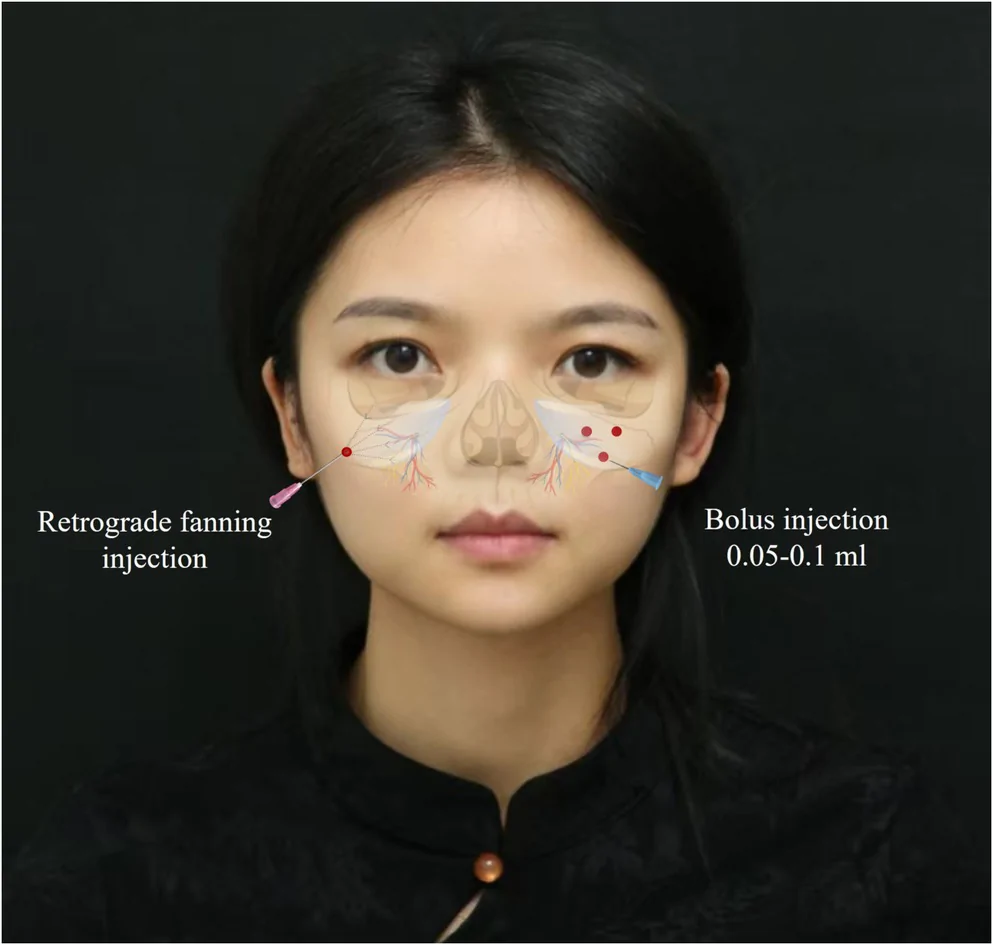
The scan settles what the resistance cue only suggests: the layer the tip is in. At the temple, in two split-face patients treated under ultrasound guidance, the subcutaneous side gained more skin and fat thickness than the fascial side, and sooner (Liu F 2025), and scans from week 4 to week 16 confirmed the plane clear of vessels while volume rose by week 8 (Arruda 2026). After injection, PLLA shows as ill-defined bright deposits that fade over the months (WFUMB 2025); 30 months after deep-dermal PLLA in the nasolabial folds of 20 patients, 16 deposits had degraded completely (Li 2022). Massage spread both PLLA products less than CaHA or PCL in 21 abdominal flaps scanned in real time (Casabona 2023).
Rescan on the label’s clock, no sooner than 3 to 4 weeks after each session, at the same marked points. Biopsies explain the wait: type I and III collagen rose at 3 and 6 months after PLLA, significantly for type I (Goldberg 2013). The label’s lipoatrophy data are skin-thickness readings: in the 30-patient Chelsea and Westminster study, three sessions 2 weeks apart raised mean skin thickness by 3.9 to 6.0 mm by week 12 (p < 0.001), and in the 50-patient VEGA study every patient gained, significantly at weeks 8 through 96. Faces without lipoatrophy move less: +0.18 mm in the mandibular dermis at week 24 (Yen 2026), less than twice the 100 µm axial resolution of a 15 MHz probe (WFUMB 2025), so the follow-up repeats the same point, gain and light pressure at every visit, as the dermis section below sets out.
Diluted Radiesse: the hyperdilution chart, planes by site and what the scan shows
Diluted Radiesse trades lift for collagen: past a 1:1 dilution the uncrosslinked carboxymethylcellulose gel disperses completely, leaving a suspension with a near-zero elastic modulus, and the CaHA microspheres serve as a biostimulator (McCarthy 2024). Each 1.5 mL syringe holds 30% CaHA microspheres of 25 to 45 µm by volume in a 70% carboxymethylcellulose gel (Radiesse label; McCarthy 2024). The global consensus calls a 1:1 mix diluted and 1:2 or more hyperdiluted (Goldie 2018). FDA premarket approval P050052 has covered Radiesse for subdermal facial wrinkles and folds since 2006, Radiesse (+) with lidocaine and Radiesse for the dorsum of the hand since 2015 and the Radiesse (+) jawline since 2021, and on March 31, 2026 it added Radiesse diluted 1:2 with saline for décolleté wrinkles, the first on-label hyperdilution in the US (S162).
| Ratio, Radiesse to diluent | Diluent added | Total volume | Onderwerp | Skin and plane | Bron |
|---|---|---|---|---|---|
| Undiluted | 0 mL | 1.5 mL | Undiluted | Volume and contour, on bone or subdermal | Lorenc 2022; Radiesse label |
| 1:1 | 1.5 mL | 3.0 mL | Diluted | Subdermal | Goldie 2018; Lorenc 2022 |
| 1:2 | 3.0 mL | 4.5 mL | Hyperdiluted | Normal skin; immediately below the dermis, at the dermal–subcutaneous junction, the plane the décolleté label calls subdermal; the on-label décolleté ratio, mixed with 0.9% saline | Lorenc 2022; Radiesse label (S162) |
| 1:3 | 4.5 mL | 6.0 mL | Hyperdiluted | Normal skin; the same junction plane | Lorenc 2022 |
| 1:4 | 6.0 mL | 7.5 mL | Hyperdiluted | Thin skin; the same junction plane; the neck trial mixed 2 mL of lidocaine with 4 mL of saline | Lorenc 2022; Trindade de Almeida 2023 |
| 1:6 | 9.0 mL | 10.5 mL | Hyperdiluted | Atrophic skin; the same junction plane | Lorenc 2022 |
The plane follows the ratio. Undiluted Radiesse goes on bone or into the subdermal plane, 1:1 goes subdermal, and 1:2 or more goes immediately below the dermis at the dermal–subcutaneous junction, by 22–25 G cannula and never as a large bolus, which compresses the vessels around it (Lorenc 2022). Undiluted gel on bone is a volume tool, placed supraperiosteally at the temple, the zygomatic arch, the anterior cheek, the pyriform aperture and the prejowl sulcus (Lorenc 2018, composite volumization). A two-step lateral-face protocol sets three arch boluses of 0.15, 0.15 and 0.10 mL with a 27 G needle, then fans 1:1 product subdermally from one cannula entry at the zygoma, 3 mL a side (Hernandez 2024).
| Locatie | Ratio | Injectielaag | Tool and technique | Dose per session | Bron |
|---|---|---|---|---|---|
| Contactvlak | 1:1 | Subdermal | Cannula retroinjection, fans or asterisk from 2–4 entries per hemiface, or short linear needle threads | 1 syringe | de Almeida 2019 |
| Jawline, Radiesse (+) | Undiluted | Subdermal or supraperiosteal | Needle, or a 27 G × 40 mm cannula | Median 1.80 mL per side in the pivotal trial | Radiesse (+) label (S129) |
| Hals | 1:2 to 1:4, by skin thickness | Subdermal, above the platysma | Cannula retroinjection from 3–5 entries | 1 syringe | de Almeida 2019; Trindade de Almeida 2023 |
| Décolleté, US label | 1:2, 4.5 mL | Subdermal | 22 G 50 mm blunt cannula through a 21 G introducer; linear threads or retrograde fans | 4.5 mL, at day 1, week 6 and week 12; up to 18 mL in total studied | Radiesse label (S162) |
| Upper arms, abdomen, buttock and thighs | 1:1 to 1:4 by site and laxity; 1:2 to 1:4 on the arms; 1:1 or 1:2 for buttock dimples | Subdermal | Cannula fans or asterisk; needle threads around the umbilicus | 0.5–1 syringe per arm; 1 syringe above and 1 below the umbilicus; 1 per buttock side or thigh area | de Almeida 2019 |
| Dorsal hand, US label | Undiluted | Between the subcutaneous layer and the superficial fascia | Needle with the skin tented; boluses starting between the 2nd and 4th metacarpals, then massage | 0.5 mL or less per bolus; up to 3 mL per hand studied | Radiesse label (S049) |
Two US pivotal trials back the label sites. On the jawline, Radiesse (+) treated 123 patients against 57 delayed-treatment controls, and 75.6% responded at week 12 (Radiesse (+) label, S129). On the décolleté, 152 women were randomized 3:1, and 71.2% of the treated arm responded at week 24 (95% CI 61.4–79.4%) against 6.3% of controls, with the treated arm followed to week 84 (Radiesse label, S162; Fabi 2026). The labeled zone covers about 100 cm² above the breast tissue, and breast imaging after treatment read without interference.
On the scan CaHA is bright, and at 18–24 MHz the shadow behind it tells the dilution: undiluted CaHA commonly casts a strong posterior acoustic shadow (WFUMB 2025); in a 21-patient series split among undiluted, diluted and HA-mixed CaHA, diluted CaHA cast a milder one, and CaHA mixed with HA a mild shadow in 57% and none in 43% (Wortsman 2023). Hyperdiluted CaHA in abdominal stretch marks reads as scattered white dots inside the dermis at a mean 0.51 mm (Kim 2025). In the neck, 1:4 CaHA shadowed the thyroid at day 15 and far less by day 60 (Faria 2022). In the dorsal hand CaHA lies as cloud-like bright bands (Gonzalez 2026).
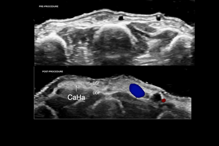
Hyperdilute CaHA runs in 2 to 3 sessions about 6 weeks apart (Lorenc 2022), with one maintenance session 12 to 18 months after the first cycle and patient massage twice a day for 3 to 7 days (de Almeida 2019). The dermis carries the result, so it is the reading to repeat; the dermis section lists the gains by study. The Lorenc chart sets 1:6 for atrophic skin; the dermatoporotic forearms in de Almeida’s trial averaged 1115 µm before one session of 1:2 Radiesse (de Almeida 2026).
HArmonyCa: HA and CaHA in one syringe, the plane and the follow-up scan
HArmonyCa puts both effects in one syringe: about 70% crosslinked HA gel at 20 mg/mL for immediate lift and about 30% CaHA microspheres of 25 to 45 µm by volume for collagen, with 3 mg/mL lidocaine, in 1.25 mL syringes (Philipp-Dormston 2025; Cavallini 2026). It is approved in more than 30 countries (Cavallini 2026), and Allergan announced its Health Canada authorization on May 25, 2026; in the US, CaHA is available as Radiesse and Radiesse (+) under FDA premarket approval P050052. The IFU places it in the deep dermis and the subdermal layer with a 27 G ½-inch thin-wall needle at about 30°, bevel down, by linear threading and cross-hatching, then gentle massage; the consensus uses it on the less mobile lateral face (Cavallini 2026).
Two published protocols set the points. The consensus marks the ligament line and the zygomatic arch, then lays 2.5 to 5 mL per session in the superficial subcutis from three entry points set off those lines, treating the lateral face first, reviewing at about 1 month and retreating after 6 months or more (Cavallini 2026). Bravo’s protocol fans 0.3 mL a side over the posterior zygoma in the superficial subcutis, then lays small boluses and retrograde threads along the mandibular body, ramus and gonial angle, 0.95 mL a side, 2.5 mL in all (Bravo 2023).
On the scan HArmonyCa reads as a bright, cloudy deposit (WFUMB 2025); at 18 MHz it forms a bright band with dark, hole-like areas inside, and the preauricular dermis above it went from 1.47 mm at baseline to 1.56 mm at day 90 and 1.68 mm at day 120, P < 0.001, on the treated side only (Bravo 2023). In a second preauricular series, elastography showed new collagen fibers from day 60, peaking between days 90 and 180 (Urdiales-Gálvez 2023). That curve sets the rescan: near day 90, when the first reading moves, and again at day 180, the end of the peak window. For misplaced product, hyaluronidase dissolves the HA part, and saline dilutes excess superficial CaHA particles (Cavallini 2026; Philipp-Dormston 2025); the HA step is in the guide to ultrasound-guided filler dissolving.
Ellansé (polycaprolactone, PCL): variants, technique by site, Ellansé vs Sculptra and the scan
Ellansé is a polycaprolactone (PCL) collagen stimulator: smooth, round PCL microspheres of 25 to 50 µm, about 30% by volume, in a carboxymethylcellulose (CMC) gel carrier of about 70%, supplied ready to inject in a prefilled syringe (Christen 2020; Ellansé IFU). The gel gives the correction on the day and is resorbed over 6 weeks to 3 months (de Melo 2017; Angelo-Khattar 2022; Christen 2020), and the microspheres carry the sustained effect through the collagen that forms around them (Christen 2020). The S, M, L and E variants share that make-up and differ in the chain length of the PCL, which sets how long each one lasts (Christen 2020; de Melo 2017).
The maker indicates Ellansé for subdermal implantation in the face for the lasting correction of wrinkles and signs of facial aging (ellanse.com), and its brochure lists the nasolabial folds, the mid-face, the temples and the forehead for both Ellansé-S and Ellansé-M (Sinclair brochure 2026). Christen’s review adds hand rejuvenation to the face in the indication (Christen 2020), and de Melo’s consensus places it subcutaneously on the back of the hand (de Melo 2017). Ellansé has carried the CE mark as a Class III device since 2009 (Christen 2020), is distributed in more than 70 countries on Sinclair’s 2025 data (ellanse.com) and is registered in China under NMPA No. 20213130100 (Hu 2025). In the US, NCT06984835 compares Ellansé-S with Radiesse in a randomized, double-blind split-face trial in the nasolabial folds, with an estimated 126 patients; ClinicalTrials.gov lists it as active, not recruiting, in its July 2026 update.
| Variant | Samenstelling | Stated duration | CE and labeled use | Bron |
|---|---|---|---|---|
| Ellansé-S (short) | PCL microspheres of 25–50 µm, about 30% by volume, in a CMC gel carrier of about 70% | Up to 12 months in the maker’s brochure, which cites the CE IFU; 1 year expected (de Melo) | CE-marked Class III, CE 0344; nasolabial folds, mid-face, temples and forehead in the brochure | Ellansé IFU; Sinclair brochure 2026; de Melo 2017; Christen 2020 |
| Ellansé-M (medium) | The same make-up, with its own PCL chain length | Up to 24 months in the maker’s brochure, which cites the CE IFU; 2 years expected (de Melo) | CE-marked Class III, CE 0344; nasolabial folds, mid-face, temples and forehead in the brochure | Ellansé IFU; Sinclair brochure 2026; de Melo 2017; Christen 2020 |
| Ellansé-L and Ellansé-E (long, extra-long) | The same make-up, each with its own PCL chain length | 3 and 4 years expected, extrapolated by de Melo from the S and M data and known PCL degradation | CE-marked Class III, CE 0344, in the S, M, L, E IFU for subcutaneous correction of wrinkles and signs of skin aging; the maker’s 2026 brochure lists S and M | Ellansé IFU; Sinclair brochure 2026; de Melo 2017; Christen 2020 |
The IFU sets the frame: subcutaneous injection through the supplied 27 G needle, bevel down at about 30°, in slow retrograde linear tunnels rather than globular deposits, a 1:1 correction with no overcorrection, and massage when an even spread needs it (Ellansé IFU). De Melo’s consensus places it subcutaneously or supraperiosteally on the face and subcutaneously on the hands, lays subcutaneous product by linear threading, fanning or cross-hatching, keeps every bolus to 0.2 mL or less at low pressure and massages the area gently straight after (de Melo 2017). For thin skin, an intradermal hyperdilute protocol mixes 0.5 mL of Ellansé-M with 0.5 mL of lidocaine and 2 mL of saline, a 1:5 dilution, and places it in the deep dermis through a 31 G five-pin injector in one session (Kim JS 2019).
| Locatie | Injectielaag | Instrument en dikte | Vectors or bolus | Volume | Bron |
|---|---|---|---|---|---|
| Voorhoofd | Supraperiosteal | 22–25 G blunt cannula from three entries: the midline 4–5 cm above the orbital rim and each temporal crest 3.5–4 cm above it | Retrograde linear threads, massaged straight after | 2–4 mL in total | de Melo 2017 |
| Lateral brow | Supraperiosteal | 25–27 G blunt cannula; a 27 G needle is acceptable | Retrograde linear threads of 0.05–0.1 mL | 0.2–0.3 mL per side | de Melo 2017 |
| Slaap | Supraperiosteal, or between the superficial and deep temporal fascia | 22–25 G rigid cannula; a needle for small corrections | Retrograde aliquots of 0.1 mL | 0.5–1 mL in total | de Melo 2017 |
| Malar area | Supraperiosteal or subcutaneous | 22–25 G cannula; a 27 G needle for precision | Retrograde injection or small pyramidal boluses | Up to 1 mL per side | de Melo 2017 |
| Malar area, 3D-measured | Supraperiosteal | 22 G × 50 mm blunt cannula | Ellansé-S on both sides after an infraorbital nerve block | 1 or 2 mL in total; at 24 months the Vectra-measured volume gain exceeded the volume injected by 50% to 150% in all 9 women | Angelo-Khattar 2022 |
| Submalar area | Subcutaneous | 25–27 G blunt cannula or 27 G needle | Retrograde linear threads with fanning and cross-hatching, 0.05–0.1 mL each | 0.5–1 mL | de Melo 2017 |
| Nasolabiale plooi | Subcutaneous along the fold; supraperiosteal for a deeper bolus at the perialar depression | 27 G needle | Retrograde fanning, plus a small perialar bolus | 0.1 mL at most per bolus | de Melo 2017 |
| Marionette lines and oral commissures | Subcutaneous | 27 G needle | Fanning, cross-hatching and microboluses of 0.05–0.1 mL | 0.5 mL per side | de Melo 2017 |
| Mental crease | Subcutaneous | 25–27 G needle | Linear threads and retrograde injection, 0.05 mL per thread | 0.3–0.4 mL | de Melo 2017 |
| Kin | Supraperiosteal, near the mental tubercle on both sides | 25–27 G needle, or a 25 or 22 G cannula | Retrograde threads or small boluses under 0.2 mL | 0.5 mL per side, 1 mL on average | de Melo 2017 |
| Prejowl sulcus, jawline and mandibular angle | Supraperiosteal at the prejowl sulcus and the angle; subcutaneous along the jawline | 27 G needle or 25–27 G cannula | Fanning at the prejowl sulcus; vectors of 0.02–0.03 mL along the jawline; boluses of 0.1 mL at the angle | 0.3–0.6 mL or more at the prejowl sulcus | de Melo 2017 |
| Back of the hand | Subcutaneous, in the most superficial layer | 25 or 22 G cannula through one entry distal to the wrist or through each interdigital space | A very small deposit at the entry, then cannula passes once the tissue distends a few minutes later | 1.0 mL per hand in Figueiredo’s 5-patient pilot | de Melo 2017; Figueiredo 2013 |
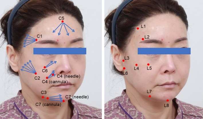
| Full face, CL Code | Temple at the STF–DTF interface; forehead submuscular; prejowl sulcus supraperiosteal; cheek, nasolabial fold and chin sub-SMAS; canine fossa in Ristow’s space; ligament points supraperiosteal or sub-SMAS | 27 G needles at the ligament points; separate cannula and needle entries at C4 and C7 | Ellansé-S in 0.3 mL aliquots at seven contour zones, then 0.1 mL of Ellansé-M at each of eight ligament points | 2.9 mL for the full face in a one-patient case report | Hong 2026 |
| Thin facial skin, hyperdilute | Deep dermis, at the injector’s 1 mm setting | Vital Injector, 31 G five-pin | About 1,000 points 5 mm apart over the face; the remainder in 20 to 40 strokes along the nasolabial fold | 0.5 mL Ellansé-M with 0.5 mL lidocaine and 2 mL saline (1:5, 3 mL), one session | Kim JS 2019 |
Ellansé vs Sculptra comes down to when the volume shows: Ellansé’s CMC gel corrects on the day, while Sculptra’s PLLA builds collagen over a course of sessions (Christen 2020; Sculptra US label).
Merz’s same-subject trial injected Ellansé, Sculptra, three Radiesse preparations, HArmonyCa and saline at separate abdominal sites in each of 10 patients, 6 months, 3 months and 2 weeks before surgery removed that tissue, and read the dermis at every site on ultrasound (NCT06093815); the table gives its Ellansé, Sculptra and saline readings. In the face, a 136-patient nasolabial cohort set Ellansé against Löviselle, a PLLA other than Sculptra, and the Ellansé group held the lower, better wrinkle score at 3, 6 and 12 months: 3.08 against 3.27, 2.23 against 2.49 and 1.92 against 2.17 (p = 0.007, 0.03 and 0.016; Hu 2025).
| Point | Ellansé (PCL) | Sculptra (PLLA) | Bronnen |
|---|---|---|---|
| Carrier and preparation | PCL microspheres of 25–50 µm in a CMC gel carrier, about 30:70 by volume, ready to inject from a prefilled syringe | 150 mg of PLLA flakes of 2–150 µm per vial, with 5 mL of sterile water hydrated for at least 2 hours, or 8 mL mixed for about a minute | Christen 2020; Ellansé IFU; Haddad 2025; McCarthy 2024; Sculptra US label; Baumann 2020 |
| Volume on the day | Immediate, from the CMC gel, which is resorbed over 6 weeks to 3 months; inject 1:1 with no overcorrection | Built over sessions; under-correct at every session: treat, wait, assess | Christen 2020; Angelo-Khattar 2022; de Melo 2017; Ellansé IFU; Sculptra US label |
| Onset and duration | New collagen visible 5–8 weeks after treatment; up to 12 months (S) and 24 months (M) in the maker’s brochure, which cites the CE IFU; in 9 women the Ellansé-S volume gain at 24 months exceeded the volume injected by 50% to 150% | Built over up to 4 monthly sessions, each reassessed no sooner than 3 to 4 weeks after the last; 71.6% responders at month 12 against 26.1% of untreated controls | de Melo 2017; Sinclair brochure 2026; Angelo-Khattar 2022; Sculptra US label; Fabi 2024 |
| Vlakken | Subcutaneous in the IFU; subcutaneous or supraperiosteal on the face and subcutaneous on the hands in the consensus, between the temporal fasciae at the temple | The deep dermis with the 5 mL preparation; the subdermal plane, subcutaneous or supraperiosteal, with the 8 mL | Ellansé IFU; de Melo 2017; Sculptra US label |
| Massage | When an even spread needs it (IFU); gently, straight after each area (consensus) | In circles after every 3 to 4 injections and for at least 2 minutes at the end, then 5 minutes, 5 times a day, for 5 days | Ellansé IFU; de Melo 2017; Sculptra US label |
| Collagen on histology | Collagen around the microspheres in human temple biopsies 13 months after Ellansé-M; thin new collagen fibers between the particles 1 year after intradermal Ellansé-M, and more at 4 years | Type I and III collagen rose at 3 and 6 months, significantly for type I | Christen 2020; Kim JS 2019; Goldberg 2013 |
| Same-subject trial, abdomen | Dermis 1.73 ± 0.25 mm at baseline, 1.85 ± 0.12 at day 14, 1.78 ± 0.27 at month 3 and 1.65 ± 0.31 at month 6, by ultrasound, 10 patients | Sculptra mixed with 5 mL of sterile saline: 1.76 ± 0.37, 1.80 ± 0.31, 1.74 ± 0.52 and 1.64 ± 0.31 mm at the same points; saline sites 1.75 ± 0.33, 1.84 ± 0.28, 1.69 ± 0.34 and 1.73 ± 0.33 | NCT06093815 |
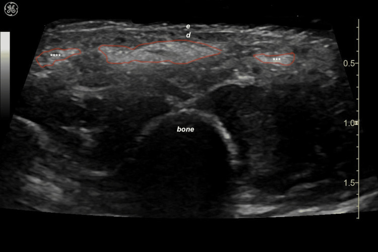
On the scan Ellansé reads as a dark (hypoechoic) matrix holding many bright spots with mini-comet tails; WFUMB adds that a PCL deposit can grow brighter over time while still reading hypoechoic (WFUMB 2025). The same pattern showed in a porcine model and in patients (Wortsman 2017), and the bright spots are the suspended microspheres (Tang 2026). That separates it from CaHA, which is bright with a posterior shadow whose strength follows the dilution, and from PLLA, which forms ill-defined bright deposits that fade (WFUMB 2025). Massage moves it: in 21 abdominal flaps scanned in real time, PCL spread further after massage, P < 0.001 (Casabona 2023).
On a Clarius L20 handheld, a 3-month-old Ellansé deposit in the deep medial cheek fat read hypoechoic, and a 0.2 mL top-up went in beside it under ultrasound guidance through a 25 G cannula, with a satisfactory result 3 months later (Wu L 2025). In the same series the scan traced a fullness to fat in one patient and to old HA in another, and read 26 syringes of Ellansé-M given over 3 years as collagen in the deep medial cheek fat; the authors add that dense new collagen in the first 6 months after Ellansé can hinder the cannula (Wu L 2025).
For a lump after Ellansé, the ultrasound work-up of filler lumps shows how PCL nodules scan and how guided treatment runs.
Cannula technique for Sculptra, Radiesse, HArmonyCa and Ellansé: gauge, entry, passes and the plane on the scan
The published cannula for Sculptra, Radiesse and HArmonyCa is 22 to 27 G, 40 or 50 mm long where a length is given, entered through a needle pre-hole at one to five points per area, and it lays retrograde threads or fans in one layer: subdermal or immediately below the dermis for diluted Radiesse, the superficial subcutis for HArmonyCa, and subdermal to subcutaneous for Sculptra on the body.
| Beslissing | What the published protocols use | Bron |
|---|---|---|
| Maat | 22 G for the labeled décolleté, the HArmonyCa protocol and Sculptra on the arms; 22–25 G for hyperdiluted Radiesse, 1:1 Radiesse (+) on the lateral face and Sculptra on the buttock (a stiff cannula); 25 or 27 G for the PLLA temple trial; 27 G for the Radiesse (+) jawline | Radiesse label (S162); Bravo 2023; Haddad 2025; Lorenc 2022; Hernandez 2024; Chang 2026; Radiesse (+) label (S129) |
| Length | 50 mm for the décolleté, the HArmonyCa protocol and the Sculptra arm trial; 40 mm, ultrathin-wall, for the Radiesse (+) jawline | Radiesse label (S162); Bravo 2023; Bravo 2026; Radiesse (+) label (S129) |
| Pre-hole | A 21 G introducer for the décolleté; the 25 G pre-hole needle packed with the jawline cannula; 21 G, 5 cm below the lateral orbital rim, for HArmonyCa; 23 G at the hairline, level with the lateral canthus, at the temple | Radiesse label (S162); Radiesse (+) label (S129); Bravo 2023; Chang 2026 |
| Entry points | One at the zygoma body and frontal process for 1:1 Radiesse (+) on the lateral face; three off the ligament line and zygomatic arch for HArmonyCa; 2 to 4 per hemiface for 1:1 Radiesse; 3 to 5 on the neck | Hernandez 2024; Cavallini 2026; de Almeida 2019 |
| Passes | Linear threads and retrograde fans on the décolleté and for HArmonyCa; fans or asterisks for diluted Radiesse, never a large bolus in one area; 0.1 mL vectors for 1:1 Radiesse (+); threading, fanning or tunneling for Sculptra on the buttock | Radiesse label (S162); Cavallini 2026; de Almeida 2019; Lorenc 2022; Hernandez 2024; Haddad 2025 |
| Laag | Subdermal for 1:1; immediately below the dermis for 1:2 and more, the plane the décolleté label calls subdermal; the superficial subcutis for HArmonyCa; subcutaneous for Sculptra on the arms, subdermal to high subcutaneous on the buttock; subcutaneous and interfascial at the temple | Lorenc 2022; Radiesse label (S162); Cavallini 2026; Bravo 2026; Haddad 2025; Chang 2026 |
For Ellansé, the IFU gives the longest 25 G and 27 G needles and cannulas, validated by extrusion tests: cannulas of 25 G up to 50 mm and 27 G up to 38 mm, and needles of 25 G up to 25 mm and 27 G up to 20 mm (Ellansé IFU). At the temple, de Melo’s consensus enters from the hairline or the temporal crest with a 22 or 25 G blunt cannula and fans under the superficial temporal fascia, over the superficial layer of the deep temporal fascia, from the posterior limit to the orbital rim and the zygomatic arch, keeping a needle for small corrections; a deep preperiosteal bolus at low pressure is the alternative and takes more product for the same result (de Melo 2017).
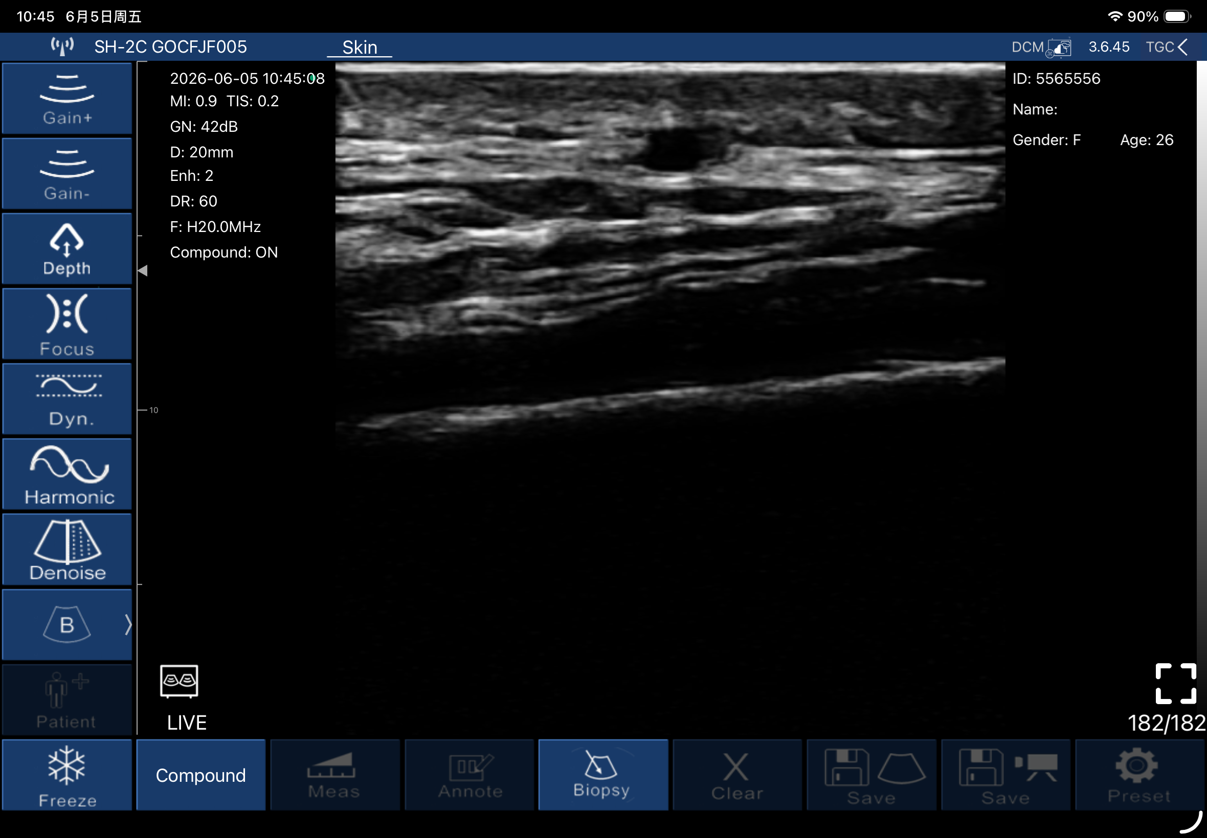
The scan sets the layer before the pre-hole and confirms it during and after the passes. On the face the target lies immediately below a dermis 0.58 to 1.30 mm thick (Meng 2022; Jeong 2023), where the medium-echo dermis meets the darker fat; a sweep along the planned track shows the vessels before the needle opens the skin. At the temple the interfascial plane is a thin layer of loose tissue, hard to see until a small first deposit goes in: the space then expands and lifts the superficial temporal fascia on the monitor, which confirms the cannula sits between the two fasciae (Desyatnikova 2022). After the passes, a rescan shows where the product lies: in 12 dorsal-hand procedures with HA, CaHA, a CaHA-HA hybrid, a PLLA-HA mix or a fat graft, every deposit sat superficial to the extensor tendons (Gonzalez 2026).
Choosing a cannula or a needle site by site is covered in cannula vs needle for filler.
Measuring the dermis before a biostimulator and at every follow-up
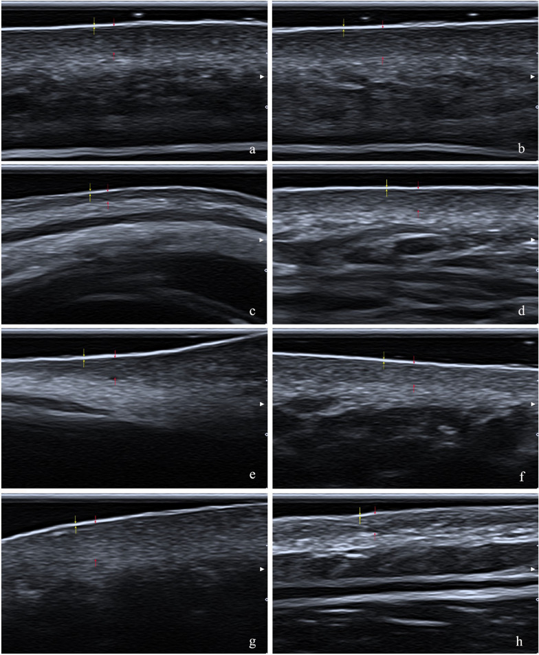
Measure the dermis peak to peak, from the entrance echo at the skin surface to the interface where the medium-echo dermis meets the dark subcutaneous fat (Alsing 2020), at the same marked point, frequency, gain and probe pressure before the first session and at every follow-up. Lay enough gel that a film shows under the probe and hold it vertical and light, so the layers stay uncompressed (Meng 2022; Santiago 2022). Keep the gain fixed: on the DermaScan C, inter-session echogenicity ICC went from 0.727 to 0.916 in a second group once the method was revised, and the authors tie repeatability to method and gain settings (Phillips 2020). Read the borders the same way at every visit and average several readings, as the HArmonyCa trial did with five measures beside an untreated control spot (Bravo 2023); on the DermaScan C, automatic border detection read about 20% thicker than manual reading of the A-mode peaks (Alsing 2020).
Measured this way the dermis repeats: intrarater ICC 0.83 to 0.94 for the whole scan and reading at 18 MHz (Santiago 2022), 0.71 to 0.83 on the DermaLab Combo (Peperkamp 2019) and 0.999 between examiners at 50 MHz (Wang Y 2025). Read the subepidermal low-echogenic band (SLEB) as well, the dark band under the epidermis that marks lax papillary dermis (Nicolescu 2023): at 20 MHz it shows at the forehead and the back of the forearm in more than half of adults over 40 (Sandby-Møller 2004), and its echogenicity shifts between morning and evening (Gniadecka 1994), so each rescan runs at the same hour.
| Locatie | Dermis, mm | Group and frequency | Bron |
|---|---|---|---|
| Voorhoofd | Men 1.17, women 0.86; central 1.019, lateral 0.978 | 118 adults at 24 MHz; 99 adults on an L18-5 linear | Meng 2022; Jeong 2023 |
| Slaap | Men 1.06, women 0.80; 1.049 | Same two groups | Meng 2022; Jeong 2023 |
| Zygoma and malar area | Men 1.14, women 0.87; malar 1.160; women 0.97 at 18–44, 0.78 at 45–59 and 0.63 at 60 and over | Same two groups | Meng 2022; Jeong 2023 |
| Nasolabial fold, mouth corner and chin | 1.276, 1.299 and 1.273, the thickest sites in that series | 99 adults on an L18-5 linear | Jeong 2023 |
| Eyelid | Men 0.67, women 0.58, the thinnest facial dermis | 118 adults at 24 MHz | Meng 2022 |
| Submandibular area | Men 1.02, women 0.91; women 1.00 at 18–44, 0.84 at 45–59 and 0.60 at 60 and over | 118 adults at 24 MHz | Meng 2022 |
| Hals | Men 0.98, women 0.99 | 118 adults at 24 MHz | Meng 2022 |
| Chest | 1.82; 1.96 | 189 adults at 22 or 75 MHz; 74 adults at 50 MHz | Wang S 2023; Wang Y 2025 |
| Upper arm | Flexor 0.92, extensor 1.34; 1.61–1.67 | Same two groups | Wang S 2023; Wang Y 2025 |
| Abdomen | 1.56; 2.39 | Same two groups | Wang S 2023; Wang Y 2025 |
| Thigh | Flexor 1.06, outer 1.32; 1.77 | Same two groups | Wang S 2023; Wang Y 2025 |
| Onderzoek | Filler and protocol | Site and patients | Instrument | Baseline to follow-up | Timepoint |
|---|---|---|---|---|---|
| Trindade de Almeida 2023 | Radiesse 1:4, sessions at day 0 and day 45 | Neck, 3 points; 22 women | DermaScan C | Pooled dermis +14.9% (95% CI 8.5–21.3%) | Day 120 |
| Bravo 2023 | HArmonyCa 2.5 mL, 1 session | Preauricular; 15 patients | 18 MHz linear | 1.47 to 1.48 mm at day 30 (no significant change), 1.56 at day 90, 1.68 at day 120 (P < 0.001); untreated side unchanged | Days 30, 90 and 120 |
| Bravo 2026 | Sculptra against Elleva, 18 mL per vial, subcutaneous | Upper arms, split; 20 patients | DermaLab Combo-4 | Sculptra 1.80 to 2.70 mm (p = 0.0039), Elleva 2.06 to 2.66 mm (p = 0.039); the two products matched (p = 0.54) | Day 120 |
| Bravo 2025 | Radiesse against Rennova Diamond Intense, 1 session | Lower face at the tragus; 30 patients | DermaLab Combo-4 | Radiesse side 1488 µm, +66 µm; comparator +210 µm; the sides matched (p = 0.29) | Day 120 |
| Munoz-Lora 2026 | Sculptra against Elleva, both planes on each hemiface, swapped at day 60 | Lateral zygoma; 20 women | 22 MHz linear | 1.27 and 1.31 mm at baseline; a rise over time at p = 0.063, with no difference by product or plane | To day 120 |
| de Almeida 2026 | Radiesse 1:2, 3.0 mL per forearm, 1 session | Forearm dermatoporosis; 6 patients | High-frequency ultrasound | 1115 to 1517 µm (+36.05%, p = 0.0007); collagen area +69.7% | Day 45 |
| Lapatina 2017 | Radiesse 1:4, 1 session, subdermal | Abdomen; 10 women | Echografie | +0.7 mm at the umbilicus, +0.4 mm at the flanks, +26.7% overall | Day 70 |
| Yen 2026 | Sculptra at weeks 0 and 6, plus week 12 when needed, by cannula | Mandibular dermis; 80 patients | High-frequency ultrasound | +0.18 mm (95% CI 0.17–0.20), P < 0.001 | Week 24 |
| Zubair 2024 | Sculptra 150 mg against saline, 3 monthly sessions | Hip dell, split body; 15 women | Echografie | Dermis +26.1% and fat +27%; saline side unchanged | To month 9 |
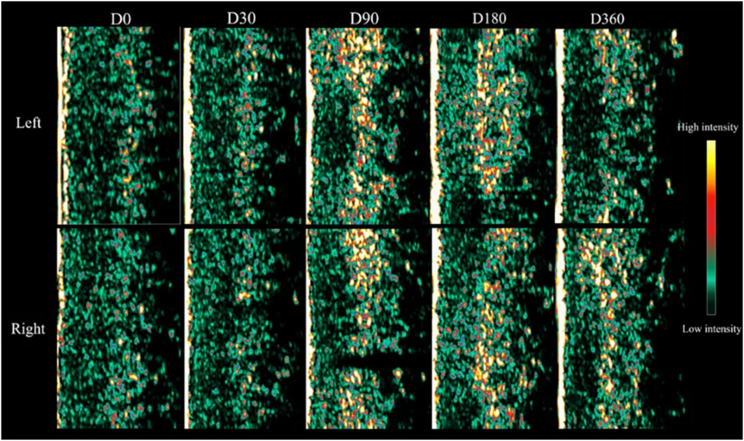
| AL-Attab 2026 | PLLA, up to 3 sessions | Midface; 12 hemifaces | DermaLab, 3 scans per site averaged | Dermal intensity peaked at day 90 and stayed above baseline | To day 360 |
| Kim JS 2019 | Ellansé-M diluted 1:5 (0.5 mL with 0.5 mL lidocaine and 2 mL saline), 1 intradermal session by 31 G five-pin injector | Temple, anterior cheek and nasolabial fold, 39 points; 13 women with thin skin | Ultrasound; temple biopsy | Dermis +21.31% on ultrasound; temple skin on biopsy 1781.11 µm against 1412.41 µm on the untreated side (+26.74%, P < 0.001) | 1 jaar |
| NCT06093815 (Merz, 2026) | Radiesse (+), Radiesse 1:1 and 1:2, Sculptra, Ellansé, HArmonyCa and saline at separate sites in each patient, injected 6 months, 3 months and 2 weeks before the tissue was removed | Abdomen; 10 patients | Echografie | Month 6 against baseline: Ellansé 1.73 to 1.65 mm, Sculptra 1.76 to 1.64, Radiesse (+) 1.75 to 1.89, Radiesse 1:1 1.76 to 2.04, Radiesse 1:2 1.67 to 1.78, HArmonyCa 1.76 to 1.92, saline 1.75 to 1.73 | Day 14, month 3 and month 6 |
Read the result at day 120, the timepoint five of these twelve studies share: HArmonyCa had not moved at day 30 (Bravo 2023), and the midface PLLA reading still sat above baseline at day 360 (AL-Attab 2026). Bring the same probe, preset, gain and site map to every visit, because the gains in these trials run from a median 66 µm (Bravo 2025) to a mean 0.90 mm (Bravo 2026), and every site in the same-subject abdomen trial read within 0.3 mm of its baseline at month 6 (NCT06093815).
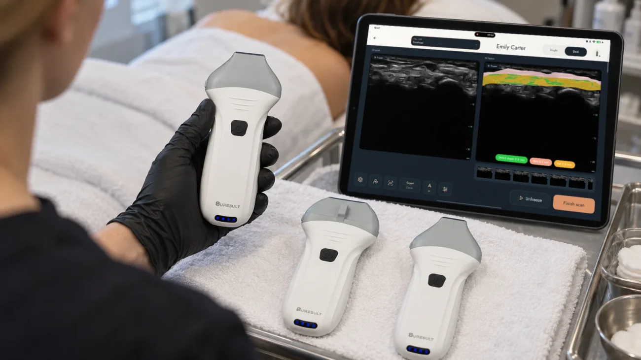
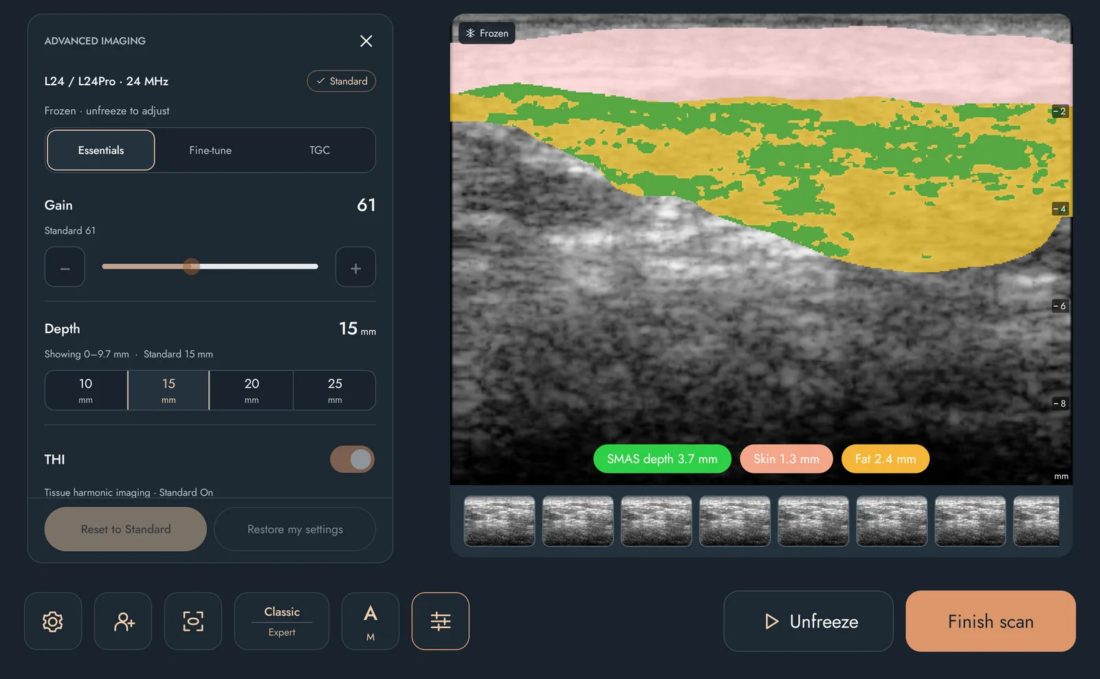
On the Suresult L-series probes, FaceAtlas AI turns this method into a visit record: it reads skin thickness, subcutaneous fat thickness and depth to SMAS in millimeters, files every scan in the client’s record, and puts this visit beside the last in one click across the same 21 standard sites. Dual View shows the raw scan beside the AI layers, and each visit exports as a PDF report. A Ø35 × 4 mm aesthetic ultrasound gel pad fixes the stand-off at 4 mm, so the readings stay comparable visit after visit. The L14Pro, L20Pro and L24Pro include 3 months of it; the L14, L20 and L24 run it on a license.
DermaScan and DermaLab vs a handheld ultrasound
The DermaScan C and the DermaLab Combo are skin-measurement consoles: they read dermal thickness, echogenicity and the low-echogenic band at 20 to 50 MHz in the first millimeters of skin, while an 18/24 MHz handheld linear reads the same dermis, follows the cannula planes below it and maps the vessels on Doppler. The tpm DUB SkinScanner sits with the consoles at 22 or 33 MHz.
| Spec | Cortex DermaScan C | Cortex DermaLab Combo | tpm DUB SkinScanner | Suresult L24Pro handheld |
|---|---|---|---|---|
| Frequentie | Broadband transducers from 20 to 50 MHz | 20 MHz probe; 10 MHz subcutaneous probe | 22 or 33 MHz applicators | 18/24 MHz lineair |
| Image depth | 14 or 23 mm at 20 MHz; 7 mm at 50 MHz | 3.4 mm at 20 MHz; 40 mm at 10 MHz | 6–8 mm | Presets at 10, 15, 20 and 25 mm |
| Beeldvormingsmodi | A- and B-mode | Skin ultrasound plus TEWL, elasticity, hydration and other probes | B-scan, HF mode, A-scan and Sum-A | B-mode with Color, Power and PW Doppler |
| Thickness readout | Average, maximum and minimum skin thickness by edge detection; ROI and intensity segmentation | Skin thickness, intensity score and the low-echogenic band | Automatic skin thickness and density | FaceAtlas AI skin, fat and SMAS depth in mm |
| Regulatory | FDA-cleared under 510(k) K983945 | “The DermaLab® Combo is an investigational laboratory instrument for measuring skin parameters.” | Medical CE, per the tpm brochure | met 510(k) K211321-clearance |
| Where sold | By quote from Cortex Technology and cyberDERM inc. | By quote from Cortex Technology and cyberDERM inc. | By quote from tpm taberna pro medicum | $4,005 at the Suresult store |
Doppler splits the two kinds. Penetration runs to about 10 mm at 20 MHz and 3 to 4 mm at 50 MHz (Mlosek 2021), room for a dermis of 1 to 3 mm. The vessel map is the step the consoles leave to a linear: dedicated 30 to 70 MHz dermal scanners carry no Doppler, mapping runs on 15 to 22 MHz linear systems, and 18 to 22 MHz shows vessels of about 0.1 mm (Jaguś 2021). For the PLLA follow-up itself, 20 MHz served better than 50 MHz in a 30-month nasolabial study (Li 2022). Camera-based analyzers that photograph the skin surface are compared in the best skin analysis machines for clinics.
What frequency and depth biostimulator work needs
Biostimulator work needs 18 to 24 MHz for the dermis and the face planes, up to 40 mm of depth for the buttock, and Color and PW Doppler for the vessel map, on top of the 15 MHz linear floor that WFUMB and the 2025 Delphi panel set for aesthetic scanning (WFUMB 2025; Velthuis 2025). The table matches each task to the Suresult Pro whose spec decides it.
| Task | Depth or thickness | Handheld | Deciding spec | Bron |
|---|---|---|---|---|
| Dermis and SLEB, face and neck | Dermis 0.58–1.30 mm on the face, 0.98–0.99 mm on the neck | L24Pro | 24 MHz and a 10 mm shallowest preset; the SLEB reads at 20 and 24 MHz | Meng 2022; Jeong 2023; Sandby-Møller 2004; WFUMB 2025 |
| Dermis on the body | 0.92–2.39 mm across the chest, upper arm, abdomen and thigh | L24Pro or L20Pro | Both bands sit above the 15 MHz floor, for a dermis within the first 3 mm | Wang S 2023; Wang Y 2025; WFUMB 2025 |
| Face planes, dermis to bone | About 7 mm of skin and fat over the temporal fascia, about 6 mm over the mentalis, about 1 mm of skin over the zygomatic arch | L24Pro | The 25 mm preset holds the temporal fascia, the mentalis and the zygoma down to bone | Meng 2022; Jeong 2023; Iyengar 2018; Suresult L24Pro page |
| Deep temple, on bone under the temporalis | Deep temporal arteries 1.3–2.1 mm above the bone | L20Pro | 30 and 40 mm presets keep the bone line on screen | Bae 2023; Zhou 2023 |
| Buttock, thigh and abdomen | Superficial fascia 6.1–38.4 mm deep at the buttock | L20Pro; L14Pro for deeper filler | The 40 mm preset covers the buttock range; the L14Pro reaches 80 mm | Cotofana 2020 (superficial fascia study); Ducati 2025 |
| Vessel map | Superficial temporal artery frontal branch, 2.7 mm deep on the forehead at the lateral canthal line; zygomatico-orbital artery 5.61 mm deep; facial artery 2.5–15.0 mm deep at its mandibular origin | L24Pro | Color, Power and PW Doppler with PRF, wall filter and steer; 15.0 mm sits inside the 25 mm window | Phumyoo 2020; Choi 2018; Ten 2021 |
De Suresult L20Pro lineaire probe is the single probe for a practice that splits face and body: 16/20 MHz reads the body dermis, and the 30 and 40 mm presets hold the deep temple and the full buttock range. The L14Pro handheld ultrasound carries body work past 40 mm with presets to 80 mm. Face numbers for the two top bands are in L20 vs L24 for aesthetic ultrasound, and the wider case for high frequency is in the high-frequency ultrasound machine guide.
The 6 best scanners for biostimulator injections and dermal follow-up, ranked
The Suresult L24Pro ranks first for biostimulator work at 90.3/100, followed by the Clarius L20 HD3 at 64.8, the GE Vscan Air SL at 43.3, the Butterfly iQ3 at 42.1, the Cortex DermaScan C at 39.8 and the tpm DUB SkinScanner at 29.0. The weights and formulas are under How I scored.
Scoreoverzicht
Weights, declared on October 30, 2026 before scoring any rival: dermal resolution 20 · Doppler 20 · dermal-thickness and follow-up software 20 · five-year cost 15 · depth 10 · needle and cannula guidance 10 · records 5. Hoe ik de scores bepaalde gives the formula behind each criterion’s points.
| Rang · fabrikantenkaart | Dermal /20 | Diepte /10 | Doppler /20 | Software /20 | Naaldgeleiding /10 | Verslaglegging /5 | Kosten /15 | Totaal /100 |
|---|---|---|---|---|---|---|---|---|
| 1 · Suresult L24Pro | 20, scoort het hoogst op dit criterium | 6.25 | 20, scoort het hoogst op dit criterium | 20, scoort het hoogst op dit criterium | 10, scoort het hoogst op dit criterium | 5, scoort het hoogst op dit criterium | 9.08 | 90.3, scoort het hoogst op dit criterium |
| 2 · Clarius L20 HD3 | 14.286 | 10, scoort het hoogst op dit criterium | 17 | 7 | 7.5 | 3 | 5.962 | 64.8 |
| 3 · GE Vscan Air SL | 2.857 | 10, scoort het hoogst op dit criterium | 13 | 0 | 5 | 3 | 9.416, scoort het hoogst op dit criterium | 43.3 |
| 4 · Butterfly iQ3 | 2.857 | 10, scoort het hoogst op dit criterium | 10 | 0 | 7.5 | 3 | 8.718 | 42.1 |
| 5 · Cortex DermaScan C | 20, scoort het hoogst op dit criterium | 5.75 | 0 | 14 | 0 | 0 | 0 | 39.8 |
| 6 · tpm DUB SkinScanner | 20, scoort het hoogst op dit criterium | 2 | 0 | 7 | 0 | 0 | 0 | 29.0 |
- Dermal: 20 of 20 each for the Suresult L24Pro at 24 MHz, the Cortex DermaScan C at 50 MHz and the tpm DUB SkinScanner at 33 MHz.
- Diepte: 10 of 10 each for the Clarius L20 HD3, de GE Vscan Air SL en de Butterfly iQ3, which image to 4 cm or more.
- Doppler: de Suresult L24Pro, 20 of 20: Color, Power and PW Doppler ship with the probe.
- Software: de Suresult L24Pro, 20 of 20: FaceAtlas AI reads skin, fat and SMAS depth and compares each visit with the last.
- Naald: de Suresult L24Pro, 10 of 10: Needle Enhance plus the in-plane and out-of-plane puncture guide.
- Verslaglegging: de Suresult L24Pro, 5 of 5: a 1,000-frame cine, DICOM export and the FaceAtlas AI PDF report.
- Kosten: de GE Vscan Air SL, 9.416 of 15: $4,999 covers every scored tool (GE HealthCare US shop, checked Nov 16, 2026).
- Totaal: de Suresult L24Pro, 90.3 of 100.

01 Suresult L24Pro — Best Overall for Biostimulator Work
18/24 MHz · presets at 10, 15, 20 and 25 mm · Color, Power and PW Doppler with the probe · FaceAtlas AI skin, fat and SMAS depth in mm · Needle Enhance · $4,005 (Suresult store, checked Nov 16, 2026)
Score: 90.3/100
145 g · 4,400 mAh for up to 4 h · IP67 and drop-tested · imaging with no subscription · $4,005 (Suresult store, checked Nov 16, 2026)
See the Suresult L24Pro handheld ultrasound: $4,005 (Suresult store, checked Nov 16, 2026)
Oordeel: Buy the L24Pro if your biostimulator list centers on the face and neck. It resolves a dermis thinner than 1.3 mm (Meng 2022) at 24 MHz, maps the temple and facial arteries on three Doppler modes it ships with, shows the cannula in the plane and keeps each patient’s millimeter readings for the rescan (Suresult store, checked Nov 16, 2026).
- Dermis and planes: 256 elements; at 24 MHz the dermis, SMAS and periosteum read as separate layers (Suresult store, checked Nov 16, 2026)
- Vessel map: Color, Power and PW Doppler in the price, with PRF, wall filter, steer and gain (Suresult store, checked Nov 16, 2026)
- Follow-up software: FaceAtlas AI reads skin thickness, fat thickness and depth to the SMAS in millimeters, puts this visit beside the last in one click, names PLLA, CaHA and PCL among seven materials, and exports each visit as a PDF report (Suresult FaceAtlas AI page, checked Nov 16, 2026)
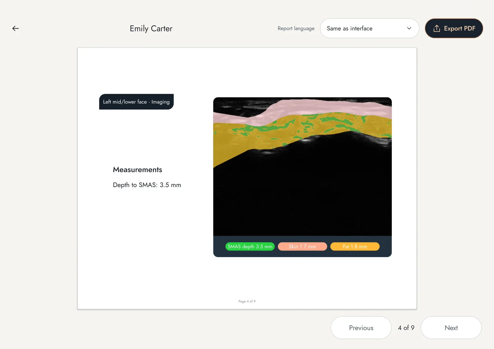
A FaceAtlas AI report page: depth to the SMAS by site, with the Export PDF control for the visit record. 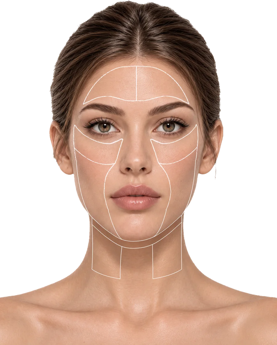
The FaceAtlas AI 21-point facial map, forehead to jawline: the fixed sites every follow-up scan repeats. - FaceAtlas AI-licentie: 3 months come with each Pro; on the L14, L20 and L24 the same software runs on a license from the first day, at $50 monthly, $500 yearly or $999 once for lifetime use (Suresult store, checked Nov 16, 2026), sold until 11:59 PM PT, December 31, 2026
- Needle and cannula tools: Needle Enhance, the in-plane and out-of-plane puncture guide, Flip and Grid (Suresult store, checked Nov 16, 2026)
- Verslaglegging: cine up to 1,000 frames, about 165 s; JPEG, PNG, MP4 and DICOM export with no subscription (Suresult store, checked Nov 16, 2026)
Waarom deze positie in de ranglijst. 24 MHz, the frequency of Meng’s facial norms, takes the full dermal credit (Meng 2022). The 25 mm deepest preset holds the dermis, the cannula planes and the zygoma’s bone line, and the deepest facial-artery reading, 15.0 mm (Ten 2021), and gives up depth credit only because the criterion runs to 40 mm for the buttock. Doppler, software, needle tools and records take full marks, and the five-year total, $5,004 with the lifetime license sold until 11:59 PM PT, December 31, 2026, is scored against the field’s lowest, the Suresult L14’s $3,046 (Suresult store, checked Nov 16, 2026).
Sub-scores: dermal resolution 20, depth 6.25, Doppler 20, dermal-thickness and follow-up software 20, needle and cannula guidance 10, records 5, five-year cost 9.08.
Every Suresult product is FDA 510(k)-cleared, FaceAtlas AI and the gel pads included (FDA 510(k) K211321).
L24Pro, L20Pro of L14Pro? The three Pros share 256 elements, three Doppler modes, Needle Enhance, up to 4 hours per charge and 3 months of FaceAtlas AI, so depth decides: the L24Pro holds the face and neck to 25 mm; the Suresult L20Pro handheld ultrasound ($3,560) adds 30 and 40 mm presets for the deep temple, buttock and thigh; and the Suresult L14Pro lineaire probe ($3,293) reaches 80 mm for deeper body filler (Suresult store, checked Nov 16, 2026).
Het meest geschikt voor: injectors who map the vessels, place Sculptra, Radiesse or HArmonyCa and measure the dermis at every follow-up on one 24 MHz probe (Suresult store, checked Nov 16, 2026). Kies een model uit deze productfamilie als: the 24 MHz band at $3,293 with FaceAtlas AI on a license suits you (the L24), or the 16/20 MHz band at $2,670 covers your sites (the L20; Suresult store, checked Nov 16, 2026). Vergelijk zorgvuldig als: you want Clarius’s published six-zone skin protocol on an Essentials membership (the Clarius L20 HD3), or a 50 MHz console for dermal readings alone (the Cortex DermaScan C; Cortex page, checked Nov 16, 2026).
Ook in deze productfamilie
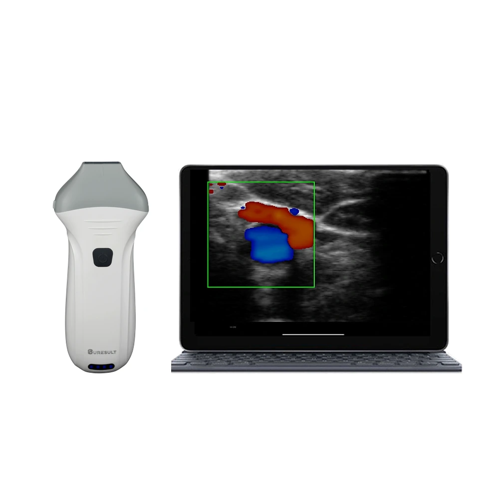
Suresult L20Pro high-frequency ultrasound probe · $3,560 (Suresult store, checked Nov 16, 2026) · 89.3/100. 16/20 MHz with presets to 40 mm: the body dermis and the full buttock range on one probe, with the full Pro kit (Suresult store, checked Nov 16, 2026).
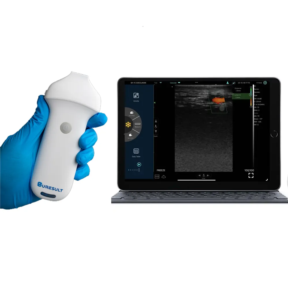
Suresult L24 high-frequency linear probe · $3,293 (Suresult store, checked Nov 16, 2026) · 81.9/100. The L24Pro’s band and 25 mm window on 192 elements, with a 2-hour battery, all three Doppler modes and Needle Enhance; FaceAtlas AI on a license (Suresult store, checked Nov 16, 2026).
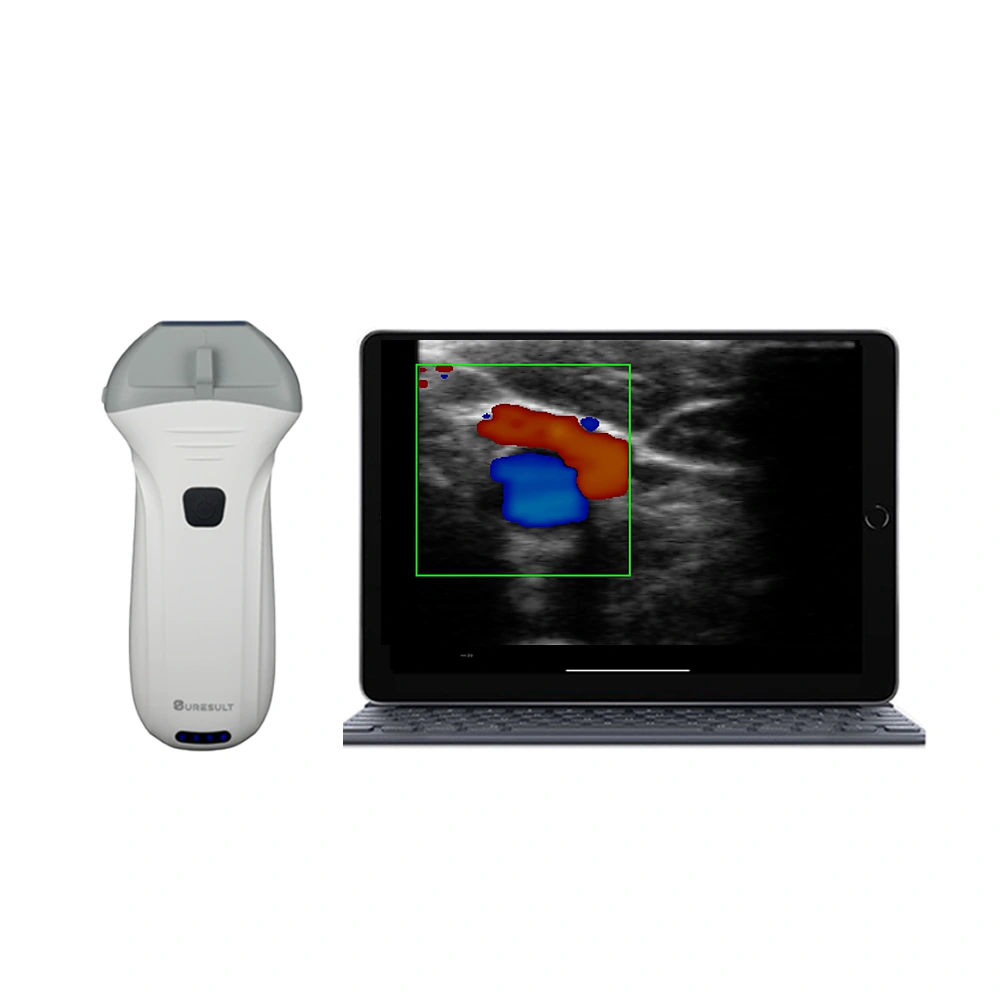
Suresult L14Pro handheld echoprobe · $3,293 (Suresult store, checked Nov 16, 2026) · 81.3/100. 10/14 MHz with presets to 80 mm for deeper filler on the buttock, thigh and abdomen, with the full Pro kit (Suresult store, checked Nov 16, 2026).
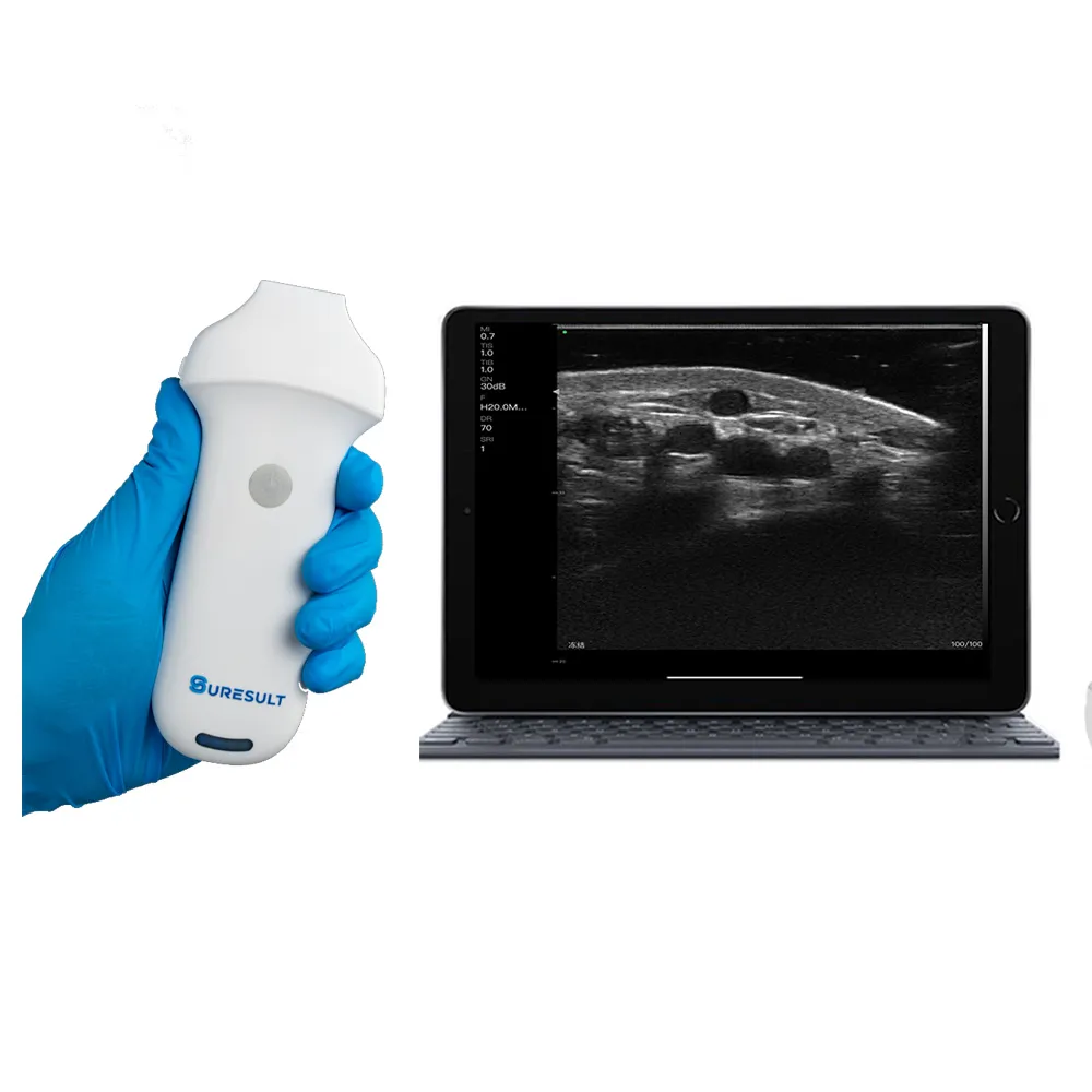
Suresult L20 wireless linear ultrasound · $2,670 (Suresult store, checked Nov 16, 2026) · 81.7/100. 16/20 MHz from 10 to 40 mm on 192 elements, a 2-hour battery, all three Doppler modes and Needle Enhance; FaceAtlas AI on a license (Suresult store, checked Nov 16, 2026).
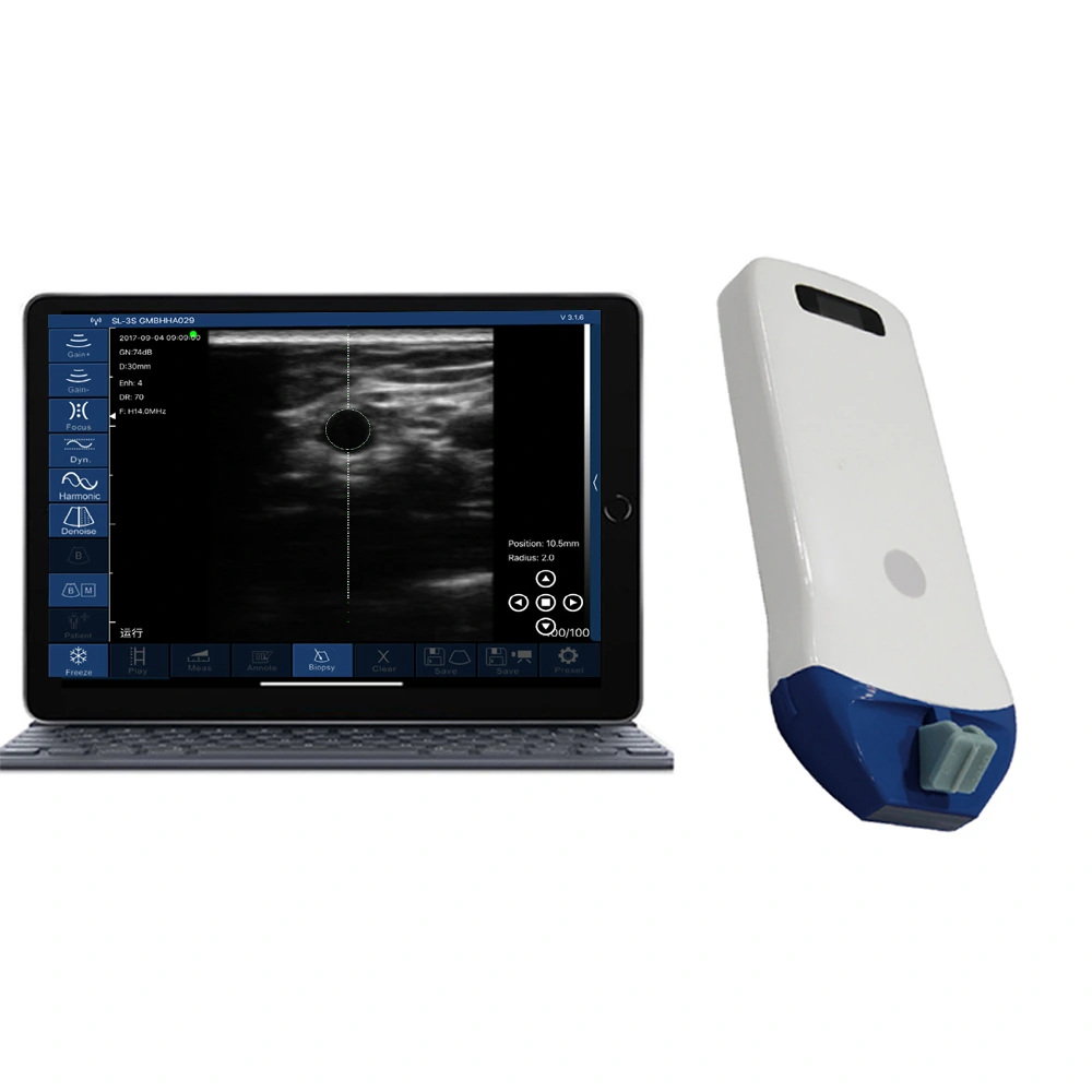
Suresult L14 wireless linear ultrasound · $2,047 (Suresult store, checked Nov 16, 2026) · 70.7/100. The lowest five-year total in the line, $3,046 with the lifetime license, sold until 11:59 PM PT, December 31, 2026: 10/14 MHz to 80 mm, 192 elements, all three Doppler modes and puncture guides (Suresult store, checked Nov 16, 2026).
Suresult Ø35 × 4 mm aesthetic gel pads (Suresult store, checked Nov 16, 2026) · Suresult handhelds for aesthetics · Suresult handhelds for dermatology
02 Clarius L20 HD3 — Best for a Published Biostimulation Protocol at 20 MHz
8–20 MHz linear · images to 4 cm · 25 mm field of view · Color and Power Doppler with the scanner · Auto Aesthetic Calipers · $5,395 plus Essentials (Clarius store, checked Nov 16, 2026)
Score: 64.8/100
$5,395 · 290 g · ~60 min scan time · IP67 · drop-tested at 1 m · 3-year warranty (Clarius store and aesthetics page, checked Nov 16, 2026)
Oordeel: Buy the L20 HD3 for a 20 MHz cordless linear that its maker has set up for biostimulator follow-up, with a six-zone skin protocol for dermal thickness and the SLEB, if you will keep the Essentials membership for PW Doppler, Needle Enhance, storage and DICOM (Clarius aesthetics page, checked Nov 16, 2026).
- Follow-up tools: Auto Aesthetic Calipers for skin, fat and tissue thickness; echogenicity Histograms; the MKTG-00355 protocol, recording dermal thickness, the SLEB, echogenicity and homogeneity in six zones at 1, 3 and 6 months (Clarius MKTG-00355 protocol, checked Nov 16, 2026)
- Doppler: Color and Power with the scanner, PW with Essentials (Clarius store, checked Nov 16, 2026)
- Naaldfuncties en beeldopslag: Needle Enhance with Essentials and a center guide Clarius describes for injections; 30 s cine; storage and DICOM with Essentials (Clarius store, checked Nov 16, 2026)
- Prijs en abonnement: $5,395 plus Essentials at $2,500 one-time, $695 a year or $1,785 every three years (Clarius store, checked Nov 16, 2026)
Waarom deze positie in de ranglijst. 20 MHz sits below the 24 MHz that takes the full dermal credit, while the 4 cm image takes the full depth credit (Clarius store, checked Nov 16, 2026). PW Doppler comes with Essentials, the Auto Aesthetic Calipers are the one software tool credited, and $7,895 over five years is the highest total among the priced rivals (Clarius store, checked Nov 16, 2026).
Sub-scores: dermal resolution 14.286, depth 10, Doppler 17, dermal-thickness and follow-up software 7, needle and cannula guidance 7.5, records 3, five-year cost 5.962.
Beperkingen. PW Doppler, Needle Enhance, storage and DICOM sit in the membership, cine runs 30 seconds, and the software measures thickness without a visit-comparison view or material naming (Clarius store, checked Nov 16, 2026).
Het meest geschikt voor: injectors who want 20 MHz with images to 4 cm, a maker-published biostimulation protocol and live thickness calipers, and who will stay on Essentials (Clarius aesthetics page, checked Nov 16, 2026). Sla dit over als: you want PW Doppler, Needle Enhance and DICOM in the purchase price, or software that compares visits and names the product in the tissue.
Ook in deze productfamilie
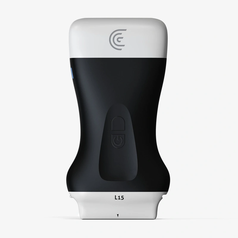
Clarius L15 HD3 · $3,795 plus Essentials (Clarius store, checked Nov 16, 2026) · 52.1/100. 5–15 MHz with a 50 mm field of view and images to 7 cm, on the same Essentials tiers; five years cost $6,295 with the one-time license (Clarius store, checked Nov 16, 2026).
Clarius aesthetics tools · Clarius L20 HD3 in de Clarius-winkel · Clarius scanner prices and Essentials costs
03 GE Vscan Air SL — Best for Color and PW Doppler at the Lowest Five-Year Rival Cost
Linear 3–12 MHz to 8 cm · 40 × 7 mm lens · Color and PW Doppler in the price · Centerline Marker · $4,999 (GE HealthCare US shop, checked Nov 16, 2026)
Score: 43.3/100
$4,999 · 218 g · 50 min of continuous scanning · IP67 · 3-year warranty (GE HealthCare US shop, checked Nov 16, 2026)
Oordeel: Buy the Vscan Air SL if you want Color and PW Doppler for the vessel map at the lowest five-year cost among the rivals, with nothing more to buy for any scored tool, and your biostimulator work centers on the subcutis and the deeper planes.
- Beeldvorming en doppler: a 3–12 MHz linear head centered at 7.7 MHz; Color and PW Doppler in the price (GE HealthCare data sheet, checked Nov 16, 2026)
- Naaldfuncties en beeldopslag: a Centerline Marker in the Vscan Air app; up to 500 exams on the mobile device; DICOM server and shared-folder export (GE HealthCare data sheet, checked Nov 16, 2026)
- Prijs en abonnement: $4,999; the optional Innovations subscription, $500 a year or $1,299 for three years, adds tools outside the score (GE HealthCare US shop, checked Nov 16, 2026)
Waarom deze positie in de ranglijst. The 12 MHz top frequency earns little dermal credit, while the 8 cm image takes the full depth credit (GE HealthCare data sheet, checked Nov 16, 2026). Color and PW come in the price, GE names no thickness, comparison or material tool, and $4,999 with every scored tool included takes the best rival cost score (GE HealthCare US shop, checked Nov 16, 2026).
Sub-scores: dermal resolution 2.857, depth 10, Doppler 13, dermal-thickness and follow-up software 0, needle and cannula guidance 5, records 3, five-year cost 9.416.
Beperkingen. 12 MHz sits under the 15 MHz aesthetic-scanning floor (Velthuis 2025), Power Doppler is off the mode list, and the needle aid is a centerline marker.
Het meest geschikt voor: clinics that want the vessel map on Color and PW Doppler to 8 cm for $4,999 with no plan to add (GE HealthCare US shop, checked Nov 16, 2026). Sla dit over als: you measure the dermis, or want Power Doppler and needle enhancement on the probe.
Productpagina van de GE Vscan Air SL · Buy at the GE HealthCare US shop
04 Butterfly iQ3 — Best Whole-Body Probe With a Facial-Vessel Preset
One 1–12 MHz probe · Vascular: Superficial preset, 1 to 4 cm · Advanced unlocks Color, Power and PW Doppler, Needle Viz and Biplane · $3,899 plus a plan (Butterfly-winkel, geraadpleegd op 6 november 2026)
Score: 42.1/100
$3,899 (Butterfly-winkel, geraadpleegd op 6 november 2026) · 300 g · up to 2 h · IPX7 waterproof, IP6X dustproof · 3-year warranty, extendable by 2 years (Butterfly iQ3 specs, checked Nov 16, 2026)
Oordeel: Buy the iQ3 if one probe has to scan the whole body as well as the face, you want Butterfly’s preset for small, slow-flow facial vessels, and you will buy Advanced for Color, Power and PW Doppler, Needle Viz and Biplane.
- Vaatfuncties: the Vascular: Superficial preset from 1 to 4 cm; Color, Power and PW Doppler with Advanced (Butterfly iQ3 specs and pricing page, checked Nov 16, 2026)
- Beeldvorming: one 1–12 MHz probe for every preset, nerve imaging to 8 cm; up to 4 linear calipers (Butterfly iQ3 specs, checked Nov 16, 2026)
- Naaldfuncties en beeldopslag: Needle Viz and Biplane with Advanced and a midline marker; unlimited cloud storage with Advanced; Basic DICOM available for purchase (Butterfly iQ3 specs and pricing page, checked Nov 16, 2026)
- Prijs en abonnement: $3,899; Advanced at $420 a year or $1,500 once for five years (Butterfly-winkel, geraadpleegd op 6 november 2026)
Waarom deze positie in de ranglijst. The 12 MHz top frequency earns little dermal credit, while the 8 cm reach takes the full depth credit (Butterfly iQ3 specs, checked Nov 16, 2026). Every Doppler mode sits behind a plan for half credit, Butterfly names no thickness, comparison or material tool, and five years with Advanced total $5,399 (Butterfly-winkel, geraadpleegd op 6 november 2026).
Sub-scores: dermal resolution 2.857, depth 10, Doppler 10, dermal-thickness and follow-up software 0, needle and cannula guidance 7.5, records 3, five-year cost 8.718.
Beperkingen. The probe tops out at 12 MHz, under the 15 MHz linear floor (Velthuis 2025); every Doppler mode needs Advanced, and DICOM is a quoted extra.
Het meest geschikt voor: clinicians who scan the whole body, with nerve imaging to 8 cm, and want biplane needle views and a facial-vessel preset on one probe (Butterfly iQ3 specs, checked Nov 16, 2026). Sla dit over als: you read the dermis, or want Doppler without a plan.
Specificaties van Butterfly iQ3 · Butterfly-abonnementen en prijzen
05 Cortex DermaScan C — Best for Dermal Measurement at 50 MHz
Broadband transducers from 20 to 50 MHz · 7 mm at 50 MHz, 23 mm at 20 MHz · A- and B-mode · FDA-cleared (FDA 510(k) K983945) · by quote (Cortex DermaScan C page, checked Nov 16, 2026)
Score: 39.8/100
Laptop-driven console · 2.44 kg main unit · mains power · 1-year warranty with an extended upgrade program (Cortex DermaScan C page, checked Nov 16, 2026)
Oordeel: Buy the DermaScan C if dermal thickness is the number you report and you want 50 MHz detail in the first 7 mm, with the vessel map and the injection on a separate linear; it read the neck after 1:4 Radiesse in Trindade de Almeida’s 2023 trial.
- Dermal tools: average, maximum and minimum skin thickness by edge detection; ROI and intensity segmentation, with areas in mm² (Cortex DermaScan C manual, checked Nov 16, 2026)
- Visit comparison: four split-screen images; a saved scan reopens beside a new one at the saved gain (Cortex DermaScan C manual, checked Nov 16, 2026)
- Imaging and records: 20 to 50 MHz, 30 × 60 µm at 50 MHz, 6 to 8 frames per second; a cineloop of the last 100 images, about 15 s; JPEG and TIFF files (Cortex DermaScan C page and manual, checked Nov 16, 2026)
- Prijs: by quote from Cortex Technology and cyberDERM inc., with the measurement software (Cortex DermaScan C page, checked Nov 16, 2026)
Waarom deze positie in de ranglijst. 50 MHz takes the full dermal credit, and edge-detection thickness with the saved-image split screen earns most of the software points (Cortex DermaScan C manual, checked Nov 16, 2026). The A- and B-mode system carries no Doppler or needle aid, its records score 0 without DICOM, and a price by quote scores 0 for cost.
Sub-scores: dermal resolution 20, depth 5.75, Doppler 0, dermal-thickness and follow-up software 14, needle and cannula guidance 0, records 0, five-year cost 0.
Beperkingen. The vessel map and the guided pass need a second, Doppler-equipped scanner, the console runs on mains power through a laptop, and the price comes by quote.
Het meest geschikt voor: clinics and trial sites that report dermal thickness at 20 to 50 MHz and map vessels on a separate linear (Cortex DermaScan C page, checked Nov 16, 2026). Sla dit over als: one instrument has to map, guide and measure.
Ook in deze productfamilie
Cortex DermaLab Combo · price on request · 38.3/100. A modular skin console: a 20 MHz probe to 3.4 mm reads skin thickness, an intensity score and the low-echogenic band, a 10 MHz probe reaches 40 mm for fat, and TEWL, elasticity and hydration probes join it (Cortex DermaLab Combo page and probe datasheets, checked Nov 16, 2026). Its Combo-4 version carried the Sculptra arm and Radiesse face trials on this page (Bravo 2026; Bravo 2025).
Image: Cortex Technology
Cortex DermaScan C page · cyberDERM, Cortex’s North American distributor
06 tpm DUB SkinScanner — Best for Automatic Skin-Thickness Readings at 33 MHz
22 or 33 MHz applicators · 6–8 mm penetration · 48 µm axial resolution at 33 MHz · 12.8 mm linear scan width · by quote (tpm DUB SkinScanner page, checked Nov 16, 2026)
Score: 29.0/100
Main unit on USB 2.0 to a Windows PC · 3.9 kg · 100–240 V AC (tpm DUB SkinScanner brochure, checked Nov 16, 2026)
Oordeel: Buy the DUB SkinScanner if you want automatic skin-thickness and density readings at 33 MHz on a PC-based system, with the vessel map and cannula work on a separate linear (tpm DUB SkinScanner page, checked Nov 16, 2026).
- Dermal tools: automatic skin-thickness and skin-density measurement; length, area, width and depth tools (tpm DUB SkinScanner page, checked Nov 16, 2026)
- Imaging and records: B-scan, HF mode, A-scan and Sum-A; 7 color scales; ScanLoop playback (tpm DUB SkinScanner page, checked Nov 16, 2026)
- Options: skin elasticity with the Vacu Elasto Pump; X-Scan with volume measurement (tpm DUB SkinScanner page, checked Nov 16, 2026)
- Prijs: by quote from tpm taberna pro medicum GmbH, with the measurement software (tpm DUB SkinScanner page, checked Nov 16, 2026)
Waarom deze positie in de ranglijst. 33 MHz takes the full 20 for the dermis, 8 mm of penetration earns 2 of 10 for depth, and automatic skin thickness earns 7 of 20 for software; with no Doppler, needle aid, named ScanLoop length or DICOM export, and a price by quote, the other criteria score 0 (tpm DUB SkinScanner page, checked Nov 16, 2026).
Sub-scores: dermal resolution 20, depth 2, Doppler 0, dermal-thickness and follow-up software 7, needle and cannula guidance 0, records 0, five-year cost 0.
Beperkingen. It images 6 to 8 mm deep with no Doppler, so the deeper planes and the vessel map run on another scanner, and the price comes by quote (tpm DUB SkinScanner page, checked Nov 16, 2026).
Het meest geschikt voor: skin clinics that track dermal thickness and density at 33 MHz (tpm DUB SkinScanner page, checked Nov 16, 2026). Sla dit over als: you need Doppler, the subcutaneous plane or a guided pass from the same instrument.
Five-year cost of each instrument with the software its scored tools need
Over five years, with the software each scored tool needs, the Suresult L24Pro costs $5,004; its one purchase after the probe is the lifetime FaceAtlas AI license, on sale until 11:59 PM PT, December 31, 2026. At prices checked November 16, 2026, Butterfly’s on November 6, the GE Vscan Air SL totals $4,999, the Butterfly iQ3 $5,399 with five years of Advanced and the Clarius L20 HD3 $7,895 with one-time Essentials, and Cortex and tpm sell by quote. I count the lifetime license because one payment covers the years after the 3 included months.
| Instrument | Prijs | Software the scored tools need | Five-year total, software bought once | Five years on a yearly plan |
|---|---|---|---|---|
| Suresult L24Pro |
$4,005 | FaceAtlas AI from month 4; imaging, Doppler and needle tools come with the probe | $5,004 with the lifetime license | $6,505: five yearly licenses at $500 |
| Clarius L20 HD3 |
$5,395 | Essentials for PW Doppler, Needle Enhance, storage and DICOM: $2,500 one-time | $7,895 | $8,870 with Essentials at $695 a year |
| GE Vscan Air SL |
$4,999 | In the price: Color and PW Doppler, the Centerline Marker, storage and DICOM | $4,999 | $4,999; the optional Innovations plan, $500 a year, adds tools outside the score |
| Butterfly iQ3 |
$3,899 | Color, Power and PW Doppler and Needle Viz come with Advanced: $1,500 for five years | $5,399 | $5,999 with Advanced at $420 a year |
| Cortex DermaScan C |
Op offertebasis | Measurement software comes with the system | By quote from Cortex Technology and cyberDERM inc. | Sold without a plan |
| tpm DUB SkinScanner |
Op offertebasis | Measurement software comes with the system | By quote from tpm taberna pro medicum | Sold without a plan |
De kostengids voor echografie in de esthetische geneeskunde sets these totals beside the wider field of aesthetic probes and software.
Every Suresult price is the final price, with No Hidden Fees: delivery worldwide with customs clearance and duties covered, orders processed within 24 hours on business days, then delivered 1–6 business days after shipment, worldwide. One FaceAtlas AI code activates one tablet. Claim the 3 months included with an L14Pro, L20Pro or L24Pro on the aanvraagpagina van FaceAtlas AI; they run from the day you press Start. After them, and from day one on the L14, L20 and L24, FaceAtlas AI runs on the maandelijks, jaarlijks of levenslang license, the lifetime one sold until 11:59 PM PT, December 31, 2026.
Hoe ik de scores bepaalde
Each maker’s card carries a score out of 100 on seven weighted criteria that I declared on October 30, 2026, before scoring any rival:
- Dermal resolution (20): 20 × (top frequency in MHz − 10) ÷ 14, held between 0 and 20, so 24 MHz and above score 20, 20 MHz 14.286 and 12 MHz 2.857.
- Depth (10): 10 × the deepest image depth in mm, counted up to 40 mm, ÷ 40.
- Doppler (20): Color 7, Power 7 and PW 6; a mode in the purchase earns its full points, a mode behind a paid tier half.
- Dermal-thickness and follow-up software (20): thickness or layer measurement in mm 7, a visit comparison or before-and-after view 7, and naming CaHA, PLLA or PCL in the scan 6; included tools earn full points and paid ones half. FaceAtlas AI scores as included on the three Pros, each sold with 3 months of it, and as paid on the L14, L20 and L24; a tool with no named tier scores as included.
- Needle and cannula guidance (10): needle enhancement 5, and 5 more for an in-plane or out-of-plane guide, biplane, or a needle center line named by the maker; a paid tool earns half.
- Records (5): cine of 60 s or more in a maker figure for the exact model 2; storage plus DICOM export 3.
- Five-year cost (15): 15 × the lowest five-year total among all 13 scored instruments ÷ this one’s total. Suresult totals add the FaceAtlas AI lifetime license to the October 30, 2026 price, with the L20 at the $2,670 its page prints; Clarius adds one-time Essentials and Butterfly five years of Advanced; the Suresult L14 sets the reference at $3,046. A system sold by quote scores 0.
Every card’s points by criterion sit in Scoreoverzicht, directly above the first ranked card.
Family models are scored inside their maker’s card: Suresult L20Pro 89.3, L24 81.9, L20 81.7, L14Pro 81.3 and L14 70.7; Clarius L15 HD3 52.1; Cortex DermaLab Combo 38.3. The Suresult card leads with the L24Pro, whose 24 MHz band takes the full dermal credit, and the Cortex card with the DermaScan C, whose 50 MHz transducer keeps it ahead of the DermaLab Combo in every run.
Seven declared reruns and twelve reading checks test the order, and the Suresult card leads every one; the table shows the seven reruns and RC1 and RC12, the two readings least favorable to it. Rival prices were read on October 30, 2026, and rival plans and tool lists on October 27 to 30.
| Berekening | Suresult-productkaart | Clarius L20 HD3 | GE Vscan Air SL | Butterfly iQ3 | DermaScan C | DUB SkinScanner |
|---|---|---|---|---|---|---|
| S1, no software credit | 70.33 | 57.75 | 43.27 | 42.08 | 25.75 | 22.0 |
| S2, cost removed | 81.25 | 58.79 | 33.86 | 33.36 | 39.75 | 29.0 |
| S3, depth removed | 84.08 | 54.75 | 33.27 | 32.08 | 34.0 | 27.0 |
| S4, card scored on the L20Pro | 89.26 | 64.75 | 43.27 | 42.08 | 39.75 | 29.0 |
| S5, card scored on the L14Pro | 81.31 | 64.75 | 43.27 | 42.08 | 39.75 | 29.0 |
| S6, Doppler van concurrenten als inbegrepen meegerekend | 90.33 | 67.75 | 43.27 | 52.08 | 39.75 | 29.0 |
| S7, quote-only makers at the median rival total, $5,399 | 90.33 | 64.75 | 43.27 | 42.08 | 48.47 | 37.72 |
| RC1, FaceAtlas AI counted as paid on the Pros | 80.33 | 64.75 | 43.27 | 42.08 | 39.75 | 29.0 |
| RC12, RC1 plus Clarius Split Screen as a paid before-and-after view and S6 | 80.33 | 71.25 | 43.27 | 52.08 | 39.75 | 29.0 |
Sculptra, Radiesse, Ellansé and biostimulator ultrasound questions
Can you reconstitute Sculptra with bacteriostatic water?
The US label specifies sterile water for injection: 5 mL hydrated for at least 2 hours for the deep dermis by 26 G needle, or 8 mL shaken for about a minute and injected at once into the subdermal plane by 25 G needle, with 1 mL of 2% lidocaine as an option.
Which plane does Sculptra go in, and how does ultrasound confirm it?
The deep dermis with the 5 mL preparation, or the subdermal plane, subcutaneous or supraperiosteal, with the 8 mL preparation (Sculptra US label); the scan shows the layer that holds the tip and each deposit.
How do you massage after Sculptra (the 5-5-5 rule)?
Five minutes of massage, five times a day, for five days after each session. In the session itself the label adds circular massage after every 3 to 4 injections and at least 2 minutes at the end with a moisturizer.
Can Sculptra be injected under the eyes?
The periorbital area sits outside the US label’s studied sites, the nasolabial fold, other facial wrinkles and the cheek, and the label asks for special care in thin skin.
What do you dilute Radiesse with, and at what ratio?
With 0.9% saline: 1:2 for décolleté wrinkles, the dilution on the US label since March 31, 2026, and by skin thickness in the consensus, 1:2 to 1:3 for normal skin, 1:4 for thin and 1:6 for atrophic (Lorenc 2022), so measure the dermis first.
What is the difference between Radiesse and hyperdilute Radiesse?
The ratio, the plane and the aim. Undiluted Radiesse adds volume subdermally or on bone; 1:1 counts as diluted and 1:2 or more as hyperdiluted (Goldie 2018), placed immediately below the dermis to build collagen (Lorenc 2022).
How do you reconstitute (mix) Radiesse?
On the décolleté label, 1.5 mL of Radiesse and 3.0 mL of 0.9% saline pass between two 5 mL syringes at least 20 times, giving 4.5 mL at 1:2, used within 30 minutes.
Does diluted Radiesse thicken the dermis on ultrasound?
Yes, in the neck, forearm and abdomen. Two sessions of 1:4 Radiesse thickened the neck dermis by 14.9% at day 120 (Trindade de Almeida 2023), 1:2 took a dermatoporotic forearm from 1115 to 1517 µm by day 45 (de Almeida 2026), and 1:4 added 0.7 mm around the umbilicus by day 70 (Lapatina 2017).
How long does diluted Radiesse last?
The course runs 2 to 3 sessions about 6 weeks apart, with a maintenance session 12 to 18 months after the first cycle (Lorenc 2022; de Almeida 2019); collagen I stood above baseline at month 7 after 1:2 to 1:6 Radiesse (Yutskovskaya 2017).
Is HArmonyCa different from Sculptra on ultrasound and in the plane?
Yes. HArmonyCa pairs crosslinked HA with CaHA microspheres for the deep dermis and the subdermal layer (HArmonyCa IFU) and scans as a bright band with dark, hole-like areas (Bravo 2023); Sculptra is PLLA and scans as ill-defined bright deposits that fade (WFUMB 2025). Hyaluronidase dissolves HArmonyCa’s HA, and excess superficial CaHA is diluted with saline (Philipp-Dormston 2025), the dissolving step covered in the ultrasound-guided filler dissolving guide.
Where can Radiesse be injected (face and body sites)?
On the US labels: moderate to severe facial wrinkles and folds such as the nasolabial folds, volume loss in the back of the hands, the jawline with Radiesse (+) and, diluted 1:2, décolleté wrinkles. The hyperdilute consensus adds the neck, upper arms, abdomen, buttocks and thighs, subdermally by cannula (de Almeida 2019).
Can ultrasound see Radiesse or Sculptra under the skin?
Yes. CaHA shows as bright deposits that cast a posterior acoustic shadow, strongest when undiluted, and PLLA as ill-defined bright deposits that fade over the months (WFUMB 2025).
Should you map facial vessels before injecting a biostimulator?
Yes. Unlike HA filler, PLLA’s effect on collagen cannot be immediately reversed (Ouyang 2025), so the Color Doppler map comes before the first pass at every site. The signs and the response to an occlusion are in the handleiding over vaatocclusie.
What MHz probe measures dermal thickness?
A linear of at least 15 MHz, the WFUMB and Delphi floor, and 18 to 24 MHz for a facial dermis of 0.58 to 1.30 mm (Meng 2022; Jeong 2023). The gids voor hoogfrequente echoapparaten compares 15, 20 and 24 MHz.
How much do skin and dermal thickness change after Sculptra?
In facial lipoatrophy, by millimeters: three sessions raised mean skin thickness by 3.9 to 6.0 mm at week 12 over baselines of 2.1 to 2.7 mm (Sculptra US label). Without lipoatrophy, the upper-arm dermis went from 1.80 to 2.70 mm by day 120 (Bravo 2026) and the mandibular dermis gained 0.18 mm by week 24 (Yen 2026).
DermaScan or DermaLab, or a handheld ultrasound?
For biostimulator work I choose a handheld linear with Doppler: the Suresult L24Pro, 90.3/100 here, reads the facial dermis at 24 MHz, maps the vessels on three Doppler modes and sets visits side by side in FaceAtlas AI. The Cortex DermaScan C (39.8) and DermaLab Combo (38.3) read the dermis at up to 50 and 20 MHz, for clinics that measure skin alone.
How long does a vessel map add to a biostimulator visit?
Under 3 minutes: Teixeira’s two-step handheld Doppler map at 20 MHz ran in 503 HA filler patients over 3 years (Teixeira 2025). In an 80-against-80 controlled study, a scan before the procedure left 70% of patients free of bruising against 28.8% with standard care (Naylor 2025).
Ellansé vs Sculptra: what is the difference?
Ellansé corrects on the day and Sculptra builds over sessions. Ellansé’s CMC gel gives immediate volume and the collagen that follows holds it, so it is injected 1:1 with no overcorrection from a ready-to-use syringe (Christen 2020; de Melo 2017; Ellansé IFU); Sculptra’s 150 mg vial is mixed with sterile water and under-corrected at every session (Haddad 2025; Sculptra US label). On the scan Ellansé shows a dark matrix with bright comet-tail spots, and Sculptra ill-defined bright deposits that fade (WFUMB 2025).
How long does Ellansé last?
Sinclair’s 2026 brochure gives up to 12 months for Ellansé-S and up to 24 months for Ellansé-M, citing the CE IFU, and de Melo’s consensus expects 1, 2, 3 and 4 years for S, M, L and E, the L and E figures extrapolated from the S and M data (de Melo 2017). In nine women treated in the malar area with Ellansé-S, the result outlasted that figure: the 3D-measured volume gain at 24 months exceeded the volume injected by 50% to 150% (Angelo-Khattar 2022).
Bronnen
- Ouyang R, Su X, Liang Y, et al. Advances in poly-L-lactic acid injections for facial and neck rejuvenation. Plast Reconstr Surg Glob Open. 2025;13(8):e7029. — doi.org, October 30, 2026
- Trindade de Almeida AR, Marques ERMC, Contin LA, et al. Efficacy and tolerability of hyperdiluted calcium hydroxylapatite (Radiesse) for neck rejuvenation: clinical and ultrasonographic assessment. Clin Cosmet Investig Dermatol. 2023;16:1341–1349. — doi.org, October 30, 2026
- Bravo BSF, de Souza ALL, Barreiro EBV, et al. Poly-L-lactic acid (PLLA) in arm contouring: a prospective and blinded trial of Rennova Elleva and Sculptra. Front Surg. 2026;13:1782368. — doi.org, October 30, 2026
- Ten B, Kara T, Kaya Tİ, et al. Beoordeling van variaties in het verloop en de diepte van de arteria facialis met dopplerechografie. J Cosmet Dermatol. 2021;20(7):2247–2258. — doi.org, October 30, 2026
- Philipp-Dormston WG, De Boulle K, Eccleston DB, et al. Prevention and management of complications with a hyaluronic acid-calcium hydroxyapatite hybrid injectable. Clin Cosmet Investig Dermatol. 2025;18:431–441. — doi.org, October 30, 2026
- Cavallini M, Braz A, Greiner-Krüger D, et al. Global recommendations for facial rejuvenation using a hyaluronic acid and calcium hydroxyapatite hybrid injectable. J Cosmet Dermatol. 2026;25(1):e70608. — doi.org, October 30, 2026
- Young SM, Goh ASC, Jeyabal P, Ooi ASH. Visual loss in biostimulator injectables: a review of incidence, risk factors, etiology and management proposal. Aesthetic Plast Surg. 2026;50(5):1994–2005. — doi.org, October 30, 2026
- Velthuis PJ, Alfageme F, Cartier H, et al. Het gebruik van echografische beeldvorming bij esthetische injectables: een aangepaste Delphi-consensus. J Plast Reconstr Aesthet Surg. 2025;104:33–37. — doi.org, October 30, 2026
- Teixeira DA. Een nieuwe tijdbesparende techniek voor echogeleide fillerinjectie: een retrospectief onderzoek over 3 jaar. Aesthet Surg J Open Forum. 2025;7:ojaf061. — doi.org, October 30, 2026
- Phumyoo T, Jiirasutat N, Jitaree B, et al. Lokalisatie en topografie van de arteriën in het middelste deel van het voorhoofd ter voorkoming van complicaties na voorhoofdsaugmentatie: conventionele kadaverdissectie en echografisch onderzoek. J Craniofac Surg. 2020;31(7):2029–2035. — doi.org, October 30, 2026
- Sigrist R, Desyatnikova S, Chammas MC, Vasconcelos-Berg R. Beste werkwijzen voor het gebruik van hoogfrequente echografie bij de geleiding van esthetische fillerinjecties—deel 1: het bovenste derde deel van het gezicht. Diagnostics (Basel). 2024;14(16):1718. — doi.org, October 30, 2026
- Galderma. Sculptra (injectable poly-L-lactic acid): physician instructions for use, PMA P030050/S039, approved April 25, 2023. — accessdata.fda.gov, October 30, 2026
- U.S. Food and Drug Administration. Premarket approval P030050, Sculptra (Q-Med AB), with supplements. — accessdata.fda.gov, November 16, 2026
- Chang G, Tan Z, Ting W, et al. Efficacy and safety of a poly-L-lactic acid filler for temporal augmentation: a randomized, no-treatment control, evaluator-blinded, multicenter study. Aesthet Surg J. 2026;46(6):652–662. — doi.org, October 30, 2026
- de Melo F, Nicolau P, Piovano L, et al. Recommendations for volume augmentation and rejuvenation of the face and hands with the new generation polycaprolactone-based collagen stimulator (Ellansé). Clin Cosmet Investig Dermatol. 2017;10:431–440. — doi.org, October 30, 2026
- Choi DH, Eom JR, Lee JW, et al. Zygomatico-orbital artery: the largest artery in the temporal area. J Plast Reconstr Aesthet Surg. 2018;71(4):484–489. — doi.org, October 30, 2026
- Liu S, Yan W, Wang G, et al. Topographic anatomy of the zygomatico-orbital artery: implications for improving the safety of temporal augmentation. Plast Reconstr Surg. 2021;148(1):19e–27e. — doi.org, October 30, 2026
- Desyatnikova S. Echogeleide fillerinjectie in de slaap. Facial Plast Surg Aesthet Med. 2022;24(6):501–503. — doi.org, October 30, 2026
- Bae H, Choi YJ, Lee KL, et al. The deep temporal arteries: anatomical study with application to augmentations procedures of the temple. Clin Anat. 2023;36(3):386–392. — doi.org, October 30, 2026
- Zhou YH, Chen CL, Luo CE, et al. Deep temporal artery anatomy: implications for improving the safety of deep temporal injections. Aesthetic Plast Surg. 2023;47(5):2045–2050. — doi.org, October 30, 2026
- Lorenc ZP, Bass LM, Fitzgerald R, et al. Composite facial volumization with calcium hydroxylapatite (CaHA) for the treatment of aging. Aesthet Surg J. 2018;38(Suppl 1):S18–S23. — doi.org, October 30, 2026
- Lorenc ZP, Bass LM, Fitzgerald R, et al. Physiochemical characteristics of calcium hydroxylapatite (CaHA). Aesthet Surg J. 2018;38(Suppl 1):S8–S12. — doi.org, October 30, 2026
- Koziej M, Polak J, Wnuk J, et al. The transverse facial artery anatomy: implications for plastic surgery procedures. PLoS One. 2019;14(2):e0211974. — doi.org, October 30, 2026
- Vasconcelos-Berg R, Desyatnikova S, Bonavia P, et al. Aanbevolen werkwijzen voor het gebruik van hoogfrequente echografie bij het begeleiden van esthetische fillerinjecties—deel 2: het middelste derde deel van het gezicht, de neus en de traangroeven. Diagnostics (Basel). 2024;14(22):2544. — doi.org, October 30, 2026
- Hernandez C, Viscomi B, Faria G, et al. Multilayer technique using calcium hydroxylapatite biostimulation with different dilutions in the lateral face. Aesthet Surg J Open Forum. 2024;6:ojae049. — doi.org, October 30, 2026
- Munoz-Lora VRM, Dias D, Bezerra F, et al. A comprehensive evaluation of patient satisfaction and clinical outcomes in poly-L-lactic acid treatments—a randomized, interventional, and comparative clinical study. J Cosmet Dermatol. 2026;25(5):e70891. — doi.org, October 30, 2026
- Pourani MR, Ebrahimzade M, Goudarzi E, et al. Beoordeling van variaties in het verloop, de diameter en de diepte van de aangezichtsslagader met dopplerechografie: een systematische review en meta-analyse. J Cosmet Dermatol. 2025;24(9):e70431. — doi.org, October 30, 2026
- Vasconcelos-Berg R, Desyatnikova S, Bonavia P, et al. Aanbevolen werkwijzen voor het gebruik van hoogfrequente echografie bij het geleiden van esthetische fillerinjecties—deel 3: het onderste derde deel van het gezicht. Diagnostics (Basel). 2025;15(7):921. — doi.org, October 30, 2026
- Pistoia F, Camerino PL, Ioppi A, et al. Echografie met hoge resolutie van de aangezichtsvaten met nieuwe oriëntatiepunten voor de vena facialis bij reconstructieve chirurgie en dermale injecties. Eur Radiol Exp. 2023;7(1):49. — doi.org, October 30, 2026
- Wu BQ, Lin YJ, Chang CC, et al. A novel six-point supraperiosteum injection with calcium hydroxyapatite for jawline refining and facial anti-aging in Asian patients. Aesthetic Plast Surg. 2025;49(19):5316–5324. — doi.org, October 30, 2026
- Bravo BSF, de Almeida TSC, Carvalho RM, et al. Dermal thickness increase and aesthetic improvement with hybrid product combining hyaluronic acid and calcium hydroxyapatite: a clinical and sonographic analysis. Plast Reconstr Surg Glob Open. 2023;11(6):e5055. — doi.org, October 30, 2026
- Yen CW, Lin C, Chien Y, Huang HW. Poly-L-lactic acid skin-tightening protocol for improvement of lower-face laxity in Asian patients: a prospective three-dimensional imaging and ultrasound assessment study. J Craniofac Surg. 2026 Oct 23. — doi.org, October 30, 2026
- Tansatit T, Phumyoo T, Jitaree B, et al. Investigation of the presence and variation of the ascending mental artery: conventional dissections and ultrasonographic study. J Cosmet Dermatol. 2019;18(6):1821–1829. — doi.org, October 30, 2026
- Cotofana S, Hexsel D, Avelar LE, et al. Calculating the thickness of the superficial fatty layer of the body using age, gender, and body mass index. J Drugs Dermatol. 2020;19(1):36–44. — doi.org, October 30, 2026
- de Almeida AT, Figueredo V, da Cunha ALG, et al. Consensus recommendations for the use of hyperdiluted calcium hydroxyapatite (Radiesse) as a face and body biostimulatory agent. Plast Reconstr Surg Glob Open. 2019;7(3):e2160. — doi.org, October 30, 2026
- Haddad A, Avelar L, Fabi SG, et al. Injectable poly-L-lactic acid for body aesthetic treatments: an international consensus on evidence assessment and practical recommendations. Aesthetic Plast Surg. 2025;49(5):1507–1517. — doi.org, October 30, 2026
- Merz North America. Radiesse injectable implant: instructions for use, PMA P050052/S162, approved March 31, 2026. — accessdata.fda.gov, October 30, 2026
- Merz North America. Radiesse diluted 1:2 for décolleté wrinkles: summary of safety and effectiveness data, PMA P050052/S162. — accessdata.fda.gov, October 30, 2026
- U.S. Food and Drug Administration. Premarket approval P050052, Radiesse and Radiesse (+) (Merz North America), with supplements. — accessdata.fda.gov, November 16, 2026
- Wang Y, Tian M, Guo R, et al. Quantification of normal skin thickness using very high-frequency ultrasound: a clinical study in Chinese adults. Quant Imaging Med Surg. 2025;15(6):5218–5231. — doi.org, October 30, 2026
- Ducati EPJ, Magacho-Vieira FN, Câmara CBF, et al. Reviewing the ultrasound anatomy of the gluteal region and the mapping of fillers. J Cosmet Dermatol. 2025;24(10):e70499. — doi.org, October 30, 2026
- Lee W, Moon HJ, Kim MS, et al. Pre-injection ultrasound scanning for treating temporal hollowing. J Cosmet Dermatol. 2022;21(6):2420–2425. — doi.org, October 30, 2026
- Suwanchinda A, Rudolph C, Hladik C, et al. The layered anatomy of the jawline. J Cosmet Dermatol. 2018;17(4):625–631. — doi.org, October 30, 2026
- Merz North America. Radiesse (+) lidocaine injectable implant: instructions for use, PMA P050052/S129, approved September 1, 2021. — accessdata.fda.gov, October 30, 2026
- Merz North America. Radiesse (+) for jawline contour: summary of safety and effectiveness data, PMA P050052/S129. — accessdata.fda.gov, October 30, 2026
- Sigrist RMS, Gonzalez C, Schelke L, et al. Dopplerechografische bevindingen bij fillergerelateerde vasculaire bijwerkingen in het gezicht: een internationale multicentrische studie. Diagnostics (Basel). 2026;16(11):1587. — doi.org, October 30, 2026
- Chammas MC, Sigrist R, Alfageme F, et al. WFUMB-standpunt: consensus over de beste werkwijze bij echografie in de esthetische dermatologie. Ultrasound Med Biol. 2025;51(11):2173–2193. — doi.org, October 30, 2026
- Bustamante FQF, Peyneau PD, Verner FS, et al. Diepte en hemodynamiek van de arteria angularis in de piriforme ruimte: een dwarsdoorsnedeonderzoek met dopplerechografie. Imaging Sci Dent. 2025;55(3):310–317. — doi.org, October 30, 2026
- McCarthy AD, Hartmann C, Durkin A, et al. A morphological analysis of calcium hydroxylapatite and poly-L-lactic acid biostimulator particles. Skin Res Technol. 2024;30(6):e13764. — doi.org, October 30, 2026
- Baumann K, Alm J, Norberg M, Ejehorn M. Immediate use after reconstitution of a biostimulatory poly-L-lactic acid injectable implant. J Drugs Dermatol. 2020;19(12):1199–1203. — doi.org, October 30, 2026
- Fabi S, Hamilton T, LaTowsky B, et al. Effectiveness and safety of Sculptra poly-L-lactic acid injectable implant in the correction of cheek wrinkles. J Drugs Dermatol. 2024;23(1):1297–1305. — doi.org, October 30, 2026
- Lam SM, Azizzadeh B, Graivier M. Injectable poly-L-lactic acid (Sculptra): technical considerations in soft-tissue contouring. Plast Reconstr Surg. 2006;118(3 Suppl):55S–63S. — doi.org, October 30, 2026
- AL-Attab R, Li J, Lo C, et al. Longitudinal multimodal imaging assessment of tissue changes following poly-L-lactic acid treatment for midfacial rejuvenation: an exploratory pilot study. J Cosmet Dermatol. 2026;25:e71178. — doi.org, October 30, 2026
- Zubair R, Ishii L, Loyal J, et al. SPLASH: split-body randomized clinical trial of poly-L-lactic acid for adipogenesis and volumization of the hip dell. Dermatol Surg. 2024;50(12):1155–1162. — doi.org, October 30, 2026
- Liu F, Qi G, Liu Q. Advancing techniques for temporal hollowing correction: insights from ultrasound-guided PLLA injection. Aesthetic Plast Surg. 2025;49(17):4754–4758. — doi.org, October 30, 2026
- Arruda S, Swearingen A, Elmadany Z, et al. Prospective single-site open-label study assessing safety and efficacy of poly-L-lactic acid for temple volume loss. J Drugs Dermatol. 2026;25(1):43–47. — doi.org, October 30, 2026
- Li A, Long A, Fang R, et al. High-frequency ultrasound for long-term safety assessment of poly-L-lactic acid facial filler. Dermatol Surg. 2022;48(10):1071–1075. — doi.org, October 30, 2026
- Casabona G, Alfertshofer M, Kaye K, et al. Ex vivo product distribution of injectable biostimulator substances. Aesthet Surg J. 2023;43(5):NP348–NP356. — doi.org, October 30, 2026
- Goldberg D, Guana A, Volk A, Daro-Kaftan E. Single-arm study for the characterization of human tissue response to injectable poly-L-lactic acid. Dermatol Surg. 2013;39(6):915–922. — doi.org, October 30, 2026
- Goldie K, Peeters W, Alghoul M, et al. Global consensus guidelines for the injection of diluted and hyperdiluted calcium hydroxylapatite for skin tightening. Dermatol Surg. 2018;44 Suppl 1:S32–S41. — doi.org, October 30, 2026
- Lorenc ZP, Black JM, Cheung JS, et al. Skin tightening with hyperdilute CaHA: dilution practices and practical guidance for clinical practice. Aesthet Surg J. 2022;42(1):NP29–NP37. — doi.org, October 30, 2026
- Merz North America. Radiesse injectable implant for the dorsum of the hand: instructions for use, PMA P050052/S049, approved June 4, 2015. — accessdata.fda.gov, October 30, 2026
- Fabi S, Moradi A, Hill D, et al. Diluted calcium hydroxylapatite carboxymethylcellulose (Radiesse) for décolleté wrinkles: safety and effectiveness in adult females. Aesthet Surg J. 2026 Jul 9:sjag142. — doi.org, October 30, 2026
- Wortsman X, Quezada N, Peñaloza O, et al. Ultrasonographic patterns of calcium hydroxyapatite according to dilution and mix with hyaluronic acid. J Ultrasound Med. 2023;42(9):2065–2072. — doi.org, October 30, 2026
- Kim J. Sonographic analysis of CureJet injection depth consistency after abdominal stretch mark treatment using hyperdiluted calcium hydroxylapatite. Plast Reconstr Surg Glob Open. 2025;13(4):e6654. — doi.org, October 30, 2026
- Eustaquio de Lima Faria G, Queiroga Bastos A, Lima Vilela C, et al. Assessment of the interference of hyperdiluted calcium hydroxyapatite for neck rejuvenation in the ultrasonographic evaluation of thyroid. J Drugs Dermatol. 2022;21(11):1243–1248. — doi.org, October 30, 2026
- Gonzalez C, Duque-Clavijo V, Suárez S, et al. High-resolution ultrasound evaluation of common dorsal hand rejuvenation techniques: a multicenter study. Cureus. 2026;18(1):e101017. — doi.org, October 30, 2026
- de Almeida ART, de Sousa Marins Barbosa MC, Michalany A, McCarthy AD. Calcium hydroxylapatite-carboxymethylcellulose as a treatment for forearm dermatoporosis: a prospective, delayed-start, contralateral case series. J Cosmet Dermatol. 2026;25(4):e70859. — doi.org, October 30, 2026
- Allergan Aesthetics. HArmonyCa with lidocaine: instructions for use, GR-HAR-230045, November 2023. — res.cloudinary.com, October 30, 2026
- Allergan Aesthetics. Health Canada authorization of HArmonyCa with lidocaine: press release, May 25, 2026. — newswire.ca, October 30, 2026
- Urdiales-Gálvez F, Braz A, Cavallini M. Facial rejuvenation with the new hybrid filler HArmonyCa: clinical and aesthetic outcomes assessed by 2D and 3D photographs, ultrasound, and elastography. J Cosmet Dermatol. 2023;22(8):2186–2197. — doi.org, October 30, 2026
- Christen MO, Vercesi F. Polycaprolactone: how a well-known and futuristic polymer has become an innovative collagen-stimulator in esthetics. Clin Cosmet Investig Dermatol. 2020;13:31–48. — doi.org, October 30, 2026
- AQTIS Medical BV. ELLANSÉ S, M, L, E: instructions for use e.00560056-001-000 (Switzerland, version 02), March 29, 2022. — sinclairpharma.qarad.eifu.online, October 31, 2026
- Angelo-Khattar M. Objective assessment of the long-term volumizing action of a polycaprolactone-based filler. Clin Cosmet Investig Dermatol. 2022;15:2895–2901. — doi.org, 31 oktober 2026
- Sinclair. Ellansé: indication, safety and availability. — ellanse.com, October 31, 2026
- Sinclair. Ellansé brochure for healthcare professionals and distributors, 2026. — ellanse.com, October 31, 2026
- Hu K, Wang S. Comparing the safety and effectiveness of PCL and PLLA injections for nasolabial fold correction. J Cosmet Dermatol. 2025;24(9):e70414. — doi.org, 31 oktober 2026
- AQTIS Medical B.V. Safety and effectiveness of ELLANSÉ-S dermal filler in the treatment of nasolabial folds (NCT06984835). ClinicalTrials.gov. — clinicaltrials.gov, October 31, 2026
- Kim JS. Changes in dermal thickness in biopsy study of histologic findings after a single injection of polycaprolactone-based filler into the dermis. Aesthet Surg J. 2019;39(12):NP484–NP494. — doi.org, 31 oktober 2026
- Figueiredo VM. A five-patient prospective pilot study of a polycaprolactone based dermal filler for hand rejuvenation. J Cosmet Dermatol. 2013;12(1):73–77. — doi.org, 31 oktober 2026
- Hong GW, Wong IKJ, Wan J, et al. A dual-phase approach to facial rejuvenation integrating contouring and ligamentous (CL Code) anchoring with a polycaprolactone-based collagen stimulator. J Cosmet Dermatol. 2026;25(4):e70796. — doi.org, 31 oktober 2026
- Merz North America. Prospective, single site, controlled comparative study to identify and assess tissue responses after injection with various biostimulatory products (NCT06093815), results posted September 22, 2026. ClinicalTrials.gov. — clinicaltrials.gov, October 31, 2026
- Wortsman X, Quezada N. Ultrasound morphology of polycaprolactone filler. J Ultrasound Med. 2017;36(12):2611–2615. — doi.org, October 30, 2026
- Tang Y, Wang Z, Bi M, Jia Y. Clinical applications of high-frequency ultrasound in filler identification and complication management. Semin Ultrasound CT MR. 2026;47(3-4):239–247. — doi.org, 31 oktober 2026
- Wu L, Salti G, Cotofana S, Vercesi F. Point-of-care ultrasound (POCUS) for precision management in Ellanse-treated patients. J Cosmet Dermatol. 2025;24(6):e70262. — doi.org, October 30, 2026
- Meng Y, Feng L, Shan J, et al. Application of high-frequency ultrasound to assess facial skin thickness in association with gender, age, and BMI in healthy adults. BMC Med Imaging. 2022;22(1):113. — doi.org, October 30, 2026
- Jeong KM, Seo JY, Kim A, et al. Echografische analyse van de dikte van de gezichtshuid in relatie tot leeftijd, locatie, geslacht en body mass index. Skin Res Technol. 2023;29(8):e13426. — doi.org, October 30, 2026
- Alsing KK, Serup J. High-frequency ultrasound skin thickness: comparison of manual reading and automatic border detection includes assessment of interobserver variation of measurement. Skin Res Technol. 2020;26(6):832–838. — doi.org, October 30, 2026
- Santiago T, Santos EJF, Luis M, et al. Normal percentile reference curves for skin ultrasound thickness and stiffness at Rodnan sites. RMD Open. 2022;8(2):e002577. — doi.org, October 30, 2026
- Phillips J, Reynolds KJ, Gordon SJ. Dermal thickness and echogenicity using DermaScan C high frequency ultrasound: methodology and reliability testing in people with and without primary lymphoedema. Skin Res Technol. 2020;26(6):813–823. — doi.org, October 30, 2026
- Peperkamp K, Verhulst AC, Tielemans HJP, et al. The inter-rater and test-retest reliability of skin thickness and skin elasticity measurements by the DermaLab Combo in healthy participants. Skin Res Technol. 2019;25(6):787–792. — doi.org, October 30, 2026
- Nicolescu AC, Ionescu S, Ancuta I, et al. Subepidermal low-echogenic band—its utility in clinical practice: a systematic review. Diagnostics (Basel). 2023;13(5):970. — doi.org, October 30, 2026
- Sandby-Møller J, Wulf HC. Ultrasonographic subepidermal low-echogenic band, dependence of age and body site. Skin Res Technol. 2004;10(1):57–63. — doi.org, October 30, 2026
- Gniadecka M, Gniadecki R, Serup J, Søndergaard J. Ultrasound structure and digital image analysis of the subepidermal low echogenic band in aged human skin: diurnal changes and interindividual variability. J Invest Dermatol. 1994;102(3):362–365. — doi.org, October 30, 2026
- Wang S, Yu RX, Fan W, et al. Detection of skin thickness and density in healthy Chinese people by using high-frequency ultrasound. Skin Res Technol. 2023;29(1):e13219. — doi.org, October 30, 2026
- Bravo BSF, Bravo LG, Gouvea BF, et al. Calcium hydroxylapatite-based fillers in facial rejuvenation: a prospective, single-center, unblinded comparative outcome study of Radiesse vs. Rennova Diamond Intense. J Clin Med. 2025;14(12):4072. — doi.org, October 30, 2026
- Lapatina NG, Pavlenko T. Diluted calcium hydroxylapatite for skin tightening of the upper arms and abdomen. J Drugs Dermatol. 2017;16(9):900–906. — pubmed.ncbi.nlm.nih.gov, 30 oktober 2026
- Mlosek RK, Migda B, Migda M. High-frequency ultrasound in the 21st century. J Ultrason. 2021;20(83):e233–e241. — pmc.ncbi.nlm.nih.gov, October 30, 2026
- Jaguś D, Skrzypek E, Migda B, et al. Het nut van dopplerechografie in de esthetische geneeskunde. J Ultrason. 2021;20(83):e268–e272. — pmc.ncbi.nlm.nih.gov, October 30, 2026
- Iyengar S, Makin IR, Sadhwani D, et al. Utility of a high-resolution superficial diagnostic ultrasound system for assessing skin thickness: a cross-sectional study. Dermatol Surg. 2018;44(6):855–864. — doi.org, October 30, 2026
- Yutskovskaya YA, Kogan EA. Improved neocollagenesis and skin mechanical properties after injection of diluted calcium hydroxylapatite in the neck and décolletage: a pilot study. J Drugs Dermatol. 2017;16(1):68–74. — pubmed.ncbi.nlm.nih.gov, 30 oktober 2026
- Naylor D, Velthuis P. Integrating facial ultrasound into medical esthetics practice. J Am Acad Dermatol. 2025;93(6):1526–1528. — doi.org, October 30, 2026
- Clarius L20 HD3: store page with specifications and Essentials plans. — store.clarius.com, November 16, 2026
- Clarius Essentials one-time license: store listing. — store.clarius.com, November 16, 2026
- Clarius Classroom: center guide. — clarius.com, November 16, 2026
- Clarius for aesthetics: Auto Aesthetic Calipers and histograms. — clarius.com, November 16, 2026
- Clarius blog: quantifying biostimulation and skin remodeling with Clarius ultrasound. — clarius.com, November 16, 2026
- Clarius biostimulation scanning protocol, MKTG-00355 Rev 1. — clarius.com, November 16, 2026
- Clarius L15 HD3: winkelpagina. — store.clarius.com, November 16, 2026
- GE HealthCare Vscan Air SL: US shop with Innovations subscriptions. — gehealthcare.com, November 16, 2026
- GE HealthCare Vscan Air SL: productpagina. — gehealthcare.com, November 16, 2026
- GE HealthCare Vscan Air: specificaties. — vscanair-support.gehealthcare.com, November 16, 2026
- GE HealthCare Vscan Air: configuration and Centerline Marker. — vscanair-support.gehealthcare.com, November 16, 2026
- GE HealthCare Vscan Air: scan time per charge. — vscanair-support.gehealthcare.com, November 16, 2026
- Butterfly Network: prijzen en lidmaatschappen. — butterflynetwork.com, November 16, 2026
- Specificaties van de Butterfly iQ3. — butterflynetwork.com, November 16, 2026
- Cortex Technology DermaScan C: product page. — cortex.dk, November 16, 2026
- Cortex Technology DermaScan C: instruction manual. — assets.ctfassets.net, November 16, 2026
- U.S. Food and Drug Administration. 510(k) K983945, Cortex DermaScan C ultrasonic system. — accessdata.fda.gov, November 16, 2026
- cyberDERM inc.: Cortex Technology instruments in North America. — cyberderm-inc.com, November 16, 2026
- Cortex Technology DermaLab Combo: product page. — cortex.dk, November 16, 2026
- Cortex Technology DermaLab ultrasound probe V2: datasheet. — assets.ctfassets.net, November 16, 2026
- Cortex Technology DermaLab subcutaneous probe. — cortex.dk, November 16, 2026
- Cortex Technology DermaLab Combo 4: instruction manual. — assets.ctfassets.net, November 16, 2026
- tpm taberna pro medicum: DUB SkinScanner. — tpm-online.de, November 16, 2026
- tpm taberna pro medicum: DUB SkinScanner brochure, 2022. — eotech.fr, November 16, 2026
- Suresult L24Pro. — suresultmed.com, November 16, 2026
- Suresult L20Pro. — suresultmed.com, November 16, 2026
- Suresult L14Pro. — suresultmed.com, November 16, 2026
- Suresult L24. — suresultmed.com, November 16, 2026
- Suresult L20. — suresultmed.com, November 16, 2026
- Suresult L14. — suresultmed.com, November 16, 2026
- FaceAtlas AI. — suresultmed.com, November 16, 2026
- Maandlicentie voor FaceAtlas AI. — suresultmed.com, November 16, 2026
- Jaarlicentie voor FaceAtlas AI. — suresultmed.com, November 16, 2026
- Levenslange licentie voor FaceAtlas AI. — suresultmed.com, November 16, 2026
- Suresult echogelpads voor de esthetische geneeskunde. — suresultmed.com, November 16, 2026
- Suresult-certificeringen en conformiteit. — suresultmed.com, November 16, 2026
Choose your Pro by the deepest plane you treat
For the face and neck, where the dermis, the vessels and the cannula plane fit on one 24 MHz screen, my pick is the L24Pro at $4,005. When the buttock and thighs share your list with the face, I choose the L20Pro at $3,560, with presets to 40 mm. For deeper filler on the body, the L14Pro at $3,293 reaches 80 mm. Each one leaves within 24 hours with customs and duties paid, arrives with a 30-day worry-free trial and 2 years of warranty, extendable to 3 or 5, and costs exactly what the checkout shows: No Hidden Fees.