

A blunt filler cannula and a sharp needle reach the same planes by different routes, and the handheld I pick to watch both tips under the skin is the Suresult L20Pro at $3,720: 16/20 MHz across a 25 mm footprint, depth presets from 10 to 40 mm, Color, Power and PW Doppler, and Needle Enhance with in-plane and out-of-plane guides in the price. In Pavicic’s 2019 cadaver study a 22G cannula needed 1.50 N to enter a facial or superficial temporal artery against 1.08 N for a 22G needle, and at 27G the difference vanished, 0.78 vs 0.70 N; in Alam’s 2021 survey of 370 US dermatologists, cannula injections carried 77.1% lower adjusted odds of vascular occlusion. Gauge, entry point and plane decide the rest, and the 2025 Delphi panel makes a vessel map mandatory before filler in the forehead, glabella, temple, nose and pyriform fossa. Below: what the force and placement studies show, the tool and gauge each facial area calls for, what ultrasound shows at a cannula or needle tip, a guided cannula pass step by step, and the five handhelds ranked for the work.


Reikwijdte van de beoordeling: I am an independent device reviewer with deep ultrasound knowledge, and for this ranking I rated needle and cannula tracking tools, top linear frequency, image depth, Doppler modes, filler and vessel software, records and five-year cost for the Suresult L20Pro, L24Pro, L14Pro, L20, L24 and L14, the GE Vscan Air SL, the Clarius L20 HD3 and its L15 HD3 sibling, the Butterfly iQ3 and the Philips Lumify L12-4, measured against the published force, placement and tip-visibility studies on filler cannulas and needles, reading the Suresult product pages on October 30, 2026 and the rival stores, manuals and PubMed records on October 29 and 30, 2026.
Quick answer: filler cannula or needle, and which handheld shows the tip?
Use a blunt filler cannula of 25G or wider to lay volume along long tracks through fat and between fasciae, in the cheek, temple, tear trough, jawline and across the forehead, and a needle for small, exact depots on bone, at the chin point and in shallow dermal work. At 22G and 25G a cannula needs more force than a needle of the same gauge to enter an artery, and the gap closes at 27G (Pavicic 2019); to keep occlusion risk down, the best-practice papers for the upper face and midface set 25G as the narrowest cannula (Sigrist 2024; Vasconcelos-Berg 2024). Cannula injections carried 77.1% lower adjusted odds of vascular occlusion in Alam’s 2021 survey, and a vessel map of the track before the pass, with the shaft held in-plane under the probe during it, shows where the tip sits against each vessel.
The Suresult L20Pro ($3,720) is the handheld I rank first for filler cannula and needle work, at 89.3/100. Its 16/20 MHz band runs from a 10 mm preset for the dermis and superficial fat to a 40 mm preset that reaches the supraperiosteal plane over the zygoma and maxilla, so one probe follows a cannula from the entry point to the deepest filler planes in the face. Needle Enhance and the in-plane and out-of-plane guides keep the shaft and tip on screen, Color, Power and PW Doppler map the vessels on the track, and FaceAtlas AI, with 3 months included, names old filler before the next pass. Five years with the FaceAtlas AI lifetime license, on sale until December 31, 2026, come to $4,719.
The Clarius L20 HD3 is second at 69.8, an 8–20 MHz linear to 4 cm whose Needle Enhance, PW Doppler and Vessel Depth AI come with the Clarius Essentials membership.
Five handhelds compared for filler cannula and needle guidance
The Suresult L20Pro leads the five at 89.3/100, with Needle Enhance, the in-plane and out-of-plane guides and all three Doppler modes in its price; all five image the face with a linear array and show Color Doppler. For cannula and needle work they part ways on seven points: the tools that keep a shaft and tip visible, the top frequency for the shallow planes, the depth that reaches bone, the Doppler modes that come with the purchase, software that reads old filler and layer depth, the length of clip the records keep, and the five-year cost of the whole kit.
| Scanner | Lineair frequency · depth | Needle and cannula tools | Doppler | Filler and vessel software | Clips and records | Weight · scan time | Five-year cost · subscription | Best for cannula work |
|---|---|---|---|---|---|---|---|---|
 Suresult L20Pro · No. 1 · 89.3/100 | 16/20 MHz · presets at 10, 20, 30 and 40 mm · 256 elements across a 25 mm footprint | Needle Enhance plus in-plane and out-of-plane puncture guides, with flip and grid | Color, Power and PW in the purchase | FaceAtlas AI with 3 maanden in the price: Filler Scan identifies prior filler among 7 materials and gives its depth and size; live skin, fat and SMAS depths; 21 mapping points | Cine buffer up to 1.000 frames (about 165 s); stored on the device; DICOM, MP4 and PNG export; FaceAtlas AI PDF report with visit comparison | 145 g · up to 4 h · its own Wi-Fi | $4,719 ($3,720 plus the FaceAtlas AI lifetime license); imaging and Doppler free of subscription | Cannula tracks from the dermis to bone with the vessels in view, on one probe |
 Clarius L20 HD3 · No. 2 · 69.8/100 | 8–20 MHz · 4 cm · 25 mm field of view | Needle Enhance in Essentials, which Clarius describes for needles or cannulas; Center Guide line | Color and Power with the scanner; PW in Essentials | Vessel Depth AI gives the shallowest vessel’s depth live, in Essentials; Auto Aesthetic Calipers for skin, fat and tissue thickness | 30-second cine; storage, DICOM and PDF reports in Essentials | 290 g · about 60 min | $7,895 ($5,395 plus the one-time Essentials plan at $2,500) | Een 20 MHz cordless scanner with a live vessel-depth number |
| GE Vscan Air SL · No. 3 · 48.3/100 | Lineair 3–12 MHz tot 8 cm behind a 40 mm lens; a sector head to 24 cm at the other end | Centerline marker in B-mode and color; the manual calls for B-mode alone during guided procedures | Color and PW with the purchase | Caption AI, cardiac and bladder tools sit in the optional Innovations plan | Up to 500 exams on the phone or tablet; DICOM export; OB report | 218 g · about 50 min | $4,999, every scored tool included without a subscription | Filler visits plus heart and lung scans on one dual-head probe |
 Butterfly iQ3 · No. 4 · 47.6/100 | 1–12 MHz on one probe; Vascular: Superficial preset from 1 to 4 cm | Needle Viz for 20–40° in-plane passes, Biplane and an out-of-plane needle preset in Advanced, listed for MSK, nerve and vascular-access presets; midline marker in the base app | Color in Core; Power and PW in Advanced | iQ Slice and iQ Fan in Advanced; no filler or facial-layer tool | Clips to the Butterfly cloud; Basic DICOM sold by quote | 300 g · up to 2 h | $5,399 (the $3,899 probe plus five years of the Advanced plan at $1,500) | Biplane needle checks on a whole-body probe |
| Philips Lumify L12-4 · No. 5 · 41.4/100 | 12–4 MHz tot 12 cm · 34.5 mm field of view | Center line marker; biopsy capable | PW and steerable color with Fast Flow and Slow Flow, included | Every Lumify feature in the purchase; no filler or facial-layer tool | Loops of 1 to 10 s; export by email, to PACS, a network share or a local folder | Powered by the phone or tablet over its cable | $10,307.95 outright at PartsSource with no subscription; Philips sells it by quote | Practices already on Lumify adding a linear head |
Each five-year figure adds the software that every scored tool needs to the handheld itself: for Suresult the FaceAtlas AI lifetime license that follows the 3 included months, for Clarius one-time Essentials, and for Butterfly Advanced for five years. The rival prices are the makers’ US list prices, plus the PartsSource listing for the Lumify, read on October 31, 2026.
Beste keuze per toepassing: snel overzicht
- Best overall for cannula and needle filler work: Suresult L20Pro, 89.3/100. A 16/20 MHz band with presets from 10 to 40 mm follows a cannula from the dermis to the bone plane, and Needle Enhance with in-plane and out-of-plane guides holds the tip on screen.
- Best for a live vessel-depth readout at 20 MHz: Clarius L20 HD3. Vessel Depth AI in Essentials prints the depth of the shallowest vessel on the image as you scan, and Clarius describes its Needle Enhance for needles and cannulas alike.
- Best for a dual-head probe that also scans the heart and lungs: GE Vscan Air SL. Linear and sector arrays share one wireless body, and Color and PW Doppler come in the $4,999 price.
- Best for biplane tip checks on a whole-body probe: Butterfly iQ3. The Advanced plan adds Biplane, Needle Viz and an out-of-plane needle preset across 1–12 MHz.
- Best for Lumify practices adding a linear head: Philips Lumify L12-4. A battery-free linear sold outright with every Lumify feature, steerable color included.
Where to buy each handheld
- Suresult L20Pro: $3,720 with three Doppler modes, Needle Enhance and 3 maanden of FaceAtlas AI; after month 3, FaceAtlas AI continues on a monthly ($50), yearly ($500) or lifetime ($999) license, the lifetime one on sale until 11:59 PM PT, December 31, 2026. Buy the Suresult L20Pro high-frequency linear probe: $3,720
- Clarius L20 HD3: $5,395 from the Clarius store; Essentials is chosen at checkout as $695 a year, $1,785 for three years or $2,500 once. Kopen bij Clarius
- GE Vscan Air SL: $4,999 from the GE HealthCare US shop, covered by a 3-year warranty. Kopen bij GE HealthCare
- Butterfly iQ3: $3,899 with a 3-year warranty; Core at $299 a year brings Color Doppler, and Advanced at $420 a year or $1,500 for five years brings Power and PW Doppler and the needle tools. Kopen bij Butterfly
- Philips Lumify L12-4: priced by quote through Philips; PartsSource lists it outright at $10,307.95. See the Lumify L12-4 at Philips
Is a filler cannula safer than a needle?
At 22G and 25G, yes: a blunt tip needs more force to enter an artery than a sharp one, and the margin shrinks as the cannula narrows. Pavicic’s team pushed needles and cannulas into facial and superficial temporal arteries 294 times: a 22G cannula needed 1.50 N against 1.08 N for a 22G needle, a 25G cannula 1.04 N against 0.69 N, and at 27G the two measured 0.78 vs 0.70 N, with no significant difference (Pavicic 2019). Siperstein’s 2023 bench test of three cannula brands at 22, 25, 27 and 30G found more force for every cannula than for the needle of the same size, and arterial entry took numerous attempts. In practice, Alam’s 2021 survey of 370 US dermatologists found 77.1% lower adjusted odds of vascular occlusion with a cannula; the signs, the Doppler findings and the first hour of treatment are in the vascular occlusion guide.
| Gauge | Needle | Cannula | Onderzoek |
|---|---|---|---|
| 22G | 1.08 ± 0.25 N | 1.50 ± 0.31 N | Pavicic 2019, 294 penetrations of facial and superficial temporal arteries |
| 25G | 0.69 ± 0.24 N | 1.04 ± 0.36 N | Pavicic 2019 |
| 27G | 0.70 ± 0.29 N | 0.78 ± 0.35 N, no significant difference (p = 0.558) | Pavicic 2019 |
| 22 to 30G, three cannula brands | Less force at every gauge | More force at every gauge; arterial entry took numerous attempts | Siperstein 2023 |
The hand cannot tell an artery from tissue at the tip. In Tansatit’s 2017 dissections of 100 arterial segments, a 25G cannula meeting the frontal branch of the superficial temporal artery at a right angle perforated it in specific situations, and the resistance of a fibrous septum in the track feels the same at the cannula tip as the resistance of an artery wall; the authors run the track away from the main artery. Ugradar’s 2019 study of 20 hemifaces drew the line at 22G: 18G and 22G microcannulas never entered the facial artery, while 23G, 25G and 27G cannulas went in at 0.72–0.81, 0.43–0.54 and 0.23–0.32 kg, less force the finer the cannula. Siperstein ties the outcome to each cannula’s friction and flexibility as well as its gauge. In the temple, tissue resistance rose with cannula diameter and with depth across 3,664 cadaver insertions from 21G to 27G (Ko 2026), and length caps force: an 18G 70 mm cannula generated at most 32.1 N in the temporal fossa, short of the 40.4 N needed to enter the skull, while a 15 mm length reached 75.3 N (Philipp-Dormston 2018).
| Bron | Gauge statement | Basis |
|---|---|---|
| Sigrist 2024 | Blunt cannulas of 25G or larger for the forehead, glabella and temple | Best-practice paper, upper face |
| Vasconcelos-Berg 2024 | A blunt 25G or larger cannula is the safest tear trough technique | Best-practice paper, midface, nose and tear troughs |
| Goodman 2020 | Cannulas finer than 25G can behave somewhat like needles in piercing vessels | Consensus on HA embolic visual loss |
| Heydenrych 2021 | Intravascular placement is possible with a cannula, especially finer than 25G or with fibrosis or skin tethering beneath | 10-point safety plan |
| Bhojani-Lynch 2025 | 25G preferred; 27G not recommended, for the false sense of security it gives | Expert overview, infraorbital hollows |
| Pavicic 2019 | A 27G cannula is no safer than a 27G needle | 294 arterial penetrations |
| Hong 2024 | 21–23G for deep work near muscle, 25–27G for superficial planes; some facial vessels are wide enough to admit a 23G cannula | Review of vascular complications |
| Bae 2024 | Most frontal-branch segments of the superficial temporal artery measure below 1 mm inside, against 1.27 mm across an 18G cannula | Temple vessel measurements |
| Ugradar 2019 | Microcannulas finer than 22G entered the facial artery with a low amount of force | 20 hemifaces |
The consensus papers favor the cannula region by region. Signorini’s 2016 global panel held that HA can be placed safely and efficaciously by cannula or needle when used appropriately, asked for caution with needles in the glabella, alar base, nose and temple, and preferred blunt cannulas there. Goodman’s 2020 visual-loss consensus rated cannulas safer in the brow and in the lateral and anterior cheek, and asked for micro-boluses under 0.1 mL with the instrument run across the main axial vessels rather than along them. Hong and Yi’s 2026 review gives the needle fast, precise, shallow work and the cannula areas with a dense vascular network, naming the forehead and the nasolabial folds, in exchange for an entry point and a longer procedure; Kazin and Bertucci’s 2026 review of the temple, nose, lips and jawline weighs the cannula’s lower occlusion risk against the needle’s value for precise placement; and Lee’s 2026 review matches the device to the region, the indication and the operator’s skill, with ultrasound to confirm the plane. In Nikolis’s 2024 Safety Task Force of 16 experts from nine countries, 93% used cannulas for some injections and 43% used ultrasound.
Filler cannula vs needle in trials: bruising, pain and placement
Head-to-head trials favor the cannula on bruising and pain in most areas, and the infraorbital hollow is where the results split. In split-face nasolabial fold trials, the cannula side bruised less, 20.0% against 54.0% in Beer 2023, with less pain, edema, redness and hematoma in Hexsel 2012, and in Jones’s 2021 cheek trial pain scored 2.3 with the cannula against 3.1 with the needle. Under the eyes, Biesman’s 2024 trial logged more treatment-related events on the cannula arm, 20.9% against 4.9%, while Fabi’s 2021 trial counted more responders with a 27G cannula than with a 32G needle, 92.9% against 71.1%.
| Onderzoek | Area, design and size | With a needle | With a cannula |
|---|---|---|---|
| Hexsel 2012 | Nasolabial folds; double-blind split-face trial; 25 patients | More pain, edema, redness and hematoma | Less of each (p = .03, < .001, .01 and < .001) |
| Beer 2023 | Nasolabial folds; split-face trial; 50 patients; 27G ½” needle against 25G 2″ cannula | Bruising on 27 of 50 sides (54.0%) | Bruising on 10 of 50 sides (20.0%), p = 0.0004 |
| Jones 2021 | Cheeks; randomized split-face trial; 60 subjects; 25G 1.5-inch cannula | Pain 3.1; bruising on the needle side alone in 20.0% | Pain 2.3, a paired difference of −0.8; bruising on the cannula side alone in 8.3% |
| Fulton 2012 | Face, décolleté and hands; 95 patients | Pain score 6, moderate | Pain score 3, mild; the same aesthetic improvement |
| Biesman 2024 | Infraorbital hollows; multicenter randomized trial | Treatment-related events in 8 of 163 (4.9%) | Treatment-related events in 32 of 153 (20.9%) |
| Fabi 2021 | Infraorbital hollows; randomized trial; 32G needle against 27G cannula | Responders 32 of 45 (71.1%) | Responders 52 of 56 (92.9%) |
| Nikolis 2022 | Infraorbital hollows; crossover-controlled trial; 42 patients | Efficacy and safety on a par with the cannula | Efficacy and safety on a par with the needle |
| Nikolis 2023 | Infraorbital hollows; meta-analysis of prospective trials | More ecchymosis | More edema |
| Gorbea 2021 | Tear troughs; systematic review; 1,956 patients | Ecchymosis in 17% | Ecchymosis in 7% (P < 0.05) |
| Rao 2022 | Tear troughs; systematic review of 42 articles | Results, satisfaction and adverse events level with the cannula | Results, satisfaction and adverse events level with the needle |
| Spada 2023 | Tear troughs; split face; 10 adults; 31G needle against 25G cannula, scanned at 14 MHz | Tighter, more uniform deposit | Slightly less bruising; product more superficial |
Cadaver placement studies add the precision side. In ten foreheads imaged by fluoroscopy, CT and MRI, needle product changed plane in 60% of injections and cannula product in none (P = 0.003); the cannula spread 25.6 mm across against 13.5 mm for the needle and showed more backflow along its track, 90.2% against 33.3% (Pavicic 2017). On the forehead bone, Faria’s 2025 specimens received a 22G 50 mm cannula and a 27G 13 mm needle held at 45° bevel down, and both put the filler on the periosteum in every case, while the same needle at 90° spread it across several layers; in Pavicic’s 126 needle injections across the forehead, scalp, zygomatic arch and mandibular angle of seven donors, a 90° angle raised the odds of superficial spread tenfold against 10° bevel down (OR 10.0). Bone contact holds the tip for the cannula more than for the needle: in van Loghem’s cadaver series cannula product stayed in the deep layers, while needle product placed on the periosteum tracked back along the needle path into several layers, and an intra-arterial injection occurred with the needle tip in constant contact with the periosteum. Surek’s 2016 study of the deep pyriform space, where the angular artery runs in the roof, describes on-bone injection there as free of vascular concern.
Filler cannula or needle by facial area: gauge, entry and plane
Area by area, the published techniques give the cannula the long tracks through fat and between fasciae and keep the needle for bone-level depots and short, exact deposits. The 2025 Delphi consensus sets which areas to map before the injection and where to guide the injection itself; the table under it gives the entry, plane, tool and gauge each area’s sources describe, with the vessel depths the probe has to reach on that track.
| Recommendation | Facial areas |
|---|---|
| Map before injecting: mandatory | Forehead, glabella, temple, nose and pyriform fossa |
| Map before injecting: recommended | Lips, lower eyelids, zygomatic arch, chin and pre-auricular region |
| Guide the filler injection with ultrasound: recommended | Forehead, glabella, temple, pre-auricular region, mandible, chin, nose, zygomatic arch, lower eyelid and pyriform fossa |
| Oppervlakte | Entry and plane | Tool and gauge | What the scan shows on the track | Bronnen |
|---|---|---|---|---|
| Forehead and glabella | Cannula from an entry at the frontalis crest, run on the periosteum; or a needle at 45° bevel down onto bone | Cannula 22G 50 mm, or 25G or 23G 5 cm; needle 27G 13 mm | Supratrochlear and supraorbital arteries 3.34 and 3.54 mm from the skin at rest; arteries wider than 0.8 mm voor 34.7% of glabellar injection sites; mapping mandatory | Faria 2025; Jung 2025; Cotofana 2020; Teixeira 2025 |
| Temple | 21G pilot over the posterior third of the zygomatic arch, then the cannula between the superficial and deep temporal fasciae (Desyatnikova 2022); within 2 cm above the arch, along the floor of the superficial temporal fat pad, clear of the middle temporal vein (Hong 2025); for a deep depot, a needle vertical to bone inside a 1 cm circle where the arch meets the lateral orbital rim | Cannula 22G 50 mm, 0.5–1.0 mL per side; needle for the submuscular bone depot | Frontal branch of the superficial temporal artery 2.7 mm from the skin at the lateral canthal line; middle temporal vein, 5–10 mm wide, about 2 cm above the arch; deep temporal arteries 1.3–2.1 mm from bone | Desyatnikova 2022; Hong 2025; Phumyoo 2020; Bae H 2023 |
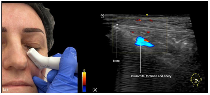
| Tear trough and infraorbital hollow | Lateral entry 1–2 cm below the lateral canthus, cannula at 60°, on the periosteum or deep to the orbicularis; needle micro-aliquots of 0.05 mL along the rim with the tip on periosteum | Cannula 25G; 27G not recommended; needle 29–30G on bone; at most 0.5 mL per eye | Angular vein inside the orbicularis 3.8–4.6 mm above the orbital rim, so a cannula deep to the muscle passes under it; display depth set to 1.5 cm voor 20–23 MHz | Bhojani-Lynch 2025; Vasconcelos-Berg 2024; Calomeni 2022; Bernardini 2025 |
| Cheek and midface | Lateral port 1.5 cm inferolateral to the lateral canthus and medial port 1.5 cm inferolateral to the alar base, into the deep cheek fat, the prezygomatic space and the SOOF; a 23G introducer to bone at 90° | Cannula 22G, or 25G 38 mm; an optional 25G needle depot at the zygomaticomalar junction | A 22G cannula under ultrasound in the intended deep compartment in all 188 midfaces; deep compartments move 0.1 mm on smiling against 3.7 mm for the superficial fat | Surek 2015; Galadari 2022; Jones 2021; Schelke 2023; Schelke 2021 |
| Deep pyriform and perialar spaces | On bone inside the deep pyriform space, with the angular artery in its roof; the perialar space beside it as a deep cannula target for the nasolabial fold | Bone-level deposit in the pyriform space; cannula into the perialar space | Skin to bone 9.11 mm; angular artery 3.6 mm from the skin and 5.1 mm from bone, 1 mm or closer to bone at 2.4% of sites; mapping mandatory | Surek 2016; Yoon 2026; Franceschelli 2024 |
| Nasolabiale plooi | Puncture lateral to the fold, with the cannula taken deeper or shallower to clear the mapped artery | Cannula 25G 2″ against a 27G ½” needle in the split-face trial; 22G cannula in the mapped series | Arteria facialis 8.04 mm deep at the lateral ala in pooled Doppler studies; arteries wider than 0.8 mm voor 13.2% of fold injection sites | Beer 2023; Teixeira 2025; Pourani 2025 |
| Nose | Dorsum: cannula on the periosteum from a single entry, the anterior nasal spine to the nasion; radix and tip: needle micro-aliquots on bone or cartilage | Large-diameter cannula, a 25G 5 cm dome tip, or 22G 50 mm through a 21G pilot; needle 29G or 31G | Dorsal nasal artery in the midline in 20% of 50 scanned patients and subdermal in 14%; subcutis 2.02 mm at the rhinion and 5.21 mm at the sellion; mapping mandatory | Lee W 2019; Radulesco 2022; Bravo 2024; Vasconcelos-Berg 2024; Yi 2024 |
| Marionette lines and prejowl | From the paragonion entry, subcutaneous retrograde threads along the mandible and a fan for the labiomandibular groove; a cannula for the deeper fat layers | Cannula 25G 5 cm | Arteria facialis 6.27 mm deep at the mandibular border in pooled Doppler studies | Chen 2022; Hong 2025b; Pourani 2025 |
| Kin | Needle depots on bone at the paragonion and at the pogonion, 15–20 mm above the inferior border in the midline; cannula through the rhombic area between pogonion, menton and paragonion in the subcutaneous plane, or through three entries or one midline entry for several layers | Needle 27G; cannula 25G 3 cm or 23G 30 mm | A 25G cannula in the deep fat on Doppler in the lower-face best-practice paper | Chen 2022; Adel 2024; Sahan 2020; Vasconcelos-Berg 2025 |
| Kaaklijn | Cannula along the mandibular border in the subcutaneous plane, in lines of 0.2 mL or less | Cannula 22G 50 mm in-plane; for VYC-25L, a 25–27G cannula or a 27–30G needle | A 22G cannula in the subcutaneous fat with no artery in the field at 18 MHz; scans during and after the pass at 11 MHz in a 10-patient series | Vasconcelos-Berg 2025; Alcantara 2025; Bertossi 2025; Hong 2025c |
| Lips | Points, planes and labial artery depths for every lip technique | Needle or cannula and gauge for each lip technique | Both labial arteries on Color Doppler before the first pass | Lip filler technique guide |
The rows cover hyaluronic acid. Poly-L-lactic acid and calcium hydroxylapatite biostimulators follow their own cannula protocols, with their own dilutions and planes.
The nose splits the sources by layer. Lee’s 2019 series scanned 50 patients before dorsal augmentation and found the dorsal nasal artery in the midline in 20%, just under the preperiosteal layer in 8% and subdermal in 14%, and favors a large-diameter cannula laid gently on the periosteum. Cong’s 2022 study of 79 specimens met a named artery at 1.3% to 11.4% of five midline points, and supports either a deep midline needle or a cannula from a single rhinion entry kept deep. Rosengaus and Nikolis prefer the needle held fully against bone or cartilage; the midface best-practice paper uses a 29G or 31G needle on the periosteum or perichondrium, or a 25G cannula in-plane at 20 MHz (Vasconcelos-Berg 2024). On ultrasound in 20 patients treated through an infratip entry, Jung found the tip 1.9 mm off the nasal bone on average and 0.4 mm at the closest, and Bravo’s 2024 series of 12 noses ran a 22G 50 mm cannula through a 21G pilot under an 18 MHz probe with no adverse effects at 6 months.
What ultrasound shows at the filler cannula or needle tip
Held in-plane, along the long axis of a high-frequency linear probe, a cannula or needle shows as a bright straight line with the tip at its leading end; crossed out-of-plane, the same shaft shows as a single bright dot. The best-practice papers for the upper, middle and lower face guide in-plane, so the whole shaft and tip stay on screen, and turn the probe out-of-plane near a vessel to confirm the tip sits outside it (Sigrist 2024; Vasconcelos-Berg 2024; Vasconcelos-Berg 2025), with 1–2 mm between cannula and vessel before injecting (Sigrist 2024). The angle of entry matters most: across 1,500 cadaver clips, visibility fell by 0.18 on the log-odds scale for every degree steeper, a larger effect than tissue, bevel or needle type (Dinges 2023). A 27G needle driven perpendicular to bone in the deep temple stays out of view, so that depot is mapped first and placed against the marks (Sigrist 2024).
| Factor | What the screen shows | Numbers | Bron |
|---|---|---|---|
| Probe orientation | In-plane, the whole shaft as a line with the tip at its end; out-of-plane, a dot, used to confirm the tip sits outside a vessel, with a 90° turn to check the crossing plane | The preferred approach in all three best-practice papers | Sigrist 2024; Vasconcelos-Berg 2024; Vasconcelos-Berg 2025 |
| Insertion angle | The line fades as the angle steepens; in-plane views work best below 30° and out-of-plane views above 60° | −0.18 log-odds per degree across 1,500 cadaver clips; 719 phantom measurements at 15 MHz | Dinges 2023; Schafhalter-Zoppoth 2004 |
| Gauge | Finer shafts show less | 17G to 22G, P < .001 | Schafhalter-Zoppoth 2004 |
| Bevel | Bevel down shows best and bevel to the side worst; on bone, a needle at about 30° bevel down | Bevel effect 1.45 on the log-odds scale | Dinges 2023; Kim JS 2024 |
| Imaging software | Beam steering and compound imaging bring a steep needle back on screen | Bij 45°, from not visible to visible with beam steering; compound imaging P < 0.0001 | Takatani 2012; Wiesmann 2013 |
| Koppeling | A water cushion re-displays a steep needle; abundant gel and minimal pressure keep the image and the flow | Tear trough display depth set to 1.5 cm voor 20–23 MHz | He 2025; Bernardini 2025; Sigrist 2026 |
| Probe tilt | Tilting the probe off the shaft breaks the needle-beam alignment | At a 45° tilt, redirections rose from 0 to 2 and insertion time from 16 to 39, in 49 residents | Mao 2021 |
| Shaft signature | A white straight line with stacked echoes deep to it from reverberation; fresh HA dark around a cannula shaft and tip | Reverberation shown under a needle at 14 MHz | Desyatnikova 2022; Bae 2024; Schelke 2023 (two phenomena) |
| Side opening | The outlet sits behind the tip, so the scan follows both | Over ons 1 mm behind the tip of a needle | Sigrist 2024; Kim JS 2024 |
| Lost tip | Small sliding and rotating moves of the probe bring the cannula back; a test aliquot confirms an interfascial plane the scan cannot show directly | A small test aliquot; 0.5–1.0 mL per temple side in total | Desyatnikova 2022 |
| Tip drift | Aspiration pulls the tip back and injection pushes it forward | Back 1.1–5.3 mm, forward 0.6–4.1 mm | Lin 2022 |
| Frequentie | Published cannula-tip frames outside vessels at 18, 20 and 24 MHz; vessels about 0.1 mm across resolved at 18 to 22 MHz | 18, 20 and 24 MHz | Vasconcelos-Berg 2024; Sigrist 2024; Jaguś 2021 |
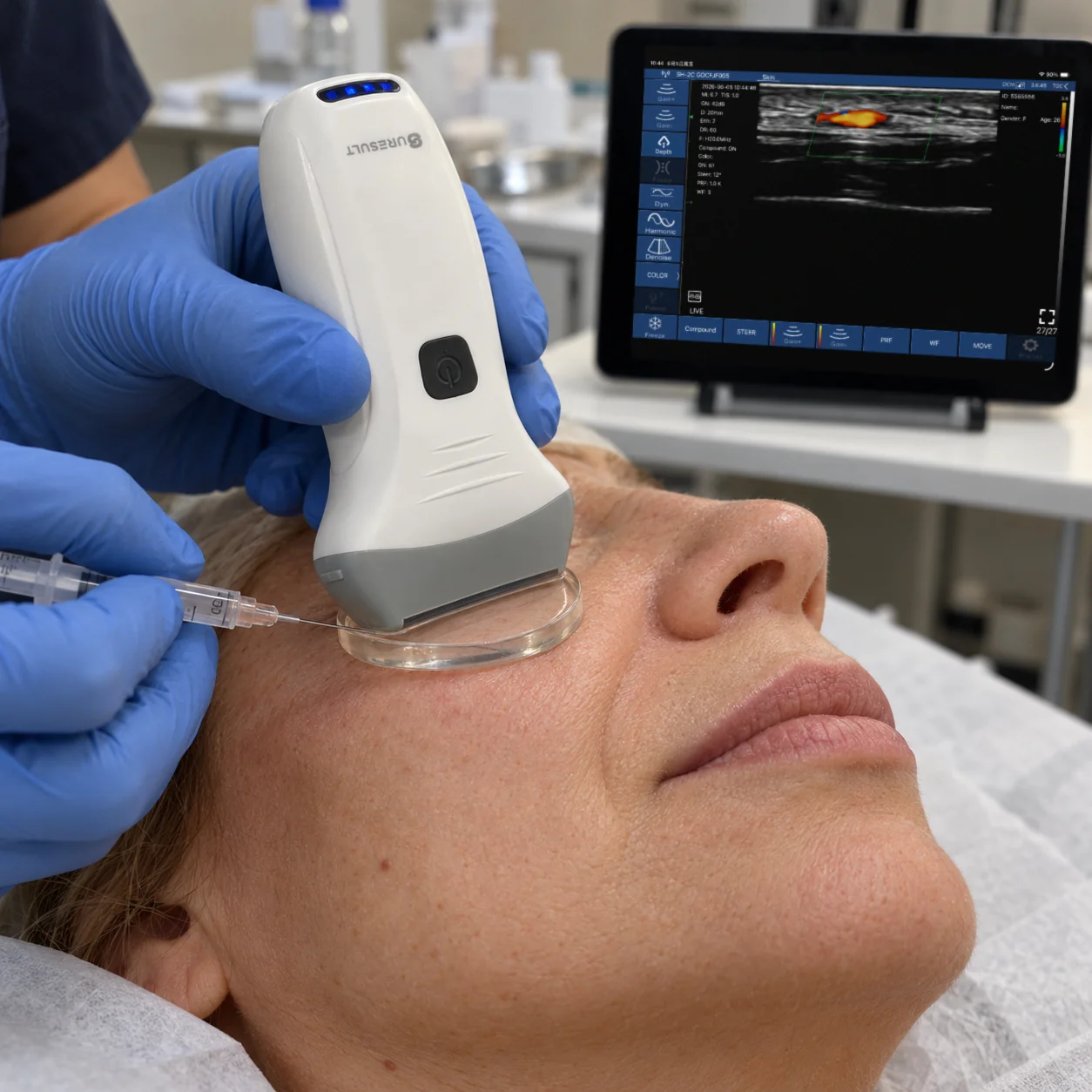
On the Suresult L20Pro, L24Pro and L14Pro, Needle Enhance highlights the needle in the image, and the in-plane and out-of-plane puncture guides, with grid overlay and flip, give real-time direction while the shaft is held in the scan plane; the L20 and L24 carry the same set, and the L14 the guides alone. Read at 24 MHz and 20 mm on a Suresult L24, the same 18/24 MHz band as the L24Pro, the two clips below show what the screen gives: a needle shaft held in-plane as one bright line from the entry to the tip, and a needle line entering a bolus of hyaluronic acid that spreads as a dark, anechoic pool around the tip.
A gel pad keeps the coupling steady through an in-plane pass. The Suresult aesthetic ultrasound gel pads are sterile Ø35 × 4 mm hydrogel discs: the probe sits on the pad, not on the skin, a needle goes straight through the hydrogel, and the second hand stays free to stretch the skin or inject. Centered under the 25 mm footprint, a pad leaves about 5 mm spare at each end of the probe for an in-plane entry, and the 4 mm stand-off takes 4 mm of the display depth, so the L20Pro’s 40 mm preset still shows about 36 mm of tissue. A pack of 50 costs $140, and the sterile setup for scanning during the pass is in the sterile coupling section of the guided filler injections guide.
Where the filler lands: ultrasound-checked cannula and needle passes
Scanned after the pass, cannula product stays in the plane it was aimed at in most published series: a 22G cannula put filler in the intended deep midfacial compartment in all 188 midfaces of 94 volunteers (Schelke 2023, 188 midfaces), a 25G cannula placed it deep to the orbicularis oculi in all 246 tear troughs (Bernardini 2025), and across 100 injections the plane held in about 90% at day 0, 14 and 30 (Calomeni 2023). Where the two tools went head to head, the plane decided more than the instrument: across 440 guided areas, angle, cannula or needle and gauge left the spread of HA unchanged, and product stayed superficial or deep to the SMAS where it was placed (Schelke 2026).
| Onderzoek | Area and size | Tool and imaging | Where the filler went |
|---|---|---|---|
| Schelke 2023, 188 midfaces | Midface deep fat; 94 volunteers | 22G cannula, ultrasound-assisted | The intended deep compartment in 100%, with no adverse events |
| Bernardini 2025 | Tear troughs; 246 sides in 123 patients | 25G 50 mm cannula from a lateral entry; 20–23 MHz | Deep to the orbicularis oculi and below the tear trough ligament on every side, 0.26 mL per side on average |
| Calomeni 2023 | Several facial areas; 100 injections in 8 patients | Cannula, with real-time ultrasound to day 30 | The plane held in about 90% at day 0, 14 and 30; deposit area 22.51, 15.97 and 12.9 mm² |
| Faria 2025 | Forehead; 4 specimens, 5 injectors | 22G 50 mm cannula; 27G 13 mm needle at 45° and at 90° | On the periosteum in every cannula pass and every 45° needle pass; spread across layers at 90° |
| Bravo 2021 | Zygoma, chin and temple; 1 patient, split face | 22G 5 cm cannula against a 27G needle; 8–18 MHz lineair | On bone with both tools at the zygoma and chin; above bone with the cannula in the temple |
| Ahn 2024 | Forehead and temple; 3 cadaver hemifaces and 2 volunteers | 18G or 21G cannula from a single entry; 18 MHz | Supraperiosteal in the forehead and on the deep temporal fascia in the temple, on target in all |
| Surek 2021 | Temple; 20 cadaver hemifaces | Ultrasound-guided injection into the intermediate temporal fat pad | On target in 18 of 20 (90%); the temporalis in 2 of 20 |
| Schelke 2026 | Face; 440 areas in 107 women | 27G needle and 25G cannula; 18 or 20 MHz | Spread held to its plane, superficial or deep to the SMAS; angle, tool and gauge left the flow unchanged |
Five details decide whether a cannula reaches the plane it is aimed at. A cannula that fails to pierce a thick SMAS feels bone yet leaves the filler above the SMAS, and product also runs back along the shaft (Schelke 2023, two phenomena). An entry placed too high cannot reach the deep midface compartments, and HA placed without guidance can end up inside the SMAS itself (Molinari 2026). Over the convex frontal bone, a cannula tip can turn upward and change plane (Sigrist 2024). Each of these shows on the scan during the pass.
How to run a guided filler cannula pass, step by step
A guided cannula pass runs in three parts: map the track before the entry, follow the cannula in-plane during the pass, and rescan the deposit and the flow after it. Mapping alone is quick: Teixeira’s map-first technique took 1 to 3 minutes per session in 503 patients on a 20 MHz handheld and moved the cannula into the plane away from the artery, against 5 to 20 minutes for guidance in real time (Teixeira 2025). Rocha’s three-step method, a map, guided filling and a perfusion rescan, was described across 480 patients at 18 MHz (Rocha 2022). The general setup for guided injections is in the workflows section of the ultrasound-guided filler injections guide; the steps below apply it to a cannula.
- Map the track. Before the entry, scan the whole path the cannula will take in B-mode and Color Doppler, across each vessel with the color box steered along it, and mark every artery the track crosses (Sigrist 2024); the Delphi table above lists the areas where this map is mandatory. Sweep for prior filler in the same pass: FaceAtlas AI Filler Scan names the material among seven and gives its depth and size.
- Choose the plane against the map. Where the map shows the artery superficial on the track, run the cannula deeper, and where the artery runs deep, stay more superficial (Teixeira 2025). In the midface, place the entry low enough to reach the deep compartments (Molinari 2026), and keep bone-level depots for the needle, as the area table sets out.
- Open the entry. Puncture the skin with a needle the size of the cannula or up to two sizes larger: a 23G introducer to bone for a 25G cannula in the midface (Galadari 2022), a 21G pilot for a 22G cannula in the temple and nose (Desyatnikova 2022; Bravo 2024), and a 23G needle for a 23G temple cannula (Kim JS 2024). Where the SMAS is thick, confirm on the scan that the cannula has passed through it before any product goes in (Schelke 2023, two phenomena).
- Advance in-plane. Lay the probe along the shaft so the whole cannula and its tip stay on screen, and keep the probe face level over it, because tilt breaks the needle-beam alignment (Mao 2021). On a Suresult probe, Needle Enhance highlights the shaft and the in-plane guide marks its path. A 25 mm field shows the leading half of a 50 mm cannula, so slide the probe along the track as the tip advances.

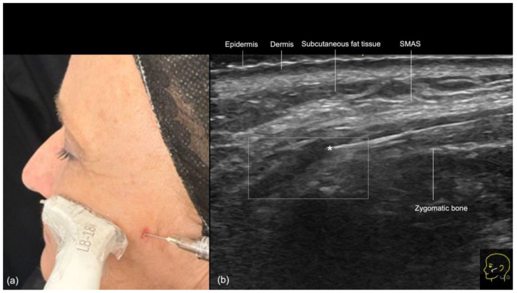
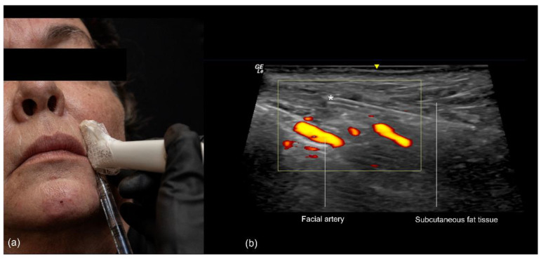
The nasolabial fold scanned while injecting: the probe held in-plane along the cannula, and on color Doppler at 18 MHz the cannula in the subcutaneous fat with its tip (*) outside the vessels and the facial artery just deep to it. Figure 10 from Vasconcelos-Berg R, Desyatnikova S, Bonavia P, et al. Diagnostics (Basel) 2024;14(22):2544. doi:10.3390/diagnostics14222544. CC BY 4.0. - Deposit in small retrograde aliquots. Inject on withdrawal in aliquots under 0.1 mL (Goodman 2020); in the temple, Ko’s 2026 study pairs a low-resistance track opened by hydrodissection with low-pressure, retrograde micro-aliquots.
- Bring back a lost tip. If the tip leaves the screen, slide or rotate the probe in small moves until the shaft shows again; where the plane itself is hard to see, as between the temporal fasciae, a small test aliquot confirms it as a dark pool between two bright fascial lines (Desyatnikova 2022).

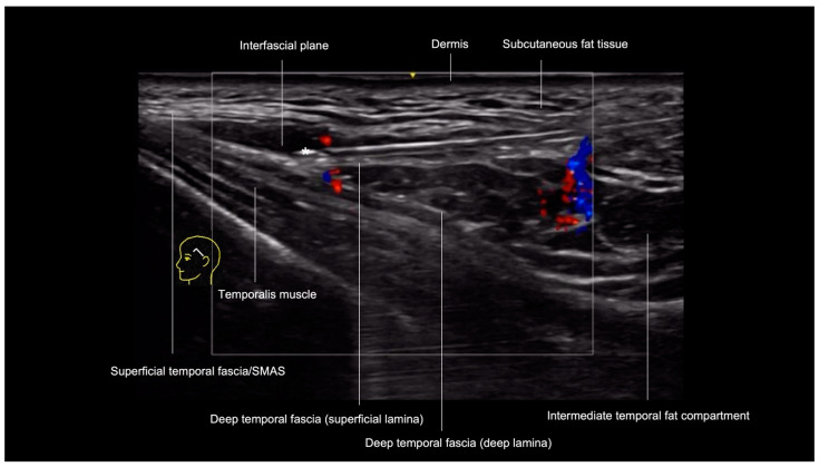
The temple on color Doppler at 20 MHz: the cannula tip (*) in the interfascial plane between the superficial temporal fascia and the superficial layer of the deep temporal fascia. Figure 3 from Sigrist R, Desyatnikova S, Chammas MC, et al. Diagnostics (Basel) 2024;14(16):1718. doi:10.3390/diagnostics14161718. CC BY 4.0. - Check out-of-plane near a vessel. Turn the probe 90° where the track nears a mapped vessel: the shaft becomes a dot, and the scan confirms the tip sits outside the lumen, with 1–2 mm between cannula and vessel before injecting (Sigrist 2024). Aspiration pulls the tip 1.1–5.3 mm back and injection pushes it 0.6–4.1 mm forward (Lin 2022), so the check follows the tip after each change of pressure.
- Rescan and save. After the last pass, image the deposit in B-mode, confirm flow on the mapped vessels in Color Doppler, and save the loop to the client’s record, the final step of Rocha’s method (Rocha 2022).
Doppler settings for the vessels on a cannula track
The arteries a cannula track crosses run slowly and close to the surface: the supratrochlear, supraorbital and angular arteries measure 0.6 to 1.0 mm inside, with peak systolic velocities of 9.2 to 24.9 cm/s (Shen 2023, 74 hemifaces). A velocity scale near 3 cm/s, with PRF and wall filter brought down to suit it, fills those vessels with color (Sigrist 2026); scan across each vessel, steer the color box along it (Sigrist 2024), and keep plenty of gel under a light hand.
| Setting | Starting point | Bron | On the Suresult app |
|---|---|---|---|
| Velocity scale | Over ons 3 cm/s, so slow facial flow fills the color box | Sigrist 2026 | Set through PRF on the color screen |
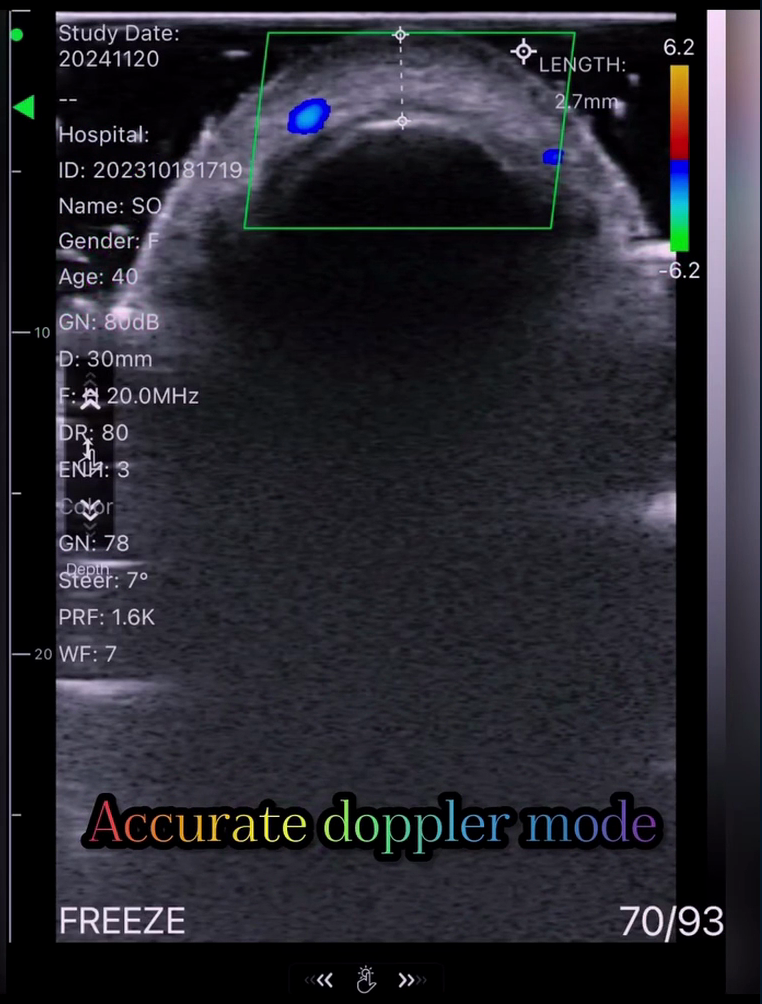
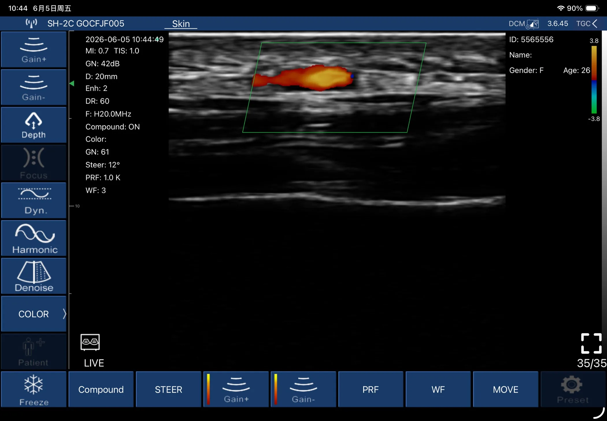
| PRF and wall filter | Brought down with the scale, since a high wall filter removes slow flow | Sigrist 2026 | PRF and WF controls; the frame below reads PRF 1.0 K and WF 3 |
| Color box | Steered along the vessel, with the probe across it | Sigrist 2024 | Steer control; 12° on the frame below |
| Koppeling | Abundant gel and minimal pressure, so shallow vessels stay open | Sigrist 2026 | The Ø35 × 4 mm gel pad carries the probe on hydrogel |
| Modi | Color to map, Power for the slowest flow, PW to confirm an arterial waveform | Delphi 2025; WFUMB 2025 | Color, Power and PW Doppler on every L-series probe |
| Frequentie | 15 MHz or more, and 18 to 24 MHz on shallow tracks | Delphi 2025; WFUMB 2025; Sigrist 2024 | 20 MHz on the L20Pro, 24 MHz on the L24Pro |
| Registratie | Stills and a loop of the map before the entry | Delphi 2025 | Cine to 1.000 frames; DICOM export |
Power Doppler picks up slower flow than color (WFUMB 2025), PW Doppler tells an artery from a vein by its waveform, and probe compression separates the two as well (Lee 2023). The full Doppler setup for guided filler, mode by mode, is in the Doppler setup section of the guided filler injections guide. On the Pro probes, a track setting saves as one of 3 customizable user sets.
The rescan after the last cannula pass
After the last pass, rescan the whole track. B-mode shows where the product sits against the plane you planned, and the published series show how closely a guided cannula holds it: all 188 midfaces in the intended deep compartment (Schelke 2023, 188 midfaces) and about 90% at day 14 and day 30 in Calomeni’s 100 injections. Color Doppler then confirms flow on every vessel mapped before the entry, the last step of Rocha’s method. Saved with the map, the loop becomes the baseline for the next visit, and FaceAtlas AI keeps it in the client’s file for a one-click comparison; old product found at a later visit is read in the filler identification section of the dissolving guide.
What frequency shows a filler cannula and the vessels around it
At least 15 MHz, with 18 to 24 MHz on shallow tracks: a cannula pass needs enough frequency to resolve the shallow vessels and layers and enough depth to reach the plane where the track ends. The 2025 Delphi panel and the WFUMB 2025 position paper set 15 MHz as the minimum for aesthetic scanning, and the published cannula-tip frames read 18, 20 and 24 MHz. Frequency trades against reach: at 20 MHz the image goes about 10 mm deep and at 8 MHz about 50 mm (Lee 2023). Most vessels on a cannula track lie inside those first 10 mm, among them the supratrochlear and supraorbital arteries at 3.34 and 3.54 mm from the skin, the frontal branch of the superficial temporal artery at 2.7 mm, the angular artery at 3.6 mm over the pyriform space and the facial artery at 6.27 mm over the mandibular border and 8.04 mm at the ala; the bone plane under the cheek calls for the deeper presets.
| Area and plane | Depth to reach | Frequency in the studies | Suresult setting that frames it |
|---|---|---|---|
| Forehead and glabella, supraperiosteal | Supratrochlear and supraorbital arteries 3.34 and 3.54 mm from the skin | 18 and 20 MHz | L20Pro at 20 MHz on the 10 or 20 mm preset; L24Pro at 24 MHz on 10 or 15 mm |
| Temple, subdermal and interfascial | Frontal branch of the superficial temporal artery 2.7 mm from the skin | 14 to 24 MHz | L20Pro at 20 MHz on 10 or 20 mm; L24Pro at 24 MHz on 10 or 15 mm |
| Tear trough, deep to the orbicularis | Angular vein 3.8–4.6 mm above the orbital rim, on a 15 mm display depth | 20 to 23 MHz | L24Pro at 24 MHz on the 15 mm preset; L20Pro at 20 MHz on 20 mm |
| Deep pyriform space, on bone | Arteria angularis 3.6 mm from the skin, with bone 9.11 mm down | 20 MHz | L20Pro at 20 MHz on 20 mm |
| Cheek and zygoma, deep fat and supraperiosteal | The bone plane over the zygoma and maxilla, under the deep fat compartments | 18 MHz | L20Pro at 16 MHz on 30 or 40 mm |
| Nasolabiale plooi | Arteria facialis 7.95 mm deep at the cheilion and 8.04 mm at the ala, pooled | 18 MHz | L20Pro at 20 MHz on 20 mm |
| Nose, on bone and cartilage | Subcutis 2.02 mm thick at the rhinion and 5.21 mm at the sellion | 18 to 20 MHz | L24Pro at 24 MHz on 10 mm; L20Pro at 20 MHz on 10 mm |
| Jawline and prejowl, subcutaneous | Arteria facialis 6.27 mm deep at the mandibular border, pooled | 18 MHz; 11 MHz in a 10-patient series | L20Pro at 20 MHz on 20 mm |
On a Suresult probe the cannula areas split by band. The L20Pro covers the whole list at 16/20 MHz: 20 MHz on the 10 and 20 mm presets for the forehead, temple, nose, fold and jawline tracks, and 16 MHz on the 30 and 40 mm presets for the bone plane under the cheek. The L24Pro adds 24 MHz on 10 to 25 mm presets for the tear trough and the temple subdermis, and the L14Pro at 10/14 MHz reaches 80 mm for body work beside the face. The frequency window by depth en de L14, L20 and L24 chooser for aesthetics set the bands side by side.
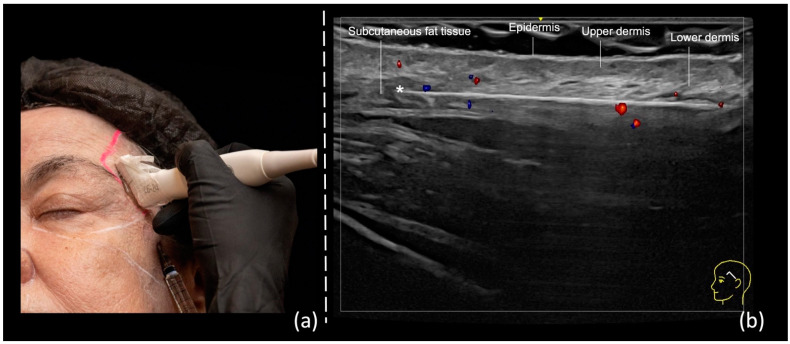
The 5 best handheld ultrasounds for cannula and needle filler work, ranked
The Suresult L20Pro ranks first for cannula and needle filler work at 89.3/100, ahead of the Clarius L20 HD3 (69.8), the GE Vscan Air SL (48.3), the Butterfly iQ3 (47.6) and the Philips Lumify L12-4 (41.4). I scored the handhelds on what a cannula pass asks of a probe: a tip that stays on screen in-plane, enough frequency for the shallow vessels and layers, enough depth for the bone plane, Color, Power and PW Doppler to map the track and confirm flow, software that reads prior filler and the facial layers, records for the rescan, and the five-year total for all of it. The weights and formulas are under How I scored.

01 Suresult L20Pro — Best Overall for Cannula and Needle Filler Work
16/20 MHz · 10–40 mm · 25 mm footprint · 256 elements · Needle Enhance and puncture guides · Color, Power and PW Doppler · FaceAtlas AI with 3 months in the price · $3,720
Score: 89.3/100
$3,720 · 145 g · up to 4 h a charge · own Wi-Fi · IP67 · imaging free of subscription
Buy the Suresult L20Pro high-frequency linear probe: $3,720
Oordeel: Buy the L20Pro if your filler list runs from the temple and tear trough to the cheek, jawline and nose, and you want every track mapped and every cannula followed on one probe: 20 MHz for the shallow vessels and layers, 16 MHz down to 40 mm for the bone plane, three Doppler modes in the purchase, and Needle Enhance with in-plane and out-of-plane guides for the pass. It is my pick for injectors who scan the track before every cannula entry.
- Cannula and needle tools: Needle Enhance and the In-Plane & Out-Plane Puncture Guide, with Flip and Grid, track the cannula in real time while the shaft stays in the scan plane
- Depth and Doppler: 16/20 MHz over een 25 mm footprint, with presets at 10, 20, 30 and 40 mm; Color, Power and PW Doppler, with steer, gain, PRF and wall filter on the color screen and gain, steer, volume and angle on PW
- Filler software: FaceAtlas AI Filler Scan reads prior product before a new track, names it among seven materials (HA, CaHA, collagen, PLLA, PCL, PMMA and PVA) and measures its depth and size. In Dual View the raw scan and the AI layers run side by side in real time, so the probe stays in place while you follow the needle angle and the filler as it goes in. The 21-point map puts 2 points on the tear trough, 2 on the cheekbone, 2 on the apple of the cheek and 4 on the jawline, and Expert mode adds the superior temporal point; 3 maanden included, and it runs on 64-bit Android 8.0 or later

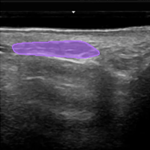
FaceAtlas AI Filler Scan on a facial B-mode frame: the read on prior product before a new cannula track is planned. 
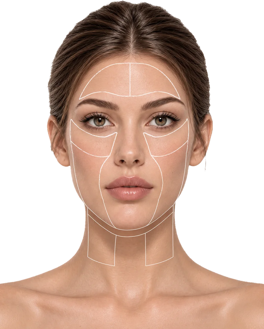
The FaceAtlas AI region map, outlining the face from the forehead to the neck: the layout each visit is scanned against. - Records: cine up to 1.000 frames, about 165 s, with shorter 100, 200 and 500-frame buffers; scans stored on the device and exported as JPEG, PNG, MP4 or DICOM; FaceAtlas AI adds a PDF report per visit and a one-click comparison with the last one
- Prijs en abonnement: $3,720 with imaging, all three Doppler modes and the needle tools free of subscription; after the 3 included months, a FaceAtlas AI license costs $500 a year or $50 a month, and the lifetime license, $999, is on sale until 11:59 PM PT, December 31, 2026
- Battery and build: 145 g and up to 4 uur a charge from a 4,400 mAh battery; pairs over its own Wi-Fi with iOS, Android and Windows phones and tablets; IP67 and drop-tested
Waarom deze positie in de ranglijst. A cannula track crosses depths from 2.7 mm, where the frontal branch of the superficial temporal artery runs at the lateral canthal line, to 9.11 mm, where bone lies under the deep pyriform space, and the L20Pro frames both on one probe: 20 MHz on the 10 and 20 mm presets for the shallow vessels and layers, and 16 MHz on the 30 and 40 mm presets for the bone plane under the cheek. Needle Enhance plus the in-plane and out-of-plane guides, all in the purchase, earn the full 20 for tip visibility. The 20 MHz top frequency scores 14.29 of 20 for superficial detail, and the 40 mm reach takes the full 10 for depth.
Color, Power and PW Doppler all come with the probe, for the full 20: color to map each vessel on the track, Power for the slowest flow, PW to confirm an arterial waveform. FaceAtlas AI takes the full 10 for software, with Filler Scan naming prior product among seven materials, the 21-point map and the layer view, and 3 months in the price. Cine to 1,000 frames, storage with DICOM export and the FaceAtlas AI PDF report make 5 of 5 for records. Five years with the FaceAtlas AI lifetime license total $4,719, 9.97 of 15 against the field’s lowest five-year total.
Sub-scores: tip visibility 20, superficial detail 14.29, depth reach 10, Doppler 20, filler software 10, records 5, five-year cost 9.97.
The L20Pro is FDA 510(k) cleared under K211321, as is every Suresult L-series probe.
L20Pro, L24Pro or L14Pro? Each Pro carries 256 elements, Color, Power and PW Doppler, Needle Enhance, a 4-hour battery and FaceAtlas AI for the first 3 months. For $465 more, the Suresult L24Pro aesthetic ultrasound probe moves the band to 18/24 MHz on presets from 10 to 25 mm, the sharpest view of the tear trough and the temple subdermis. The Suresult L14Pro lineaire probe ($3,441) reaches 80 mm at 10/14 MHz, for practices that also image joints, nerves and vessels in the body. Every cannula area on one probe: the L20Pro. The tear trough and the temple first: the L24Pro. The L14, L20 and L24 aesthetic ultrasound chooser sets the three bands side by side.
Het meest geschikt voor: injectors who map the track before each cannula entry and want every facial area, from the temple branch at 2.7 mm to the bone plane under the cheek, on one 16/20 MHz probe.Kies een model uit een productfamilie als: the tear trough and temple subdermis lead your list (the L24Pro), you scan the body as well (the L14Pro), you want the 24 MHz band for less (the L24 at $3,441), or you want Needle Enhance and the 16/20 MHz band for less, with FaceAtlas AI bought as a license (the L20 at $3,000).Vergelijk zorgvuldig als: you want a live vessel-depth readout in a membership (the Clarius L20 HD3), or a dual-head probe that also scans the heart and lungs (the GE Vscan Air SL).
Ook in deze productfamilie
Suresult L24Pro aesthetic ultrasound probe · $4,185 · 86.6/100. The highest frequency in the line, 18/24 MHz on 10, 15, 20 and 25 mm presets, for tracks inside the first 25 mm such as the tear trough and the temple subdermis; 256 elements, 4 hours a charge and 3 months of FaceAtlas AI.
Suresult L20 linear ultrasound probe · $3,000 · 86.1/100. The L20Pro’s 16/20 MHz band and 10 to 40 mm presets for less, on 192 elements and 2 hours a charge at 147 g, with all three Doppler modes and Needle Enhance; FaceAtlas AI by license, and the lowest five-year total of any Needle Enhance probe in the line.
Suresult L24 linear ultrasound probe · $3,441 · 83.1/100. The L24Pro’s band at a lower price: 18/24 MHz on 10 to 25 mm presets, with 192 elements, 2 hours a charge, three Doppler modes and Needle Enhance; FaceAtlas AI by license.
Suresult L14Pro linear ultrasound probe · $3,441 · 81.3/100. The deepest Pro, for clinics that scan the body beside the face: 10/14 MHz to 80 mm in four presets, 256 elements and 4 hours a charge, with every Doppler mode, Needle Enhance and 3 months of FaceAtlas AI.
Suresult L14 linear ultrasound probe · $2,139 · 70.7/100. The entry point of the line: 192 elements at 10/14 MHz, imaging 20 to 80 mm, with Color, Power and PW Doppler and the in-plane and out-of-plane guide lines for needle work; 200 g, 2 hours a charge and FaceAtlas AI by license.
Suresult handheld echografie voor esthetische geneeskunde · Suresult aesthetic ultrasound gel pads · Suresult L20Pro high-frequency linear probe
02 Clarius L20 HD3 — Best for a Live Vessel-Depth Readout at 20 MHz
8–20 MHz · 4 cm · 25 mm field of view · Needle Enhance, PW Doppler and Vessel Depth AI in Essentials · $5,395 before Essentials
Score: 69.8/100
$5,395 before Essentials · 290 g · roughly an hour a charge · Color and Power Doppler in the scanner · IP67, 1 m drop rating · 3-year warranty
Oordeel: Buy the L20 HD3 if you want a cordless 20 MHz linear that prints the depth of the shallowest vessel live on screen, with cheek, chin and jaw, pyriform and lips among its listed applications, and you will keep the Essentials membership that carries its needle and vessel tools.
- Cannula and needle tools: Needle Enhance in Essentials, which Clarius describes for needles or cannulas; the Center Guide, a dotted line down the middle of the image for lining up the entry
- Depth and Doppler: 8–20 MHz tot 4 cm over een 25 mm field of view; Color and Power Doppler with the scanner and PW Doppler in Essentials; Vessel Depth AI prints the depth of the shallowest vessel and updates it in real time, in Essentials
- Layer software: skin, fat and tissue thickness by Auto Aesthetic Calipers; filler volume estimates in Split Screen; facial layer labels in T-Mode AI Aesthetics with Essentials
- Records: cine playback of 30 seconds; local and cloud storage, DICOM and PDF reports with Essentials
- Prijs en abonnement: $5,395 from Clarius (read October 31, 2026); Essentials billed at $695 a year, $1,785 per three years or $2,500 paid once
- Battery and build: 290 g; roughly an hour of scanning a charge; IP67 with a 1 m drop rating; 3-year warranty
Waarom deze positie in de ranglijst. Teixeira’s map-first series of 503 patients ran on a Clarius L20 at 20 MHz, and at that frequency the L20 HD3 takes 14.29 of 20 for superficial detail, with the full 10 for depth from its 4 cm reach. Needle Enhance in Essentials earns half the enhancement credit, and the Center Guide, sold with no tier named, the full guide credit: 15 of 20 for tip visibility. Color and Power Doppler come with the scanner and PW sits in Essentials, for 17 of 20.
Software scores 4.5 of 10: the Auto Aesthetic Calipers measure layers with no tier named, for the full 3, and Vessel Depth AI, sold in Essentials, takes half of the 3 for a vessel map. Records earn 3 of 5, from storage with DICOM and the PDF reports in Essentials; the cine credit asks for 60 seconds, and Clarius cine plays back 30. With Essentials bought once, five years come to $7,895, or 5.96 of 15 on cost.
Sub-scores: tip visibility 15, superficial detail 14.29, depth reach 10, Doppler 17, filler software 4.5, records 3, five-year cost 5.96.
Beperkingen. The membership carries PW Doppler, Needle Enhance, Vessel Depth AI, storage and DICOM; cine playback stops at 30 seconds; and no Clarius tool reads the filler material.
Het meest geschikt voor: injectors who want 20 MHz with a live vessel-depth readout and named aesthetic applications, and who will stay on Essentials.Sla dit over als: you want every Doppler mode, Needle Enhance and DICOM in the purchase price, or a tool that reads the filler material before a new track.
Ook in deze productfamilie
Clarius L15 HD3 · $3,795 before Essentials · 62.6/100. The 5–15 MHz linear in the HD3 line, reaching 7 cm across a 50 mm field of view; five years with Essentials bought once come to $6,295.
Clarius prices and Essentials plans, 2026 · Clarius L20 HD3 specifications · Clarius L20 HD3 store page
03 GE Vscan Air SL — Best for a Dual-Head Probe That Also Scans the Heart and Lungs
3–12 MHz linear to 8 cm and a sector array to 24 cm · Color and PW Doppler in the price · Centerline Marker · $4,999
Score: 48.3/100
$4,999 with a 3-year warranty · 218 g · 50 minutes of continuous scanning · IP67 and MIL-810G
Oordeel: Choose the Vscan Air SL if filler visits share a clinic with heart, lung and abdominal scans and one dual-head probe should serve both, with Color and PW Doppler and every scored tool in the $4,999 price.
- Cannula and needle tools: the Centerline Marker, shown in B-mode and color mode; GE runs guided procedures in B-mode
- Imaging and Doppler: a 3–12 MHz linear array of 192 elements, centered at 7.7 MHz, behind a 40 × 7 mm lens and imaging to 8 cm, plus a sector array to 24 cm; Color and PW Doppler included
- Software: Caption AI, AutoEF, Lung Guidance and Bladder Volume in the Innovations plan, $500 a year or $1,299 for three years, for cardiac, lung and bladder work
- Records: up to 500 exams stored on the mobile device; DICOM export to a server; an OB worksheet and report
- Prijs en abonnement: $4,999 at the GE HealthCare US shop, checked October 31, 2026, with a demo unit at $4,299; every scored tool in the purchase
- Battery and build: 218 g; 50 minuten of continuous scanning; IP67 and MIL-810G; 3-year warranty
Waarom deze positie in de ranglijst. The linear head’s 12 MHz top frequency takes 2.86 of 20 for superficial detail, and its 8 cm reach the full 10 for depth. The Centerline Marker earns the guide credit, 10 of 20 for tip visibility. Its Doppler set is Color and PW in the purchase, without Power Doppler in GE’s mode list: 13 of 20. Storage with DICOM export and the OB report make 3 of 5 for records. Priced at $4,999 with every scored tool in the purchase, it costs the least of the four rivals over five years: 9.42 of 15. No GE tool reads filler or measures facial layers, so software scores 0.
Sub-scores: tip visibility 10, superficial detail 2.86, depth reach 10, Doppler 13, filler software 0, records 3, five-year cost 9.42.
Beperkingen. The linear stops at 12 MHz, under the 15 MHz floor set by the 2025 Delphi panel and the WFUMB 2025 paper; GE lists no Power Doppler; and guided procedures run in B-mode, so the vessel map and the pass run as separate steps.
Het meest geschikt voor: practices that see filler clients and also scan hearts, lungs and abdomens, on one dual-head probe.Sla dit over als: your cannula list calls for 18 MHz or more, Power Doppler or a needle-enhancement mode.
Vscan Air SL at the GE HealthCare US shop · GE Vscan Air SL product page · Vscan Air SL specifications
04 Butterfly iQ3 — Best for Biplane Tip Checks on a Whole-Body Probe
1–12 MHz on one probe for the whole body · facial vessels in the Vascular: Superficial preset at 1–4 cm, with the Midline Tool · Needle Viz, Biplane, Power and PW Doppler in Advanced · $3,899 plus Core or Advanced
Score: 47.6/100
$3,899 with a 3-year warranty, plus Core or Advanced · 300 g · up to 2 hours a charge · IPX7 and IP6X · 4-ft drop testing, MIL-STD-810G
Oordeel: Choose the iQ3 if one probe should cover the whole body and you want Biplane to show the tip in two planes at once, with the Advanced membership for the needle tools and the full Doppler set.
- Cannula and needle tools: Needle Viz overlays an image tuned for needles at 20–40° in-plane; Biplane shows the long and short axes at once; the Out-of-Plane preset brings the tip into view within 1 mm of the entry point; all three in Advanced, listed for the MSK, MSK-Soft Tissue, Nerve and Vascular: Access presets; the Midline Tool in the base app
- Imaging and Doppler: 1–12 MHz tot 30 cm on one probe; facial vessels in the Vascular: Superficial preset at 1 to 4 cm, which runs B-mode, Color and Power Doppler and the Midline Tool; Color Doppler with Core, and Power and PW with Advanced
- Records: recorded clips and unlimited cloud storage with Core; Basic DICOM for PACS sold by quote
- Prijs en abonnement: $3,899 for the probe; Core costs $299 a year, and Advanced $420 a year or $1,500 for five years of access
- Battery and build: 300 g; up to 2 uur a charge; IPX7 and IP6X with 4-ft drop testing; a 3-year warranty with 2 more years as an option
Waarom deze positie in de ranglijst. Needle Viz in Advanced earns half the enhancement credit, and the Midline Tool, in the base app and enabled in Vascular: Superficial, the full guide credit: 15 of 20 for tip visibility. The 12 MHz top frequency takes 2.86 of 20 for superficial detail and the 30 cm reach the full 10 for depth. Every Doppler mode sits in a paid plan, Color in Core and Power and PW in Advanced, so each counts at half: 10 of 20. Records earn 1 of 5, for cloud storage with Basic DICOM sold by quote. Five years with Advanced come to $5,399, 8.72 of 15 on cost, and software scores 0, with no Butterfly tool for filler or facial layers.
Sub-scores: tip visibility 15, superficial detail 2.86, depth reach 10, Doppler 10, filler software 0, records 1, five-year cost 8.72.
Beperkingen. Butterfly lists Needle Viz and Biplane for the MSK, nerve and vascular-access presets rather than Vascular: Superficial, every Doppler mode needs a membership, and the top frequency is 12 MHz.
Het meest geschikt voor: clinics that scan the whole body and want biplane tip checks on one probe.Sla dit over als: the face needs 18 MHz or more, or you want Doppler and needle tools in the purchase price.
Butterfly memberships and prices · Specificaties van Butterfly iQ3 · Gebruikershandleiding Butterfly iQ3
05 Philips Lumify L12-4 — Best for Lumify Practices Adding a Linear Head
12–4 MHz linear to 12 cm across a 34.5 mm field of view · steerable color and PW Doppler in the price · center line marker · $10,307.95 at PartsSource
Score: 41.4/100
By quote through Philips, or $10,307.95 outright at PartsSource · every Lumify feature in the purchase, with no subscriptions · 5-year basic warranty on transducers bought through Philips
Oordeel: Choose the L12-4 if your clinic already scans with Lumify transducers and wants a linear head on the same app, with color and PW Doppler in the purchase.
- Cannula and needle tools: a center line marker on the image; Philips lists the L12-4 as biopsy capable
- Imaging and Doppler: 12–4 MHz tot 12 cm over een 34.5 mm field of view; color Doppler you can steer, in Fast Flow for arteries and Slow Flow for veins, plus PW Doppler
- Records: loops of 1 to 10 seconds; studies sent to PACS as DICOM, by email, to a shared network or to a local folder
- Prijs en abonnement: Philips quotes the price on request; PartsSource lists a new L12-4 outright at $10,307.95 with a 90-day warranty (checked October 31, 2026); Lumify carries no subscriptions
- Garantie: 5-year basic warranty on Lumify transducers bought through Philips
Waarom deze positie in de ranglijst. At 12 MHz the L12-4 takes 2.86 of 20 for superficial detail, and its 12 cm reach the full 10 for depth. The center line marker earns the guide credit, 10 of 20 for tip visibility. Its Doppler pair is color and PW, both in the purchase: 13 of 20. Records take 1 of 5, for storage with DICOM; loops run 1 to 10 seconds. At $10,307.95 it carries the highest five-year cost here, 4.57 of 15, and software scores 0, with no Philips tool for filler or facial layers.
Sub-scores: tip visibility 10, superficial detail 2.86, depth reach 10, Doppler 13, filler software 0, records 1, five-year cost 4.57.
Beperkingen. The top frequency is 12 MHz, under the 15 MHz consensus floor, loops stop at 10 seconds, and Philips sets the price by quote.
Het meest geschikt voor: practices on Lumify that want a linear head for occasional facial vessel maps.Sla dit over als: you want 18 MHz or more, Power Doppler, a needle-enhancement mode or a list price from the maker.
Productpagina van Philips Lumify L12-4 · PartsSource L12-4 listing
Five-year cost of each handheld with the software its tools need
Counted over five years with the software each scored tool calls for, the Suresult L20Pro with the FaceAtlas AI lifetime license totals $4,719, against the GE Vscan Air SL at $4,999, the Butterfly iQ3 at $5,399, the Clarius L20 HD3 at $7,895 and the Philips Lumify L12-4 at $10,307.95. Clarius puts PW Doppler, Needle Enhance, Vessel Depth AI, storage and DICOM in Essentials, and Butterfly sells Color with Core and Power, PW, Needle Viz and Biplane with Advanced. The L20Pro carries all three Doppler modes, Needle Enhance, the puncture guides and the imaging app in its price, and adds FaceAtlas AI by license from month 4.
| Handheld | Prijs vandaag | Software the scored tools call for | Five years, software bought once | Five years on yearly billing |
|---|---|---|---|---|
| Suresult L20Pro | $3,720 | FaceAtlas AI from month 4; imaging, Doppler and needle tools in the price | $4,719 with the FaceAtlas AI lifetime license, on sale until 11:59 PM PT, December 31, 2026 | $6,220 with the $500 yearly license from month 4 |
| Clarius L20 HD3 | $5,395 | Essentials for PW Doppler, Needle Enhance, Vessel Depth AI and DICOM, bought once at $2,500 | $7,895 | $8,870 at $695 a year for Essentials |
| GE Vscan Air SL | $4,999 | All in the purchase: storage, DICOM, the Centerline Marker, and Color and PW Doppler | $4,999 | $4,999, with Innovations at $500 a year as an option for Caption AI, outside the score |
| Butterfly iQ3 | $3,899 | Advanced, bought as five years of access for $1,500, for every Doppler mode, Needle Viz and Biplane | $5,399 | $5,999 at $420 a year for Advanced |
| Philips Lumify L12-4 | $10,307.95 outright at PartsSource | Every Lumify feature in the purchase | $10,307.95 | $10,307.95, with no subscriptions |
Each rival figure is the maker’s US list price or, for the L12-4, the PartsSource listing, all read on October 31, 2026. Probe-by-probe totals for the wider aesthetic field sit in the kostengids voor echografie in de esthetische geneeskunde.
The checkout price is the full price: worldwide delivery, customs clearance and duties are included, so every Suresult order arrives with No Hidden Fees, and orders ship within 24 hours. The 30-day worry-free trial runs from delivery, and the 2-year warranty extends to 3 or 5 years. Each FaceAtlas AI license covers one device, arrives as a key by email and renews only when you buy again. Owners of the L14Pro, L20Pro and L24Pro claim their 3 months on the FaceAtlas AI claim page, and the clock starts when they press Start; on the L14, L20 and L24, the maandelijks, jaarlijks en levenslang licenses apply from the first scan. Sterile Suresult aesthetic ultrasound gel pads for scanning during the pass cost $140 for 50.
Hoe ik de scores bepaalde
I scored each maker’s card out of 100 on seven weighted criteria, declared on October 29, 2026, before a single rival was scored: tip visibility 20, superficial detail 20, depth reach 10, Doppler 20, filler software 10, records 5 and five-year cost 15. Together they ask what a cannula pass needs from a probe: whether it keeps the tip on screen, resolves the shallow vessels and layers, reaches the bone plane, maps and confirms flow on the track, reads prior filler and the facial layers and keeps the rescan on file, and what the whole kit costs over five years.
- Tip visibility (20): 10 for a mode that brightens the needle, and 10 for a guide the maker names for needle work, whether an in-plane or out-of-plane overlay, a biplane view or a midline marker; each at half when a paid plan carries it.
- Superficial detail (20): the top linear frequency, scored 20 × (MHz − 10) ÷ 14 and held between 0 and 20: 20 at 24 MHz, 14.29 at 20 MHz and 2.86 at 12 MHz.
- Depth reach (10): 10 × (maximum image depth in mm − 20) ÷ 20, held between 0 and 10, so 40 mm or more scores the full 10 and 25 mm scores 2.5.
- Doppler (20): Color 7, Power 7 and PW 6; a mode sold with the handheld earns its full weight, and a mode behind a paid plan half.
- Filler software (10): 4 for naming the filler material, 3 for a facial map of injection points or vessels and 3 for layer measurement, halved when a paid plan carries the tool. The 3 maanden of FaceAtlas AI on the Pro probes count as included, and so does a rival tool sold with no tier named.
- Records (5): 2 for 60 seconds of cine or more, in a figure the maker gives for this exact model; 1 for storage on the device or in the app with DICOM export; 2 for a visit report or a side-by-side comparison, credited in full whichever plan carries them.
- Five-year cost (15): 15 × the lowest five-year total among the scored models ÷ this model’s total. Suresult totals add the FaceAtlas AI lifetime license to the price listed on October 30, 2026; Clarius totals add Essentials bought once, Butterfly totals five years of Advanced, and the Philips total is the PartsSource price. The Suresult L14 with the lifetime license, at $3,138, is the reference.
| Rank and maker card | Tip /20 | Superficial /20 | Depth /10 | Doppler /20 | Software /10 | Records /5 | Cost /15 | Totaal /100 |
|---|---|---|---|---|---|---|---|---|
| 1 · Suresult L20Pro | 20 | 14.29 | 10 | 20 | 10 | 5 | 9.97 | 89.3 |
| 2 · Clarius L20 HD3 | 15 | 14.29 | 10 | 17 | 4.5 | 3 | 5.96 | 69.8 |
| 3 · GE Vscan Air SL | 10 | 2.86 | 10 | 13 | 0 | 3 | 9.42 | 48.3 |
| 4 · Butterfly iQ3 | 15 | 2.86 | 10 | 10 | 0 | 1 | 8.72 | 47.6 |
| 5 · Philips Lumify L12-4 | 10 | 2.86 | 10 | 13 | 0 | 1 | 4.57 | 41.4 |
Family members sit inside their maker’s card: Suresult L24Pro 86.6, L20 86.1, L24 83.1, L14Pro 81.3 and L14 70.7; Clarius L15 HD3 62.6. The Suresult card leads with the L20Pro because a cannula list spans the shallow vessels and the bone plane, and the L20Pro covers both at 16/20 MHz on 256 elements, with a 4-hour battery and FaceAtlas AI for the first 3 months.
Six reruns test the order: dropping the software credit (79.3 against 65.3 for the Clarius L20 HD3), dropping cost (79.3 against 63.8), dropping depth (79.3 against 59.8), scoring the Suresult card on the L24Pro (86.6) or on the L14Pro (81.3), and counting every rival needle tool and Doppler mode as included (89.3 against 77.8). Each keeps Suresult first and the Clarius L20 HD3 second. Under the combination least favorable to the L20Pro, with FaceAtlas AI at half credit, map credit only for a vessel map and every rival needle tool and Doppler mode counted as included, it still leads, 82.8 to 77.8. The middle places move in two runs: without cost, the GE Vscan Air SL and the Butterfly iQ3 tie at 38.9, and with rival needle tools and Doppler counted as included, the Butterfly iQ3 rises to third at 62.6. The Philips Lumify L12-4 stays fifth in each of them. Every rival figure was read from the maker’s US pages or the PartsSource listing on October 29 and 30, 2026, and every Suresult figure from its product pages.
Filler cannula and needle questions
Is a filler cannula safer than a needle?
At 22G and 25G, yes on the bench: a 22G cannula needed 1.50 N to enter a facial or superficial temporal artery against 1.08 N for a 22G needle, and 1.04 N against 0.69 N at 25G, while at 27G the two showed no significant difference, 0.78 vs 0.70 N (Pavicic 2019). Across 370 US dermatologists surveyed by Alam in 2021, cannula use carried 77.1% lower adjusted odds of vascular occlusion. The consensus papers set the line at 25G or larger, and a scan of the track shows where the artery runs before the entry; the signs and first steps after an occlusion are in the vascular occlusion guide.
Can a filler cannula enter an artery?
Yes, and the finer the cannula, the easier it goes in. In Ugradar’s 2019 study of 20 hemifaces, 18G and 22G microcannulas never entered the facial artery, while 23G, 25G and 27G cannulas did, at less force the finer they were. Siperstein’s 2023 bench test of three brands at 22 to 30G found every cannula needed more force than the matching needle, with arterial entry taking numerous attempts, and Tansatit’s 2017 dissections showed a 25G cannula perforating the frontal branch of the superficial temporal artery in specific situations, when it met the vessel at a right angle. A fibrous septum and an artery wall feel the same at the cannula tip, so the scan, not the hand, shows where the artery runs.
Which facial areas carry the most filler risk, and where is a filler cannula preferred?
The glabella, alar base, nose and temple: Signorini’s 2016 global panel names them as areas associated with higher risks of vascular complications and prefers blunt cannulas there. The 2025 Delphi panel lists the forehead, glabella, temple, nose and pyriform fossa as the areas where a vessel map before filler is mandatory, and the area table above gives the entry, plane and gauge for each.
What gauge filler cannula should you use?
25G or larger for most of the face. The upper-face and midface best-practice papers recommend blunt cannulas of 25G or larger for the forehead, glabella, temple and tear trough (Sigrist 2024; Vasconcelos-Berg 2024); Goodman’s 2020 consensus puts cannulas finer than 25G closer to needles in how they pierce vessels; and a 27G cannula enters an artery with about the force of a 27G needle (Pavicic 2019). Hong’s 2024 review uses 21–23G for deep work near muscle and 25–27G for superficial planes. In the published techniques, a 22G cannula runs the temple and the jawline, a 25G the tear trough and the marionette lines, and either size the cheek, forehead, nose and nasolabial fold.
What needle size opens the entry for a filler cannula?
In the published pairs, the entry needle matches the cannula or runs up to two sizes larger: a 23G introducer taken to bone for a 25G 38 mm cannula in the cheek (Galadari 2022), a 21G pilot for a 22G cannula in the temple and nose (Desyatnikova 2022; Bravo 2024), a 23G needle for a 23G temple cannula (Kim JS 2024), and a needle pilot for a 21G or 23G cannula in the midcheek groove (Hong 2026). For the tear trough tangents, Wilde’s 2023 series punctured the skin with a 25G needle at 45°.
Which filler cannula size suits the cheeks?
A 22G or 25G cannula into the deep cheek fat. Schelke’s 2023 ultrasound-assisted series placed product with a 22G cannula in the intended deep midface compartment in all 188 midfaces; Galadari’s 2022 technique takes a 25G 38 mm cannula through a 23G introducer opened to bone at 90°; and Jones’s 2021 trial used a 25G 1.5-inch cannula with an optional 25G 1-inch needle depot on bone at the zygomaticomalar junction. Surek’s 2015 ports sit 1.5 cm inferolateral to the lateral canthus and 1.5 cm inferolateral to the alar base, into the deep cheek fat, the prezygomatic space and the SOOF.
Which filler cannula suits the tear trough?
A 25G cannula about 50 mm long, from a lateral entry. Bernardini’s 2025 series ran a 25G 50 mm cannula from a lateral entry to a plane deep to the orbicularis oculi in all 246 tear troughs; Bhojani-Lynch’s 2025 overview prefers 25G and advises against 27G for the false sense of security it gives; and Wilde’s 2023 cannula series of 1,452 tear trough applications in 583 patients recorded bruising in 4.3% and swelling in 10%. The area table gives the entry point and the volume per eye.
Is a filler cannula better for cheek filler?
For bruising and pain, the trial data favor it. In Jones’s 2021 randomized split-face cheek trial of 60 subjects, pain scored 2.3 on the cannula side against 3.1 on the needle side, and bruising on that side alone ran 8.3% against 20.0%. Goodman’s 2020 consensus rates cannulas safer in the lateral and anterior cheek. The cheek techniques pair the two: the cannula through the deep fat compartments, and a needle for a depot on bone at the zygomaticomalar junction.
Does a filler cannula hurt less than a needle?
In the folds and cheeks, yes: the cannula side hurt less in Hexsel’s 2012 split-face nasolabial fold trial of 25 participants (P = 0.03), pain scored 2.3 against 3.1 in Jones’s 2021 split-face cheek trial of 60 subjects, and Fulton’s 95 patients scored their pain at 3 with a cannula against 6 with a needle, with the same aesthetic improvement.
What is the difference between a filler cannula and a needle?
A needle has a sharp bevel, with its opening about 1 mm from the tip (Kim JS 2024); a filler cannula has a blunt, closed tip with its opening on the side, set back from the tip (Sigrist 2024), and enters through a puncture made first with a needle. Filler needles in the cited protocols run about 13 mm, while cannulas run 30 to 70 mm, so one entry reaches a whole track. Hong and Yi’s 2026 review gives the needle fast, precise, shallow work and the cannula the areas with a dense vascular network, in return for an entry point and a longer procedure.
What is a blunt microcannula?
A blunt microcannula is a fine filler cannula with a rounded, closed tip and a side opening set back from it, passed through a needle puncture and threaded along one plane. The facial techniques cited here use sizes from 18G to 27G in lengths of 30 to 70 mm. Gauge decides its force advantage: in Ugradar’s test, 18G and 22G microcannulas never entered the facial artery, and finer ones went in with a low amount of force.
Which facial areas take a filler cannula, and which take a needle?
The cannula takes the long tracks through fat and between fasciae: the temple between the temporal fasciae, the tear trough deep to the orbicularis, the deep cheek fat, the perialar space, the nasolabial fold, the nasal dorsum, the marionette lines, the jawline and the subcutaneous chin. The needle takes bone-level depots and short, exact deposits: the deep temple on bone, the zygomaticomalar junction, the deep pyriform space, the nasal radix and tip, and the chin points on bone. Signorini’s 2016 panel preferred blunt cannulas in the glabella, alar base, nose and temple, and Goodman’s 2020 consensus rated them safer in the brow and the lateral and anterior cheek. The area table gives the entry, plane and gauge for each.
Can you see a filler cannula on ultrasound?
Yes. On a high-frequency linear probe held along the shaft, a cannula shows as a bright straight line with the tip at its leading end and stacked reverberation echoes beneath it; with the probe turned across it, the shaft becomes a bright dot. Fresh HA spreads as a dark pool around the tip. The image fades as the entry steepens, by 0.18 on the log-odds scale per degree across 1,500 cadaver clips (Dinges 2023), and the published face frames read the tip at 18, 20 and 24 MHz. On Suresult probes, Needle Enhance and the in-plane and out-of-plane guides keep the shaft and its path on screen.
Should you scan before a filler cannula pass, or while injecting?
Scan before every pass, and during it in the areas the consensus names. Teixeira’s map-first technique took 1 to 3 minutes per session in 503 patients and steered the cannula’s plane away from the mapped artery, against 5 to 20 minutes for real-time guidance. The 2025 Delphi panel makes the map before injecting mandatory for the forehead, glabella, temple, nose and pyriform fossa, and recommends guiding the injection itself there and in the pre-auricular region, mandible, chin, zygomatic arch and lower eyelid. Rocha’s three-step method adds a rescan of perfusion after the filling.
How do you run an ultrasound-guided filler cannula pass in the tear trough?
Map the vessels along the rim first: the angular vein runs inside the orbicularis 3.8 to 4.6 mm above the orbital rim (Calomeni 2022), so a cannula deep to the muscle passes beneath it. Bernardini’s 2025 technique set the display depth to 1.5 cm at 20 to 23 MHz, entered laterally with a 25G 50 mm cannula and placed product deep to the orbicularis oculi and below the tear trough ligament in 6 to 8 passes of 0.02 to 0.04 mL, 0.26 mL per side on average, across 246 sides. On a Suresult probe, the L24Pro’s 15 mm preset at 24 MHz frames that plane, as does the L20Pro’s 20 mm preset at 20 MHz.
What MHz probe shows a filler cannula and the facial arteries?
At least 15 MHz, the floor set by the 2025 Delphi panel and the WFUMB 2025 position paper, and 18 to 24 MHz on shallow tracks, where the published cannula-tip frames were read. At 20 MHz the image reaches about 10 mm, which holds the supratrochlear and supraorbital arteries at 3.34 and 3.54 mm and the frontal branch of the superficial temporal artery at 2.7 mm, while the bone plane under the cheek calls for a deeper preset. The Suresult L20Pro covers both at 16/20 MHz on 10 to 40 mm presets, the L24Pro adds 24 MHz on 10 to 25 mm, and the high-frequency ultrasound guide sets 20 against 24 MHz.
What is the best handheld ultrasound for filler cannula work?
The Suresult L20Pro, at 89.3/100 under the protocol declared October 29, 2026, ahead of the Clarius L20 HD3 (69.8), the GE Vscan Air SL (48.3), the Butterfly iQ3 (47.6) and the Philips Lumify L12-4 (41.4). Needle Enhance and in-plane and out-of-plane guides come in its price, along with 16/20 MHz on 10 to 40 mm presets, Color, Power and PW Doppler and 3 months of FaceAtlas AI, for a five-year total of $4,719 with the lifetime license, on sale until 11:59 PM PT, December 31, 2026. The Clarius L20 HD3 places second, with a live vessel-depth readout and Needle Enhance in its Essentials membership.
How do you use a filler cannula, step by step?
Map the track in B-mode and Color Doppler and mark each artery it crosses; choose the plane against the map; open the entry with a needle that matches the cannula or runs up to two sizes larger; advance in-plane with the whole shaft and tip on screen; deposit on withdrawal in aliquots under 0.1 mL; turn the probe across the shaft near a vessel to confirm the tip sits outside it; then rescan the deposit and the flow and save the loop. The eight steps, each with its source, are in the step-by-step section above.
Do you need a sterile probe for a filler cannula pass?
Yes, for scanning during the pass: guided injection runs with sterile coupling between probe and skin, set out in the sterile coupling section of the guided filler injections guide, while the map before the entry runs on gel over intact skin. For the pass itself, Suresult’s aesthetic ultrasound gel pads are sterile Ø35 × 4 mm hydrogel discs: the probe rests on the pad and the needle goes straight through it, at $140 for 50.
Bronnen
- Alam M, Kakar R, Dover JS, et al. Rates of vascular occlusion associated with using needles vs cannulas for filler injection. JAMA Dermatol. 2021;157(2):174–180. — October 30, 2026
- Velthuis PJ, Alfageme F, Cartier H, et al. The use of ultrasound imaging in aesthetic injectables: a modified Delphi consensus. J Plast Reconstr Aesthet Surg. 2025;104:33–37. — October 30, 2026
- Pavicic T, Webb KL, Frank K, Gotkin RH, Tamura B, Cotofana S. Arterial wall penetration forces in needles versus cannulas. Plast Reconstr Surg. 2019;143(3):504e–512e. — October 30, 2026
- Sigrist R, Desyatnikova S, Chammas MC, Vasconcelos-Berg R. Best practices for the use of high-frequency ultrasound to guide aesthetic filler injections—part 1: upper third of the face. Diagnostics (Basel). 2024;14(16). — October 30, 2026
- Vasconcelos-Berg R, Desyatnikova S, Bonavia P, Chammas MC, Navarini A, Sigrist R. Best practices for the use of high-frequency ultrasound to guide aesthetic filler injections—part 2: middle third of the face, nose, and tear troughs. Diagnostics (Basel). 2024;14(22). — October 30, 2026
- Siperstein RD, Broadfield DC, Yerke Hansen P, Conger JR, Zhang-Nunes SX. Arterial wall and tissue penetration force with various cannulas and needles. Aesthet Surg J. 2023;43(12):1521–1529. — October 30, 2026
- Tansatit T, Apinuntrum P, Phetudom T. A dark side of the cannula injections: how arterial wall perforations and emboli occur. Aesthetic Plast Surg. 2017;41(1):221–227. — October 30, 2026
- Ugradar S, Hoenig J. Measurement of the force required by blunt-tipped microcannulas to perforate the facial artery. Ophthalmic Plast Reconstr Surg. 2019;35(5):444–446. — October 30, 2026
- Ko HS, Choi JW, Nam YS, Kim YS. Cannula gauge and tissue penetration forces in the temporal region: a biomechanical basis for injection safety. Aesthetic Plast Surg. 2026;50(16):6799–6811. — October 30, 2026
- Philipp-Dormston WG, Bieler L, Hessenberger M, et al. Intracranial penetration during temporal soft tissue filler injection-is it possible? Dermatol Surg. 2018;44(1):84–91. — October 30, 2026
- Goodman GJ, Magnusson MR, Callan P, et al. A consensus on minimizing the risk of hyaluronic acid embolic visual loss and suggestions for immediate bedside management. Aesthet Surg J. 2020;40(9):1009–1021. — October 30, 2026
- Heydenrych I, De Boulle K, Kapoor KM, Bertossi D. The 10-point plan 2021: updated concepts for improved procedural safety during facial filler treatments. Clin Cosmet Investig Dermatol. 2021;14:779–814. — October 30, 2026
- Bhojani-Lynch T, Berros P, Snozzi P. Optimizing infraorbital hollows treatment with hyaluronic acid fillers: overview of anatomy, injection techniques, and product considerations. Aesthet Surg J Open Forum. 2025;7:ojaf069. — October 30, 2026
- Hong GW, Hu H, Chang K, et al. Adverse effects associated with dermal filler treatments: part II vascular complication. Diagnostics (Basel). 2024;14(14):1555. — October 30, 2026
- Bae JH, Park SH, Yi KH. Consideration of the diameter of superficial temporal arteries related to filler injections in the temporal region. Skin Res Technol. 2024;30(4):e13674. — October 30, 2026
- Signorini M, Liew S, Sundaram H, et al. Global aesthetics consensus: avoidance and management of complications from hyaluronic acid fillers-evidence- and opinion-based review and consensus recommendations. Plast Reconstr Surg. 2016;137(6):961e–971e. — October 30, 2026
- Hong GW, Yi KH. Needles versus cannulas: advantages, disadvantages, and selection criteria for injecting dermal fillers. Plast Aesthet Nurs (Phila). 2026;46(1):30–36. — October 30, 2026
- Kazin R, Bertucci V. Areas of high risk and high reward: temple, nose, lip, and jawline filler. Dermatol Surg. Published online 2026. — October 30, 2026
- Lee KWA, Chan LKW, Lee CH, et al. Cannula is safer than needle in filler injection? JPRAS Open. 2026;48:415–424. — October 30, 2026
- Nikolis A, Cohen JL, Enright KM, et al. Deliberations of the Safety Task Force: risk factors and treatment of adverse events associated with aesthetic injectables. J Cosmet Dermatol. 2024;23(11):3551–3564. — October 30, 2026
- Beer K, Biesman B, Cox SE, Smith S, Picault L, Trevidic P. Efficacy and safety of resilient hyaluronic acid fillers injected with a cannula: a randomized, evaluator-blinded, split-face controlled study. Clin Cosmet Investig Dermatol. 2023;16:959–972. — October 30, 2026
- Hexsel D, Soirefmann M, Porto MD, Siega C, Schilling-Souza J, Brum C. Double-blind, randomized, controlled clinical trial to compare safety and efficacy of a metallic cannula with that of a standard needle for soft tissue augmentation of the nasolabial folds. Dermatol Surg. 2012;38(2):207–214. — October 30, 2026
- Jones D, Palm M, Cox SE, McDermott M, Sartor M, Chawla S. Safety and effectiveness of hyaluronic acid filler, VYC-20L, via cannula for cheek augmentation: a randomized, single-blind, controlled study. Dermatol Surg. 2021;47(12):1590–1594. — October 30, 2026
- Biesman BS, Green JB, George R, et al. A multicenter, randomized, evaluator-blinded study to examine the safety and effectiveness of hyaluronic acid filler in the correction of infraorbital hollows. Aesthet Surg J. 2024;44(9):1001–1013. — October 30, 2026
- Fabi S, Zoumalan C, Fagien S, Yoelin S, Sartor M, Chawla S. A prospective, multicenter, single-blind, randomized, controlled study of VYC-15L, a hyaluronic acid filler, in adults for correction of infraorbital hollowing. Aesthet Surg J. 2021;41(11):NP1675–NP1685. — October 30, 2026
- Fulton J, Caperton C, Weinkle S, Dewandre L. Filler injections with the blunt-tip microcannula. J Drugs Dermatol. 2012;11(9):1098–1103. — pubmed.ncbi.nlm.nih.gov, October 30, 2026
- Nikolis A, Berros P, Enright KM, Cordoba M, Nguyen Q. A randomized, crossover-controlled evaluator-blinded trial evaluating cannula- vs needle-assisted hyaluronic acid injections for infraorbital deformities. Aesthet Surg J. 2022;42(3):285–297. — October 30, 2026
- Nikolis A, Enright KM, Berros P, Sampalis JS. Safety of infraorbital hyaluronic acid injections: outcomes of a meta-analysis on prospective clinical trials. J Cosmet Dermatol. 2023;22(9):2382–2390. — October 30, 2026
- Gorbea E, Kidwai S, Rosenberg J. Nonsurgical tear trough volumization: a systematic review of patient satisfaction. Aesthet Surg J. 2021;41(8):NP1053–NP1060. — October 30, 2026
- Rao BK, Berger LE, Reilly C, Alamgir M, Galadari H. Tear trough filler techniques utilizing hyaluronic acid: a systematic review. Plast Reconstr Surg. 2022;149(5):1079–1087. — October 30, 2026
- Spada J, Ravera K, Schneider C. Needle versus cannula to treat tear trough: a prospective study comparing both methods. Plast Reconstr Surg Glob Open. 2023;11(11):e5327. — October 30, 2026
- Pavicic T, Frank K, Erlbacher K, et al. Precision in dermal filling: a comparison between needle and cannula when using soft tissue fillers. J Drugs Dermatol. 2017;16(9):866–872. — pubmed.ncbi.nlm.nih.gov, October 30, 2026
- Faria GEL, Nassif AD, Medina RIS, Espicalsky S, Zattar L, Boggio RF. Precise needle and cannula placement in the forehead during aesthetic procedures: can device and angulation influence accuracy? A fresh frozen specimen study. Aesthetic Plast Surg. 2025;49(2):569–575. — October 30, 2026
- Pavicic T, Mohmand HM, Yankova M, et al. Influence of needle size and injection angle on the distribution pattern of facial soft tissue fillers. J Cosmet Dermatol. 2019;18(5):1230–1236. — October 30, 2026
- van Loghem JAJ, Humzah D, Kerscher M. Cannula versus sharp needle for placement of soft tissue fillers: an observational cadaver study. Aesthet Surg J. 2018;38(1):73–88. — October 30, 2026
- Surek CK, Vargo J, Lamb J. Deep pyriform space: anatomical clarifications and clinical implications. Plast Reconstr Surg. 2016;138(1):59–64. — October 30, 2026
- Jung JY, Choi H, Han SC, et al. Recommendations on ultrasound-guided hyaluronic acid soft tissue augmentation of the upper face in Asians. J Cosmet Dermatol. 2025;24(1):e16759. — October 30, 2026
- Cotofana S, Alfertshofer M, Frank K, et al. Relationship between vertical glabellar lines and the supratrochlear and supraorbital arteries. Aesthet Surg J. 2020;40(12):1341–1348. — October 30, 2026
- Teixeira DA. A new time-saving technique for ultrasound-guided filler injection: a 3-year retrospective study. Aesthet Surg J Open Forum. 2025;7:ojaf061. — October 30, 2026
- Desyatnikova S. Ultrasound-guided temple filler injection. Facial Plast Surg Aesthet Med. 2022;24(6):501–503. — October 30, 2026
- Hong GW, Wan J, Choi W, Yi KH. An expert guide to anatomy-based filler injection for the temple: techniques and clinical insights. Life (Basel). 2025;15(2):266. — October 30, 2026
- Phumyoo T, Jiirasutat N, Jitaree B, Rungsawang C, Pratoomthai B, Tansatit T. Localization and topography of the arteries on the middle forehead region for eluding complications following forehead augmentation: conventional cadaveric dissection and ultrasonography investigation. J Craniofac Surg. 2020;31(7):2029–2035. — October 30, 2026
- Bae H, Choi YJ, Lee KL, Gil YC, Hu KS, Kim HJ. The deep temporal arteries: anatomical study with application to augmentations procedures of the temple. Clin Anat. 2023;36(3):386–392. — October 30, 2026
- Calomeni M, Alfertshofer MG, Frank K, et al. Real-time ultrasound imaging of the tear trough: lessons learned from functional anatomy. Aesthet Surg J. 2022;42(5):518–526. — October 30, 2026
- Bernardini FP, Skippen B, Cetto R, et al. Bridging the gap rather than filling the entire valley-anatomic insights when treating the medial infraorbital region. J Cosmet Dermatol. 2025;24(1):e16582. — October 30, 2026
- Surek C, Beut J, Stephens R, Lamb J, Jelks G. Volumizing viaducts of the midface: defining the Beut techniques. Aesthet Surg J. 2015;35(2):121–134. — October 30, 2026
- Galadari H, Weinkle SH. Injection techniques for midface volumization using soft tissue hyaluronic acid fillers designed for dynamic facial movement. J Cosmet Dermatol. 2022;21(3):924–932. — October 30, 2026
- Schelke L, Velthuis PJ, Lowry N, et al. Precision in midfacial volumization using ultrasound-assisted cannula injections. Plast Reconstr Surg. 2023;152(1):67–74. — October 30, 2026
- Schelke L, Velthuis PJ, Lowry N, et al. The mobility of the superficial and deep midfacial fat compartments: an ultrasound-based investigation. J Cosmet Dermatol. 2021;20(12):3849–3856. — October 30, 2026
- Yoon SE, Choi JW, Nam YS, Kim IB. Targeting the perialar space for enhanced nasolabial fold rejuvenation: a cadaveric study. J Cosmet Dermatol. 2026;25(2):e70698. — October 30, 2026
- Franceschelli A, Cazzulani M, Urso SU, et al. In vivo ultrasound study of the angular artery anatomy: practical indications for the treatment of the deep pyriform space. Aesthet Surg J Open Forum. 2024;6:ojae113. — October 30, 2026
- Pourani MR, Ebrahimzade M, Goudarzi E, et al. Evaluation of facial artery course variations, diameters, and depth using Doppler ultrasonography: a systematic review and meta-analysis. J Cosmet Dermatol. 2025;24(9):e70431. — October 30, 2026
- Lee W, Kim JS, Oh W, Koh IS, Yang EJ. Nasal dorsum augmentation using soft tissue filler injection. J Cosmet Dermatol. 2019;18(5):1254–1260. — October 30, 2026
- Radulesco T, Braccini F, Kestemont P, Winter C, Castillo L, Michel J. A safe nonsurgical rhinoplasty procedure. Plast Reconstr Surg. 2022;150(1):83e–86e. — October 30, 2026
- Bravo BSF, Carvalho RM, Elias MC, et al. Nasal filling guided by high frequency ultrasound: reducing risks. J Cosmet Dermatol. 2024;23(10):3256–3262. — October 30, 2026
- Yi KH, Kim SB, Silikovich F, Frank K, An MH, Kim HJ. Depth of the subcutaneous tissue in the nose: application to filler injection. J Cosmet Dermatol. 2024;23(9):2836–2842. — October 30, 2026
- Chen B, Ma L, Wang J. Chin augmentation with hyaluronic acid: an injection technique based on anatomical morphology. Dermatol Surg. 2022;48(7):747–751. — October 30, 2026
- Hong GW, Wong S, Yoon SE, Wan J, Yi KH. Anatomical-based diagnosis and filler injection techniques: marionette line (static labiomandibular fold). J Dermatolog Treat. 2025;36(1):2452954. — October 30, 2026
- Adel N. The three-points chin: a multilayered filler approach using a cannula. Plast Reconstr Surg Glob Open. 2024;12(5):e5772. — October 30, 2026
- Sahan A, Karaosmanoglu N, Ozdemir Cetinkaya P. Chin augmentation with the use of cannula from a single, midline entry point: evaluation of 50 patients. J Cosmet Dermatol. 2020;19(6):1301–1306. — October 30, 2026
- Vasconcelos-Berg R, Desyatnikova S, Bonavia P, Navarini A, Chammas MC, Sigrist R. Best practices for the use of high-frequency ultrasound to guide esthetic filler injections—part 3: lower third of the face. Diagnostics (Basel). 2025;15(7). — October 30, 2026
- Alcantara A, Vassoler Guerrero Puccia É, Perrella de Rezende C, Ho Kim J, Viana R. Optimizing dermal filler for chin and jawline definition as an advanced approach for natural results: a prospective case series with ultrasonographic and 3D facial imaging evaluation. Cureus. 2025;17(12):e99477. — October 30, 2026
- Bertossi D, Denkova R, Hoo AJS, et al. Structural aesthetic treatment with the hyaluronic acid filler VYC-25L: global expert considerations for safe and effective long-term outcomes. J Cosmet Dermatol. 2025;24(1):e16555. — October 30, 2026
- Hong GW, Wan J, Yoon SE, Yi KH. Anatomic-based diagnosis and filler injection techniques: chin augmentation and jawline contouring. J Craniofac Surg. 2025;36(2):688–694. — October 30, 2026
- Cong LY, Liao ZF, Zhang YS, Li DN, Luo SK. Three-dimensional arterial distribution over the midline of the nasal bone. Aesthet Surg J. 2022;42(7):784–790. — October 30, 2026
- Rosengaus F, Nikolis A. Cannula versus needle in medical rhinoplasty: the nose knows. J Cosmet Dermatol. 2020;19(12):3222–3228. — October 30, 2026
- Jung GS, Chu SG, Lee JW, et al. A safer non-surgical filler augmentation rhinoplasty based on the anatomy of the nose. Aesthetic Plast Surg. 2019;43(2):447–452. — October 30, 2026
- Dinges HC, Hoeft J, Cornelius VM, et al. Nominal logistic regression analysis of variables determining needle visibility in ultrasound images – a full factorial cadaver study. BMC Anesthesiol. 2023;23(1):369. — October 30, 2026
- Schafhalter-Zoppoth I, McCulloch CE, Gray AT. Ultrasound visibility of needles used for regional nerve block: an in vitro study. Reg Anesth Pain Med. 2004;29(5):480–488. — pubmed.ncbi.nlm.nih.gov, October 30, 2026
- Kim JS. Efficacy, safety, and longevity of hyaluronic acid filler injection in treating temple hollowness by sonographic identifying 17 soft tissue layers. Plast Reconstr Surg Glob Open. 2024;12(9):e6154. — October 30, 2026
- Takatani J, Takeshima N, Okuda K, Uchino T, Hagiwara S, Noguchi T. Enhanced needle visualization: advantages and indications of an ultrasound software package. Anaesth Intensive Care. 2012;40(5):856–860. — October 30, 2026
- Wiesmann T, Bornträger A, Zoremba M, Neff M, Wulf H, Steinfeldt T. Compound imaging technology and echogenic needle design: effects on needle visibility and tissue imaging. Reg Anesth Pain Med. 2013;38(5):452–455. — October 30, 2026
- He W. A water cushion improved needle visualization by artificial beam steering during ultrasound-guided in-plane technique: an in vitro simulation study. Quant Imaging Med Surg. 2025;15(5):4791–4795. — October 30, 2026
- Sigrist RMS, Gonzalez C, Schelke L, et al. Doppler ultrasound findings in filler-related facial vascular adverse events: an international multicenter study. Diagnostics (Basel). 2026;16(11):1587. PMID: 42279455. — October 30, 2026
- Mao Q, He H, Lu Y, et al. Ultrasound probe tilt impedes the needle-beam alignment during the ultrasound-guided procedures. Sci Rep. 2021;11(1):1599. — October 30, 2026
- Schelke L, Cotofana S, Velthuis P. Two new phenomena associated with filler injection. Aesthet Surg J. 2023;43(2):NP134–NP135. — October 30, 2026
- Lin F, Goodman GJ, Magnusson M, et al. Movement of the syringe during filler aspiration: an ultrasound study. Aesthet Surg J. 2022;42(10):1109–1116. — October 30, 2026
- Jaguś D, Skrzypek E, Migda B, Woźniak W, Mlosek RK. Usefulness of Doppler sonography in aesthetic medicine. J Ultrason. 2021;20(83):e268–e272. — October 30, 2026
- Calomeni M, Bravo BSF, Schelke LW, et al. Precision of soft-tissue filler injections: an ultrasound-based verification study. Aesthet Surg J. 2023;43(3):353–361. — October 30, 2026
- Schelke L, Harris S, Cerón Bohórquez JM, et al. Analysis of filler spread: facial retinacula play a prominent role. Dermatol Surg. 2026;52(5):e8–e17. — October 30, 2026
- Bravo BSF, Elias MC, da Rocha CRM, Carvalho RM. Supraperiosteal application of hyaluronic acid with cannula: is it possible? J Cosmet Dermatol. 2021;20(10):3177–3180. — October 30, 2026
- Ahn HS, Kim HM, Oh W, Yi KH, Kim J, Kim HJ. Singular entry point technique for forehead and temple filler augmentation: anatomical and clinical perspectives. J Cosmet Dermatol. 2024;23(10):3195–3201. — October 30, 2026
- Surek CC. A new target for temple volumization? An anatomical and ultrasound-guided study of the intermediate temporal fat pad. Aesthet Surg J. 2021;41(12):1339–1343. — October 30, 2026
- Molinari P, Urso SU, Faso C, Iacovone I, Mosti G. Mastering midface rejuvenation: a proposal for a new three-layer, ultrasound-guided technique. J Cosmet Dermatol. 2026;25(1):e70664. — October 30, 2026
- Rocha PS, Guerra TA, Teixeira DA. Description of a safe Doppler ultrasound-guided technique for hyaluronic acid filler in the face-a method to avoid adverse vascular events. J Cosmet Dermatol. 2022;21(7):2783–2787. — October 30, 2026
- Shen WW, Du JN, Ma JX, Xia YC, Cui LG. Evaluation of supratrochlear, supraorbital and angular artery course variations and depth by Doppler ultrasound. Aesthetic Plast Surg. 2023;47(2):791–798. — October 30, 2026
- Chammas MC, Sigrist R, Alfageme F, et al. WFUMB position paper: consensus on best practice in aesthetic dermatologic ultrasound. Ultrasound Med Biol. 2025;51(11):2173–2193. — October 30, 2026
- Lee W. Hyaluronic acid filler injection guided by Doppler ultrasound. Arch Plast Surg. 2023;50(4):348–353. — October 30, 2026
- Hong GW, Wong IKJ, Kim JH, Yi KH. Anatomical-based filler injection techniques for the midcheek groove and infraorbital region: narrative review. JPRAS Open. 2026;48:626–637. — October 30, 2026
- Wilde CL, Gupta A, Lee S, Ezra DG. Tear trough filler using the three-point tangent technique: lessons from 1452 tear trough applications. Plast Reconstr Surg Glob Open. 2023;11(6):e5060. — October 30, 2026
- Clarius L20 HD3: store page with Essentials plans. — store.clarius.com, October 29, 2026
- Clarius L20 HD3 specifications. — clarius.com, October 29, 2026
- Clarius aesthetics: applications, Needle Enhance, Vessel Depth AI and layer tools. — clarius.com, October 29, 2026
- Clarius Classroom: Center Guide. — clarius.com, October 30, 2026
- Clarius L15 HD3: store page. — store.clarius.com, October 29, 2026
- GE HealthCare Vscan Air SL: US shop. — gehealthcare.com, October 29, 2026
- GE HealthCare Vscan Air SL: product page. — gehealthcare.com, October 29, 2026
- GE HealthCare Vscan Air: specifications. — vscanair-support.gehealthcare.com, October 30, 2026
- GE HealthCare Vscan Air: about Vscan Air (modes and markers). — vscanair-support.gehealthcare.com, October 30, 2026
- GE HealthCare Vscan Air: configuration. — vscanair-support.gehealthcare.com, October 30, 2026
- Butterfly Network: pricing and memberships. — butterflynetwork.com, October 29, 2026
- Butterfly iQ3 specifications. — butterflynetwork.com, October 29, 2026
- Butterfly iQ3 product page. — butterflynetwork.com, October 30, 2026
- Butterfly iQ3 user manual, revision I. — manual.butterflynetwork.com, October 30, 2026
- Philips Lumify L12-4 linear array transducer. — usa.philips.com, October 29, 2026
- Philips Lumify handheld ultrasound: purchase model. — usa.philips.com, October 29, 2026
- Philips Lumify: product offering FAQ. — usa.philips.com, October 30, 2026
- Philips Lumify for anesthesiology. — usa.philips.com, October 30, 2026
- PartsSource: Philips Lumify L12-4 listing. — partssource.com, October 29, 2026
- Suresult L20Pro. — suresultmed.com, October 30, 2026
- Suresult L24Pro. — suresultmed.com, October 30, 2026
- Suresult L14Pro. — suresultmed.com, October 30, 2026
- Suresult L20. — suresultmed.com, October 30, 2026
- Suresult L24. — suresultmed.com, October 30, 2026
- Suresult L14. — suresultmed.com, October 30, 2026
- FaceAtlas AI. — suresultmed.com, October 30, 2026
- FaceAtlas AI monthly license. — suresultmed.com, October 30, 2026
- FaceAtlas AI yearly license. — suresultmed.com, October 30, 2026
- FaceAtlas AI lifetime license. — suresultmed.com, October 30, 2026
- Suresult aesthetic ultrasound gel pads. — suresultmed.com, October 30, 2026
- Suresult certifications and compliance. — suresultmed.com, October 30, 2026
Choose the probe around your cannula list
For every cannula area on one probe, from the temple branch at 2.7 mm to the bone plane under the cheek, I recommend the L20Pro at $3,720. For the tear trough and the temple subdermis at 24 MHz, I choose the L24Pro at $4,185. Practices that image the body as well take the L14Pro at $3,441, whose presets reach 80 mm. Orders ship within 24 hours; each Pro arrives with a 30-day worry-free trial and a 2-year warranty that extends to 3 or 5 years; and the checkout price already includes customs and duties: No Hidden Fees.

