

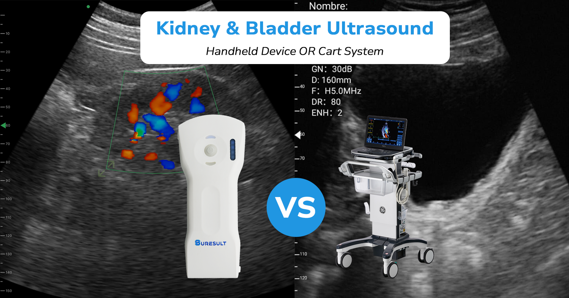
Bladder and kidney ultrasounds are among the most common point-of-care scans clinicians perform. With the increasing availability of handheld ultrasound machines focused on providing POCUS care, can they also replace full cart-based systems for these scans?
The short answer is: often, yes. But how, and what factors should be considered?
Let’s walk through the technical requirements of these scans, the device features that support them, and how handheld systems perform in real-world studies.
Buying for urology? The urology page compares the Suresult models for that work window by window, with real captures, verified specs and live pricing. See the urology page →
Bladder and Kidney POCUS: The Clinical and Technical Requirements
To determine the suitability of any ultrasound system, we must first establish the essential diagnostic parameters for bladder and kidney imaging.
Bladder scanning is often less about fine anatomic detail and more of a quantitative exercise. The main objectives are to measure bladder volume, assess post-void residual (PVR) urine, detect wall abnormalities, and identify focal masses or clots. This requires specific functionalities:
- High-Resolution Near-Field Imaging: Since the bladder is a superficial structure, a high-frequency linear or curvilinear probe (typically 3.5–6 MHz for sufficient penetration with good detail) is ideal. Excellent near-field resolution is crucial for accurately measuring:
- Wall Thickness: Assessed using manual calipers. Focal or diffuse thickening can indicate conditions like outlet obstruction, cystitis, or carcinoma.
- Mucosal Irregularities & Diverticula: Requires good contrast resolution to differentiate anechoic urine from bladder tissue and identify lesions or outpouchings.
- Adjustable Gain and Time-Gain Compensation (TGC): Proper gain settings are vital to “fill in” the anechoic bladder lumen without creating artifactual echoes, ensuring a clear demarcation of the wall and any internal debris or clots.
- Automated Volume Calculation: By tracing the bladder’s dimensions in two planes (usually transverse and sagittal), the system software applies an ellipsoid formula (Length x Height x Width x 0.52) to instantly calculate volume. This is far more efficient and reproducible than manual caliper measurements for serial monitoring of PVR.
Kidney ultrasound is a more nuanced assessment of deeper structures. It requires a probe with better penetration and high parenchymal resolution to obtain size metrics (length, cortical thickness), evaluate parenchymal appearance, and assess for obstruction (hydronephrosis) and focal lesions.
- Low-Frequency Deeper Imaging: For adult renal imaging, a convex probe in the ~2–6 MHz range is typical to achieve sufficient depth. For cortical detail and superficial segments (or in thin patients), a higher bandwidth is beneficial.
- B-mode Resolution and Dynamic Range: The system must resolve subtle echogenicities and shadows to identify hypoechoic renal pyramids from the more echogenic cortex. It should also clearly differentiate:
- Hydronephrosis
- Masses and Cysts
- Calculi
- Manual Calipers: Kidney dimensions—including length, width, and cortical thickness—are measured with calipers. Renal size is a critical marker in chronic kidney disease (CKD).
- Doppler Capability (Advanced): While not always essential for basic POCUS, spectral Doppler is needed to measure Resistive Index (RI)—a marker of renal vascular resistance—and color Doppler is used to assess renal perfusion and rule out vascular anomalies.
Functional assessment: Cart vs Handheld Ultrasound
Premium carts offer unparalleled image resolution, a vast dynamic range, and incredible processing power. However, high-end handheld devices, such as the Suresult D3Ultra (single-probe, multi-transducers) or the GE Vscan Air CL, also provide more than sufficient resolution to meet the core requirements outlined above.
The question is not whether handhelds can match carts in every metric—they cannot. The question is whether they are sufficiently accurate for clinical decision-making in bladder and kidney POCUS.
The research suggests they are.
A 2019 systematic review from Denmark published in MDPI analyzed 16 studies comparing HHUS (handheld ultrasound) with high-end systems. The review concluded that there was “good overall agreement” between the devices for evaluating hydronephrosis and ascites, among other conditions. The authors noted that for distinct clinical questions, “hand-held devices may be a valuable supplement to physical examination.”
A more recent 2024 study in the Journal of Diagnostic Medical Sonography specifically evaluated the validity of a handheld device for kidney metrics. It found moderate to excellent agreement (ICC = 0.75–0.94) between the handheld and a cart-based system for measuring kidney length, volume, and cortical thickness.
The mean absolute percentage error (MAPE) for all measurements was less than 5%, which is considered clinically acceptable. However, advanced vascular assessment, such as Renal Resistive Index (RRI), may require confirmation with a cart-based system, especially for measurements of the left kidney.

8 Most Recommended Handheld Ultrasound Machines for Kidney and Bladder Imaging
| Device Name | Best For | Probe Type | Imaging Modes | Specialized Features | Battery Life | Software/Cloud | Total Cost (3-Yr) | Key Consideration |
| Suresult D3Ultra | Maximum flexibility & value; all-in-one solution | Tri-mode: Convex, Linear, Phased | B, M, Color, PW | Renal/Urology presets, Needle assist, 8-TGC | ~2 hours | DICOM, No subscription | $2,604 (All-in) | Best feature-to-cost ratio; no recurring fees |
| Butterfly iQ3 | AI-powered automation & hybrid design | Single “Ultrasound-on-Chip” | B, M, Color, PW | AI Auto Bladder Vol., Needle Viz™, iQ Slice™ | ~2 hours | Subscription required for AI/Cloud | ~$4,598 (Device + Core Sub) | Cable can limit maneuverability; subscription model |
| Clarius C3HD3 | Reliable abdominal AI & wireless convenience | Convex | B, M, Color, Power, PW | AI Bladder Vol., Needle enhanced | ~60 minutes | Subscription required for saving images | ~$5,380 (Device + Sub) | Short battery life; subscription needed for core features |
| GE Vscan Air CL | Dual-probe efficiency & brand reputation | Dual-probe: Curved + Linear | B, M, Color, PW | SignalMax™ processing, Bladder Vol. tool | ~50 minutes | Optional sub for cloud ($430/yr) | ~$6,145 (Device + Sub) | Shortest battery life; high upfront & optional sub cost |
| Philips Lumify C5-2 | Long, uninterrupted scanning sessions | Curved | B, M, Color, PW | XRES, Harmonic Imaging, Reacts telemed | ~7.5 hrs (host device) | Optional sub for Reacts | ~$12,027 (Device) | Tethered design; highest upfront cost; iOS requires LPM |
| TodoPocus C5SH | Budget-friendly, full-featured performance | Convex | B, B/M, Color, PDI, PW | Puncture assist, 27+ measurements | ~1.5 hours | DICOM, No subscription | $2,390 (All-in) | Balanced cost with no subscriptions; strong specs |
| Mindray TE Air e5M | Comprehensive full-body scanning | Convex | B, M, Color, Power, PW | Smart Bladder tool, iWorkflows | N/A (Fast 35min charge) | No subscription (paid add-ons) | ~€3,500 (Base) | Limited to EU/UK; some advanced features are paid add-ons |
| Vave Health Universal | Flexible generalist & teaching environments | Universal PZT | B, M, Color Doppler | VaveCast™ teaching tool | Removable battery | Free unlimited cloud, DICOM | $3,299 (All-in) | US-only; generalist probe not specialized for abdomen |
Performance Comparison of Handheld vs. Cart Ultrasound
How do these two categories of devices truly compare side-by-side in a clinical environment?
We performed a side-by-side scan on a 62-year-old male with a high BMI (approximately 32) presenting with flank pain and a suspected obstructing stone to understand the potential differences in a real-world setting.
1- Image quality & resolution
- Beginning with the handheld Suresult D3Ultra using its abdominal preset, we could confidently identify moderate hydronephrosis and a 6mm stone at the ureterovesical junction (UVJ). The image was perfectly diagnostic.
- The same scan, repeated minutes later on a Philips EPIQ CVx cart, revealed slight nuances. The renal sinus fat had more defined textural detail, and the posterior acoustic shadowing of the stone was slightly sharper and more defined.
This is typically due to superior processing power, which allows for better speckle reduction, a higher dynamic range, and more advanced beamforming, resulting in a slightly “smoother” and more detailed image. So while the clinical findings were identical, the cart’s image had a higher level of polish. This could be a more important factor in technically difficult patients.
2- Depth & field of view
For renal and bladder imaging, both devices performed effectively equivalent.
- In the same patient, both the handheld and the cart-based system penetrated to the depth of the kidneys without issue.
- The field of view on the Suresult D3Ultra was more than sufficient to capture the entire length of the right kidney for measurement in a single frame.
- The depth and gain controls on the handheld’s touchscreen interface were intuitive and responsive, allowing for quick optimization to clearly differentiate the renal cortex, medulla, and collecting system.
3- Measurement precision
This is an area with high operator dependence. Personal preference for the user interface (UI), menus, and probe positioning can make a significant difference.
In my experience, handhelds hold a slight advantage here. While both platforms are highly calibrated, the streamlined, app-based measurement tools on handhelds are often more intuitive and faster to operate.
- We measured left renal length and bladder post-void residual (PVR) using the Suresult D3Ultra’s calipers. The probe positioning felt comfortable, and repeat measurements were simple.
- On the cart, the measurements were essentially the same (with no meaningful discrepancy), but the process of positioning and measuring felt faster on the handheld.
4- Workflow & integration
Most high-end handhelds and all cart systems offer DICOM compatibility, allowing you to securely send images and clips directly to a PACS. Our Suresult D3Ultra offers local storage on the connected app.
- From there, we can easily send the scan to the hospital’s PACS; simply tap “Export,” select the hospital’s PACS server from the pre-configured list, and the images are sent instantly. This process is very similar to the image export function on most cart systems.
Some handheld brands, like the GE Vscan Air CL, offer an optional subscription for cloud storage and collaboration tools through a proprietary platform. For a solo practitioner or a mobile team, this cloud-native approach could offer a slight edge in accessibility and data management outside a traditional hospital PACS environment.
Cost-Benefit Analysis: Calculating the Real-World Value
Beyond image quality lies the critical question of value, which involves far more than just the sticker price.
1. Upfront & Lifetime Cost
This is the most staggering differentiator. A premium cart system represents a major capital expenditure ($15,000 – $150,000+), often requiring service contracts, dedicated space, and high maintenance costs. Handhelds are operational expenses, with minimal overhead.
Let’s analyze the lifetime cost of two handheld options versus a cart:
| Cost Component | Suresult D3Ultra | GE Vscan Air CL | Premium Cart (e.g., Philips/GE/Siemens) |
| Upfront Hardware Cost | ~$2,800 (one-time) | ~$4,855 (one-time) | ~$80,000 (financed or capital) |
| Annual Service/Support | $0 | $0 | $8,000 – $15,000 (service contract) |
| Software/Cloud Subscription | $0 | $430/yr (Digital Tools) | $0 (typically included in service) |
| 3-Year Total Cost | $2,604 | $6,145 | ~$110,000 (lowest estimates) |
| 5-Year Total Cost | $2,604 | $7,005 ($4,855 + ($430 x 5)) | ~$155,000 (lowest estimates) |
Note: Cart costs are highly variable but consistently orders of magnitude higher. Many institutions lease carts or pay per exam, which changes the model but not the fundamental high cost.
The takeaway is that for the price of one cart, you could equip an entire department, or even a small clinic, with capable handheld devices, dramatically increasing point-of-care access.
2. Utilization Efficiency
A cart is stationary. A handheld is mobile. This simple fact transforms clinical workflow. The handheld can be carried on rounds, used in the ER bay, taken to a nursing home visit, or deployed in a crowded clinic room without moving the patient.
This eliminates logistical delays, reduces exam time from decision to diagnosis from hours to seconds, and allows for rapid serial scans (e.g., monitoring bladder volume over a shift). This isn’t just a convenience; it’s a force multiplier for clinical throughput and patient care.
3. Training & User Adaptability
Cart-trained sonographers already have core skills; so from an image interpretation standpoint, they can generally transition to handhelds quickly.
In addition, the simpler interface of a handheld can reduce the initial learning curve for the sonography technique. Especially in teaching institutions, the lower price barrier of handhelds makes them more available to residents and students for hands-on scanning experience early in their training. This exposure can result in faster skills acquisition and accelerated competency.
Handheld vs. Cart-Based Ultrasound: Comparison Table
| Premium Cart System | High-End Handheld Device |
| Dedicated radiology/imaging departments, comprehensive diagnostic exams. | Point-of-care (POCUS): bedside, clinic, rounds, remote/urgent care. |
| Unparalleled. Superior resolution, dynamic range, and processing for exceptional detail. | Diagnostically sufficient. Meets core requirements for many exams. |
| Broad selection of specialized probes | Usually 1–2 probes; some offer multi-frequency or multi-transducer designs. |
| Full Doppler suite, elastography, advanced vascular and tissue analysis. | Basic Doppler (color, power) is often included; spectral Doppler is limited or absent in many models. |
| Large, stationary unit; requires a dedicated room or space. | Pocket-sized, highly mobile; usable bedside, in clinics, or remote locations. |
| Seamless DICOM/PACS. | Most offer DICOM/ PACS. |
| High. Requires expensive service contracts and maintenance. | Very Low. Minimal service fees; some may have optional software subscriptions. |
| Complex cases, obese patients, detailed vascular studies, and comprehensive organ imaging. | Rapid diagnosis, serial monitoring, physical exam enhancement, and use in resource-limited settings. |
So, Could a Professional Realistically Choose a Handheld Device Over a Premium Cart?
The answer is a resounding yes, for a significant subset of clinical scenarios. The decision ultimately hinges on your specific practice environment, patient volume, and diagnostic needs.
Look how Dr. Brandon Rommakeo, a neuromusculoskeletal medicine specialist, uses the Suresult D3Ultra not only in his regular practice but also for training new professionals.
To make your choice, follow these considerations:
- For Comprehensive, Advanced Imaging: In radiology departments handling complex cases, detailed vascular studies, and technically challenging patients (e.g., high BMI), the premium cart’s superior image processing and advanced capabilities remain irreplaceable.
- For Point-of-Care Excellence: For hospital rounds, urgent care, sports medicine, primary care clinics, and remote settings, the handheld device is not merely adequate—it is often the superior tool due to its immediacy and portability.
The modern handheld ultrasound is a powerful, validated, and clinically capable instrument. While it does not replace the high-end cart for all applications, it successfully challenges its necessity for a wide range of renal and bladder POCUS exams, offering a compelling blend of performance, portability, and value.
By Suresult
As an international manufacturer at the forefront of handheld ultrasound technology, Suresult is committed to being a forerunner in providing industry insights and empowering medical professionals with the knowledge to make informed decisions about their diagnostic tools. This article is an effort to move beyond marketing and offer a transparent, evidence-based comparison to help you navigate the evolving landscape of medical imaging.
If you’re interested in further reading, we invite you to check our detailed review, Which Handheld Ultrasound Is the Most Accurate in 2025? Our Top Picks.
For any queries related to our products, or if you need a personalized recommendation based on your specific clinical needs, please feel free to contact our team of experts.

