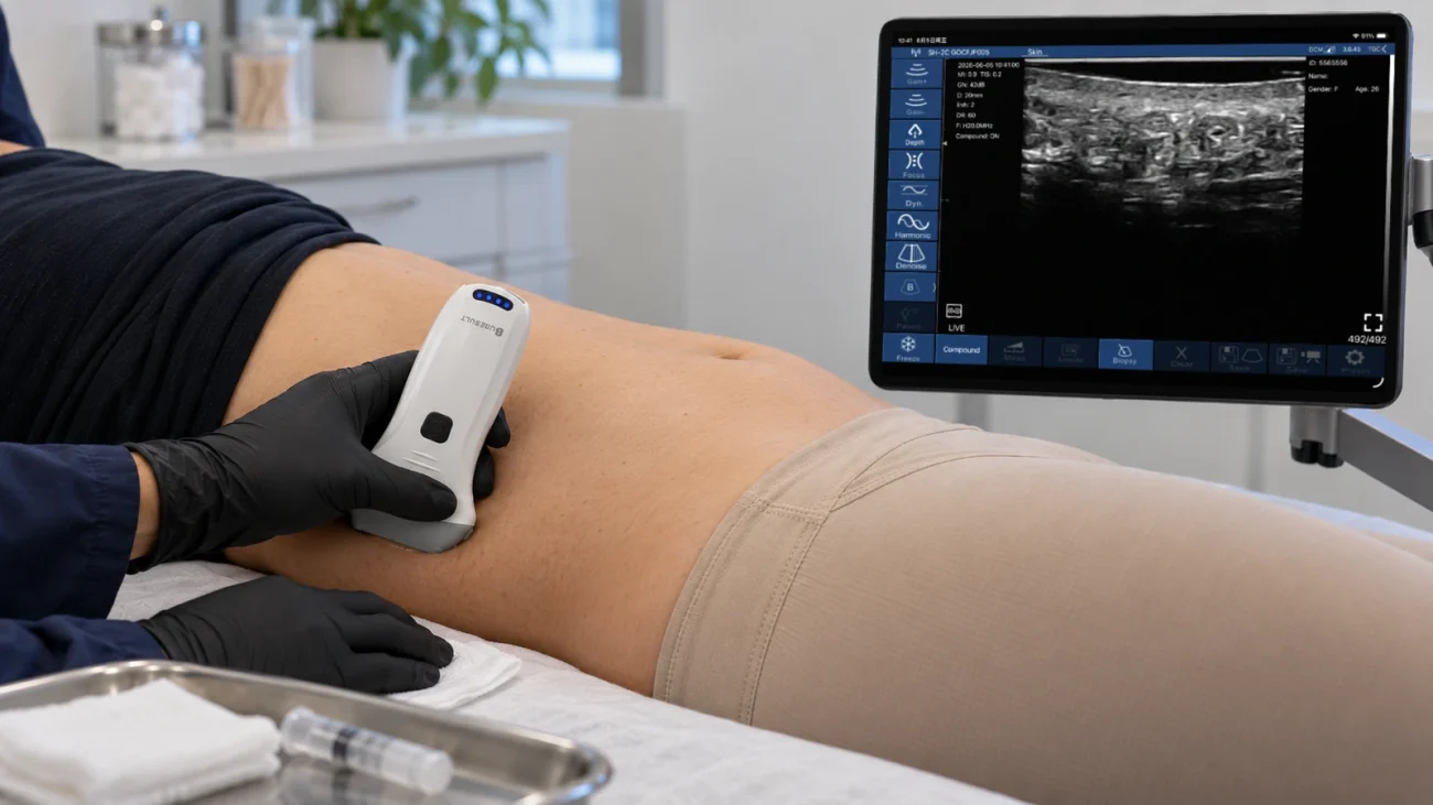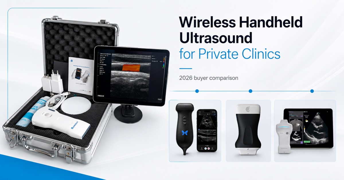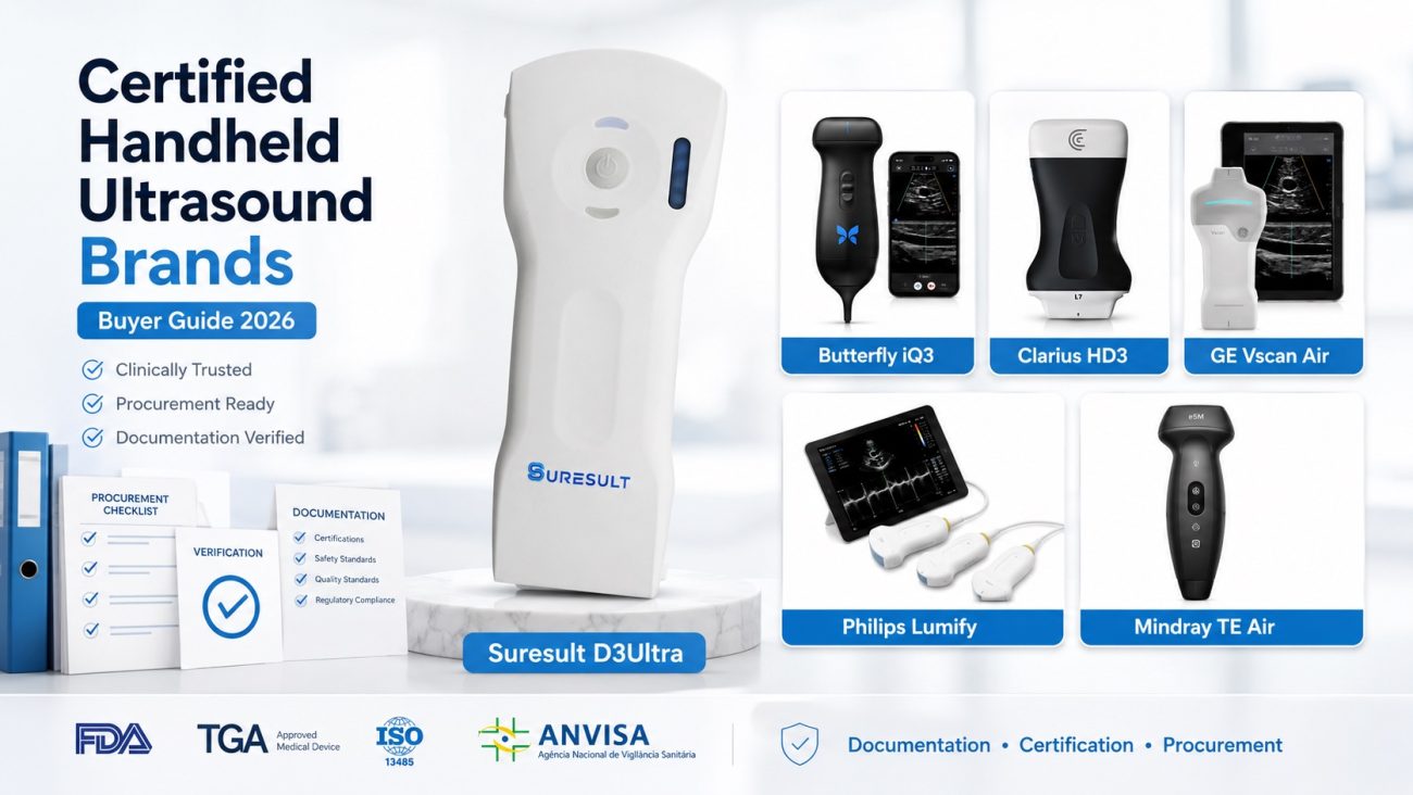
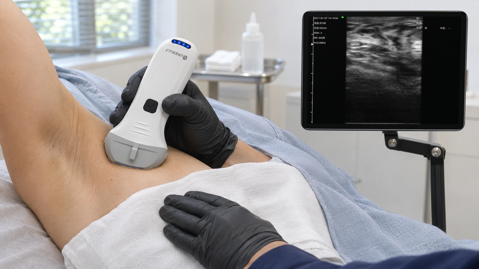
For breast implant ultrasound I pick the Suresult L14Pro at $3,293. Under the FDA’s September 2020 labeling guidance, silicone implants without symptoms get a first ultrasound or MRI 5 to 6 years after surgery and another every 2 to 3 years, a recurring scan that belongs in the surgeon’s own office. Its 10/14 MHz linear draws the shell and capsule at 14 MHz, holds the posterior shell and chest wall of implants to about 4.5 cm projection on its 80 mm preset, and carries Color, Power and PW Doppler, needle guides, calipers, volume, cine and DICOM in the price, with no subscription. Each visit comes down to six reads: an intact shell and capsule, the stepladder of intracapsular rupture, the snowstorm of free silicone, silicone in the axillary nodes, late fluid that needs a guided tap, and capsule thickness by Baker grade, each set beside the last study. For vessel mapping and the other scans of a plastic surgery office, see handheld ultrasound for plastic surgery.


Escopo da avaliação: I judge implant-surveillance scanners as an independent device reviewer with deep ultrasound knowledge. I scored ten handhelds from six makers for in-office implant surveillance, on seven criteria declared on November 12, 2026, before any rival carried a score: the Suresult L14Pro with its L20Pro, L14 and L20 siblings, the Mindray TE Air e5M, the GE Vscan Air CL, the Clarius L7 HD3 with the L15 HD3, the Butterfly iQ3 and the Philips Lumify L12-4. The criteria are depth to the posterior shell and chest wall, near-field detail for the shell and capsule, Doppler, guided aspiration of peri-implant fluid, measurement, records for serial comparison and five-year cost. Each device fact, label line and study figure on this page comes from manufacturer records, FDA device labels and PubMed, checked November 20, 2026, Butterfly’s probe and plan prices on November 12.
Quick answer: which handheld should you buy for breast implant ultrasound?
The Suresult L14Pro ($3,293) is the handheld I recommend for breast implant ultrasound, at 100.0/100 in my six-maker ranking. At 14 MHz it draws the shell lines and the capsule, and its 20, 40, 60 and 80 mm presets take in the posterior shell and chest wall of implants to about 4.5 cm projection. Color, Power and PW Doppler come with the probe, as do Needle Enhance with in-plane and out-of-plane guides for a fluid tap, calipers, volume from three diameters and text annotation. Cine runs to 1,000 frames, about 165 seconds; studies save on your own phone, tablet or PC and leave by DICOM export or DICOM send, with no subscription. In the same card, the L20Pro adds 20 MHz for the anterior shell, the capsule and the axilla.
Second place goes to the Mindray TE Air e5M at 82.4: Color, Power and PW Doppler, iNeedle, the VA Grid and B-mode volume come in one $4,499 purchase (Mindray NA store, checked Nov 20, 2026), on a single transducer with a 2.0–9.0 MHz bandwidth that images to 40 cm.
Breast implant scanners compared: six handhelds side by side
Across the six ranked handhelds, four specs decide implant follow-up: a top frequency at or above the 12 MHz the ACR names for breast ultrasound, a frame deep enough to hold the posterior shell, a needle view for tapping peri-implant fluid, and what every scored tool costs over five years.
| Scanner | Frequência máxima · profundidade | Doppler | Needle tools for a fluid tap | Medição | Cine, storage and transfer | Custo em cinco anos e assinatura | Melhor para |
|---|---|---|---|---|---|---|---|
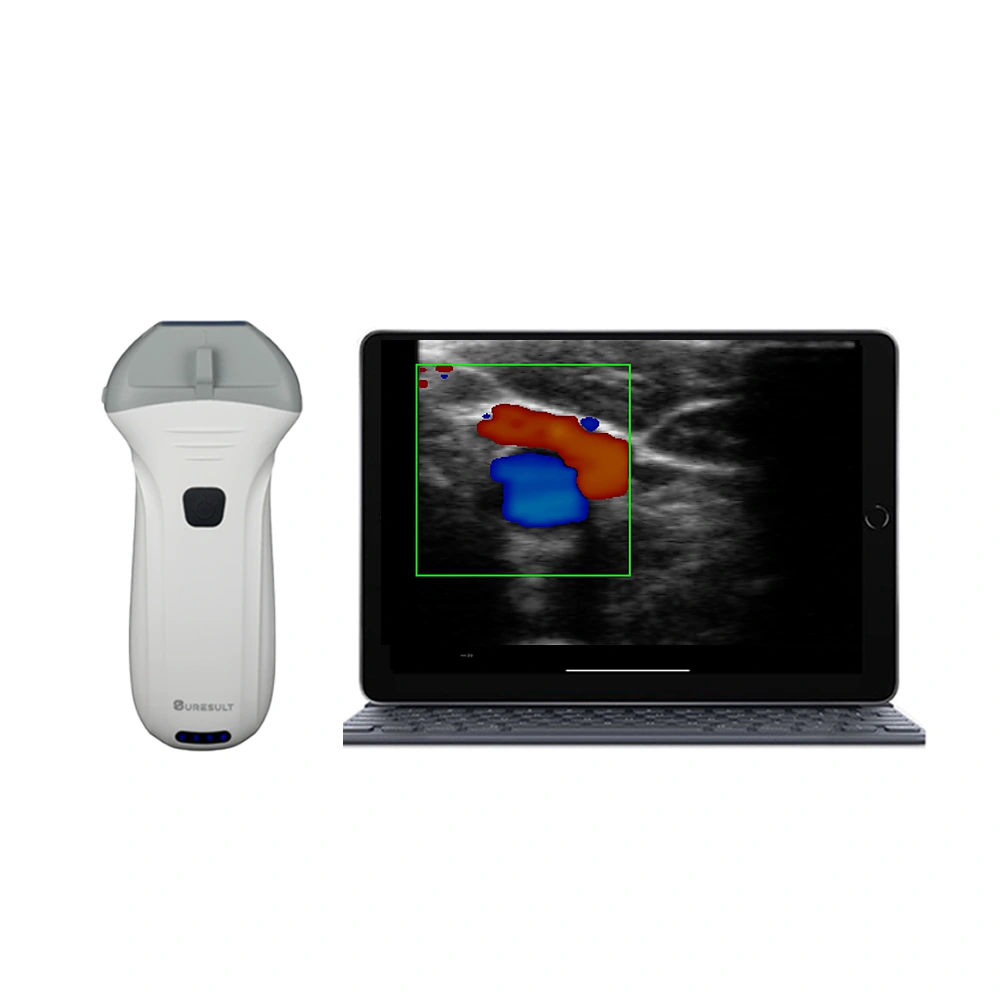 Suresult L14Pro · No. 1 · 100.0/100 |
14 MHz on a 10/14 MHz linear · presets of 20, 40, 60 and 80 mm | Color, Power and PW, every mode with the probe | Needle Enhance; in-plane line and out-of-plane target | Calipers; volume from three diameters; text comments on the image | 1,000-frame cine, about 165 s; studies kept on your own device; DICOM export and DICOM send | $3,293 one-time, no subscription | The whole implant to about 4.5 cm projection with every scored tool in the price |
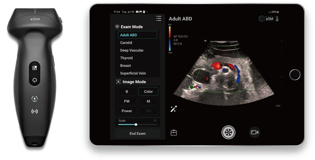 Mindray TE Air e5M · No. 2 · 82.4/100 |
9.0 MHz at the top of a 2.0–9.0 MHz bandwidth · to 40 cm | Standard Color, Power and PW Doppler | iNeedle enhancement; VA Grid | Distance and area; B-mode volume in five exam packages; Comments and 251 body marks | 125-frame cine of 2–5 s; onboard storage; DICOM 3.0 Store and Tricefy | $4,499 one-time, sold with no subscription plan | Three Doppler modes, a needle aid and volume paid for once |
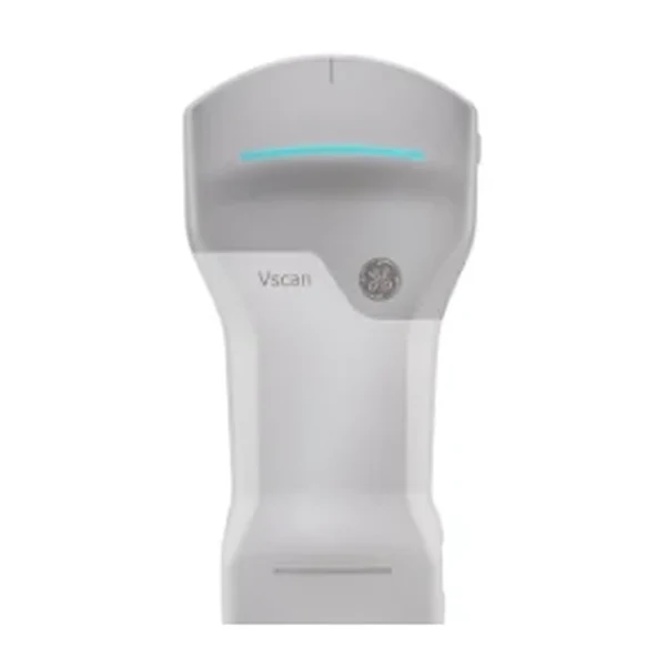 GE Vscan Air CL · No. 3 · 76.9/100 |
12 MHz on the 3–12 MHz linear head · images to 8 cm | Color and PW, both in the price | Marcador da linha central | Distance and ellipse; free text and arrows | Up to 500 exams on the phone or tablet; a configured DICOM image server | $4,999 one-time; the optional Innovations plan sits outside the score | An 8 cm linear head beside a curved head, nothing to renew |
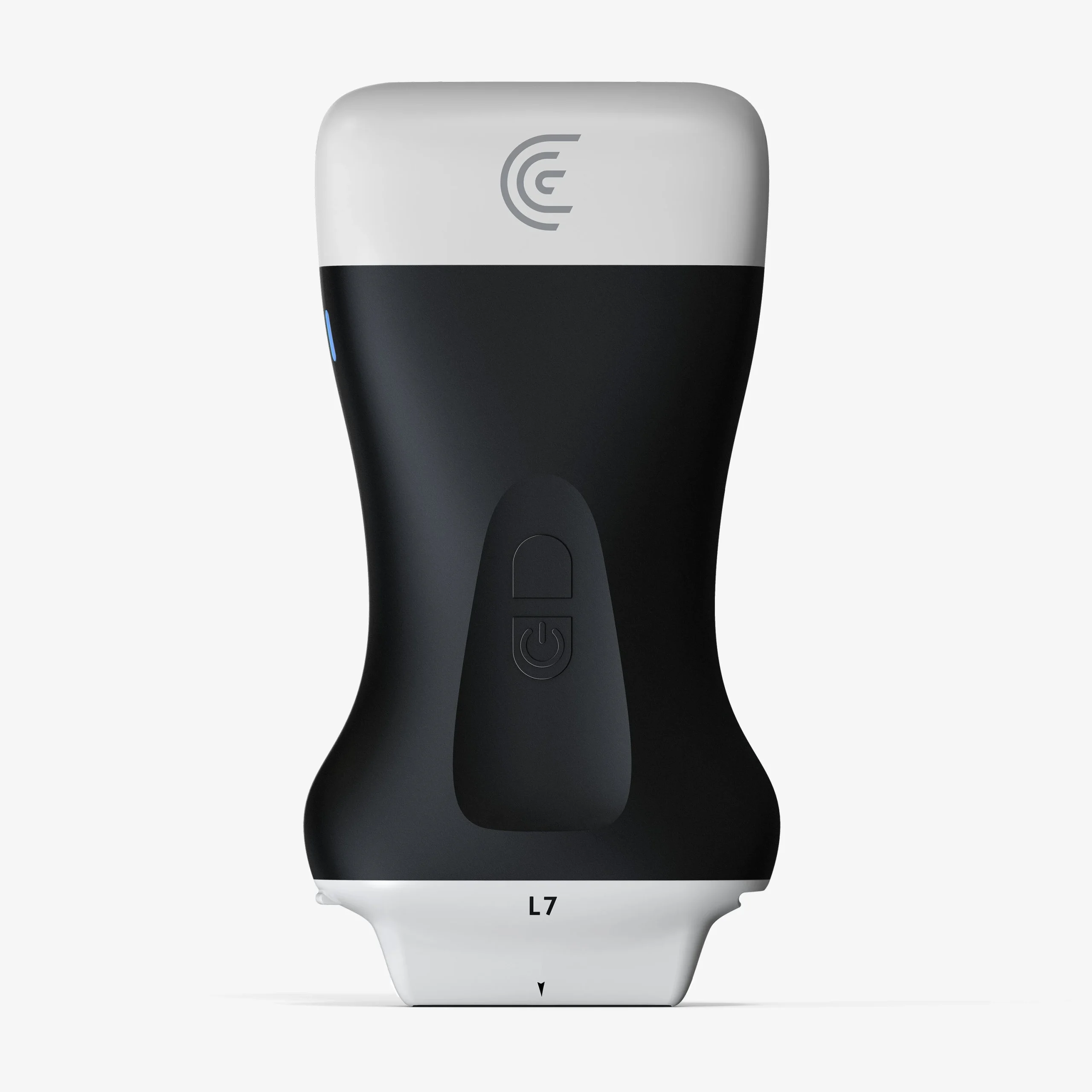 Clarius L7 HD3 · No. 4 · 75.0/100 |
13 MHz on a 4–13 MHz linear · Max Depth 15 cm | Color and Power with the scanner; PW on Essentials | Needle Enhance on Essentials; Center Guide | Distance and area calipers; Annotations | 30 s cine playback; storage, DICOM and PDF reports on Essentials | $6,295: $3,795 plus the $2,500 one-time Essentials license ($695 a year on the annual plan) | The maker’s own implant protocol, filmed on this scanner |
 Butterfly iQ3 · No. 5 · 73.6/100 |
12 MHz (1–12 MHz) · to 30 cm, MSK-Soft Tissue to 12 cm | Doppler colorido, Power e pulsado com Advanced | Needle Viz, Biplane and Needle: Out-of-Plane on Advanced; midline marker | 4 linear and 1 elliptical calipers; Generic Volume Calculation; on-screen annotation | Start-stop clips; unlimited cloud storage with Advanced; Basic DICOM as an add-on | $5,399 one-time: the iQ3 with five years of Advanced | Calipers, volume and biplane needle views on one whole-body probe |
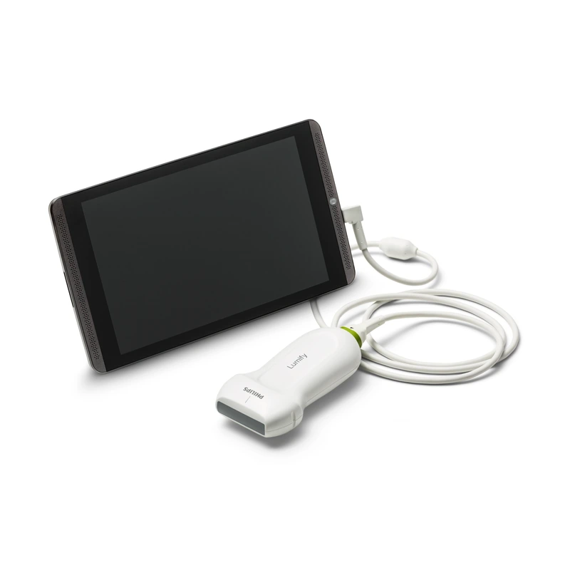 Philips Lumify L12-4 · No. 6 · 71.6/100 |
12 MHz (12–4 MHz) · scan depth to 12 cm | PW and steerable color | Marcador de linha central | Distance and ellipse; text annotation on Android | Loops of 1–10 s; hundreds of exams; email, DICOM to PACS or a shared network | $10,307.95 one-time at PartsSource, all software included | A battery-free linear owned outright |
Melhor para cada uso: escolhas rápidas
- Best Overall for Breast Implant Ultrasound: Suresult L14Pro, 100.0/100: 14 MHz, an 80 mm preset, all three Doppler modes, Needle Enhance and DICOM in one price.
- Best for Full Doppler, Needle Aid and Volume in One Purchase: Mindray TE Air e5M, 82.4/100: three Doppler modes, iNeedle and B-mode volume with no subscription plan.
- Best for Color and PW on an 8 cm Linear Head With Nothing to Renew: GE Vscan Air CL, 76.9/100: Color, PW and DICOM in the purchase, beside a curved head.
- Best for the Maker’s Own Implant Protocol to 15 cm: Clarius L7 HD3, 75.0/100: 13 MHz, 15 cm and a Classroom implant protocol filmed on it.
- Best for Calipers, Volume and Biplane on One Whole-Body Probe: Butterfly iQ3, 73.6/100: four linear calipers and an elliptical one, volume, and Biplane on Advanced.
- Best for a Battery-Free Linear Bought Outright: Philips Lumify L12-4, 71.6/100: a 12–4 MHz linear under 136 g with every software feature included.
Onde comprar cada ultrassom portátil
- Suresult L14Pro: $3,293 at the Suresult store, paid once with every mode and export unlocked, a 30-day worry-free trial and a 2-year warranty, extendable to 5 years. Order the Suresult L14Pro at $3,293
- Mindray TE Air e5M: sold direct by Mindray North America at $4,499, checked November 20, 2026, with a 3-year warranty. TE Air e5M na loja Mindray
- GE Vscan Air CL: $4,999.00 at the GE HealthCare US shop, checked November 20, 2026, with a standard 3-year warranty. Vscan Air CL na loja GE HealthCare
- Clarius L7 HD3: $3,795 at the Clarius store, checked November 20, 2026, with a 3-year warranty and a 30-day return window; Essentials, which carries PW Doppler, Needle Enhance, storage and DICOM, costs $2,500 one-time or $695 a year, and the cart shows $4,490 with the first year. Clarius L7 HD3 na loja Clarius
- Butterfly iQ3: $5,399 in one payment at the Butterfly US store, checked November 20, 2026, with Advanced and a 3-year warranty: the $3,899 probe plus $1,500 for five years of Advanced at Butterfly’s November 12, 2026 pricing. Butterfly iQ3 at the Butterfly store
- Philips Lumify L12-4: PartsSource lists it new and outright at $10,307.95 with a 90-day warranty, checked November 20, 2026; Philips also sells it through its own team. Lumify L12-4 na PartsSource
How often to scan silicone implants: the FDA schedule as an office protocol
Silicone implants without symptoms get a first ultrasound or MRI 5 to 6 years after the operation and then one every 2 to 3 years, in cosmetic and reconstructive patients alike, under the FDA’s labeling guidance of September 29, 2020 (FDA 2020). It replaced the MRI-only schedule of the earlier labels, MRI at 3 years and every 2 years after, on the 2019 advisory panel’s consensus that ultrasound is an acceptable screening test (FDA 2011; Natrelle label L034 Rev.12; FDA 2020). Symptoms, or an ultrasound that leaves the shell in doubt, send the patient to MRI; saline implants carry no screening schedule.
The new labeling, with a boxed warning and a patient decision checklist, was approved by the FDA on October 27, 2021 (FDA 2025, labeling). The same FDA page lists the approved gel implants by premarket approval (PMA) number: Natrelle round P020056, MemoryGel P030053, Sientra P070004, Natrelle 410 P040046 and Motiva P230005. The current Natrelle label (L034rev15, November 2024) names ultrasound or MRI for the schedule and advises removing an implant whose rupture shows on imaging (Natrelle label L034rev15). The checklist each patient initials gives example rates of up to 31.2% for rupture or leak, 51.7% for capsular contracture and 59.7% for reoperation (FDA 2020), and the FDA’s patient page puts it plainly: “Breast implants are not lifetime devices” (FDA 2023).
The office scan closes a compliance gap the labels themselves record. In Sientra’s core study 69.9% of the MRI cohort kept its follow-up MRIs, 56.5% at year 10, and the label tells surgeons that lifetime imaging can cost more than the operation and that insurance does not always pay for it (Sientra label DFU-0002 R7). In a survey of U.S. plastic surgeons, 37.7% recommended MRI at the FDA intervals and 55% imaged only when a problem appeared (Carr 2020); among 143 breast radiologists, 46.9% start an implant check with handheld ultrasound (Moosavi 2024).
Most ruptures are silent, so the schedule cannot wait for symptoms. In the MemoryGel core study 75 of 77 ruptured implants had none (Caplin 2021); in Sientra’s, 35 of 36 confirmed ruptures were silent (Sientra label DFU-0002 R7); and a clinical breast exam found 30% of ruptures, at 88% specificity, against MRI in 109 implants (Hölmich 2005, clinical exam). MRI over-calls as well: 39% of the implants Sientra’s study MRIs read as silently ruptured were intact at explant or on the next MRI (Sientra label DFU-0002 R7).
Rupture climbs with implant age. These are the 10-year rates from the core studies behind each PMA, with the Danish repeat-MRI cohort and a pooled explant series for the long view:
| Implant and study | Gel and cohort | Years followed | Rupture | Fonte |
|---|---|---|---|---|
| Natrelle round (P020056), Core Study | Primary augmentation, MRI cohort | 10 | 9.3% of patients (Kaplan-Meier) | Natrelle label L034rev15; Spear 2014 |
| Natrelle round, Core Study | Primary reconstruction, MRI cohort | 10 | 35.4% of patients | Natrelle label L034rev15 |
| MemoryGel (P030053), Core Study | Primary augmentation, MRI cohort | 10 | 9.81% of patients and 7.36% of implants, confirmed at explant | Caplin 2021 |
| MemoryGel, Core Study | Primary augmentation, all 552 patients | 10 | 24.2%, suspected or confirmed | Caplin 2021 |
| Sientra (P070004), Core Study | Primary augmentation, MRI cohort of 398 | 10 | 8.5% of patients | Sientra label DFU-0002 R7 |
| Sientra, Core Study | Primary reconstruction, MRI cohort | 10 | 16.5% of patients | Sientra label DFU-0002 R7 |
| Natrelle 410 (P040046), form-stable gel | MRI cohort of the 941-subject study | 10 | 5.7%, confirmed | Maxwell 2015 |
| Danish cohort, repeat MRI | 317 implants intact at least 3 years after surgery | 5 and 10 | Rupture-free: 98% at 5 years, 83–85% at 10 | Hölmich 2003 |
| Pooled explant series | 1,099 older-generation implants | 10 and 15 | Intact: 79.1% at 10 years, 48.7% at 15; median life span 16.4 years | Goodman 1998 |
The schedule also has an end: a 2026 scoping review recommends stopping routine scans of asymptomatic silicone implants after age 75 (Vernice 2026).
Run as an office protocol, the schedule turns into five kinds of visit, each saved so the next one compares like with like:
| Visit | Timing | What to scan | What to save for the next visit |
|---|---|---|---|
| First surveillance scan | 5 to 6 years after implantation, no symptoms | Both implants and both axillae: shell, gel, capsule, the space around the implant and the nodes | Six views and a sweep clip per breast, capsule thickness in mm, and the preset and depth used |
| Interval scan | Every 2 to 3 years after the first | The same coverage, view by view against the last study | The same views at the same preset and depth, labeled by clock face and distance from the nipple, sent by DICOM |
| Doubtful scan or symptoms | Any time | The finding in two orthogonal planes, then MRI without contrast | The images and clip of the finding for the MRI reader |
| Rupture confirmed | At diagnosis | How far the free silicone reaches, and every node it touches | Snowstorm extent and node status, the map for later visits |
| Late fluid | One year or more after implantation | The fluid, the capsule for a mass, and the nodes; then a guided tap | Depth and size of the collection, the tap clip and the volume sent to pathology |
The normal breast implant on ultrasound
A normal breast implant on ultrasound is an anechoic body of gel under a shell drawn as one to three parallel echogenic lines, with the fibrous capsule as one more parallel line outside it and often a thin film of fluid between the two (Kanavou 2023; Rukanskienė 2021). On a 12 MHz linear a textured Mentor shell draws two parallel white lines and a smooth Motiva shell three (Rukanskienė 2021). The capsule shows three layers, echogenic, anechoic, echogenic, and measured 0.6 mm (± 0.2) at Baker I on an 18 MHz probe (Torres-Zuniga 2022). In one series, all four implants whose lumen was completely free of echoes were intact (Palmon 1997).
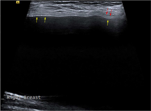
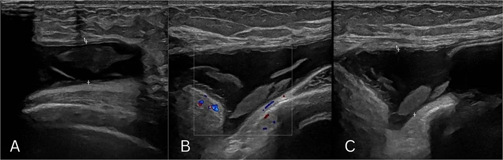
Lines inside an intact implant are common: linear echoes appeared in 57 of 64 implants (89%), and on their own they do not predict rupture (Palmon 1997). The radial fold is a normal one: an infolding of the shell that runs in from the periphery perpendicular to the capsule and stays joined to the shell; folds multiply as a capsule contracts (Kanavou 2023; Jaeger 2026). Rupture lines lie the other way, parallel to the probe in the anterior gel. Reverberation bands inside the near gel, and echoes behind the implant, imitate rupture for an inexperienced reader (Shah 2016).
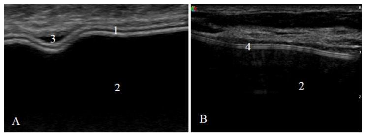
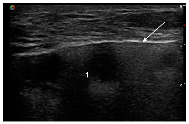
Saline and silicone look alike: both are anechoic inside an echogenic envelope, and saline rupture is a clinical diagnosis, made by the deflation the patient sees (Shah 2016; Kanavou 2023). The plane reads off the layers above the shell: a subglandular implant sits under the gland in front of pectoralis major; a subpectoral one under the muscle, which thinned from about 3.7–4.1 mm to 2.0–2.2 mm three months after augmentation in 87 women (Hong 2023); a prepectoral one after mastectomy under skin and a flap that averaged 10.1 mm (3–25 mm) at surgery in 1,663 mastectomies (Salgarello 2026). Behind the posterior shell lie pectoralis major and the ribs for a subglandular implant, and the chest wall itself for a subpectoral one, read as the distance from the capsule to the parietal pleura (Fleury 2025).
Intracapsular rupture on ultrasound: stepladder, keyhole and subcapsular line
Intracapsular rupture is a tear in the implant shell with the silicone still held inside the fibrous capsule; on ultrasound it shows as the stepladder sign, the keyhole sign or a subcapsular line, and it is the usual form: the Danish MRI studies the Natrelle label summarizes found about three quarters of ruptures intracapsular, with about 10% of those moving outside the capsule over 2 years (Natrelle label L034rev15). In 299 ruptured fourth-generation cohesive implants confirmed at surgery, 81.6% were intracapsular (Bresnick 2026). Each sign sits inside the gel or at the shell, with the capsule line intact around them:
| Sign | O que a tela mostra | O que isso significa | Fonte |
|---|---|---|---|
| Stepladder | Stacked horizontal echogenic lines in the gel, parallel to the probe in the anterior implant | The collapsed shell folded on itself and lifted off the capsule with silicone between; the ultrasound counterpart of the MRI linguine sign; the most reliable rupture sign in its first series, present in 14 of 20 ruptures | DeBruhl 1993; Seiler 2017 |
| Keyhole sign (teardrop, noose) | A radial fold whose loop fills with gel | Silicone has crossed the shell into the fold: an early sign of intracapsular rupture | Kanavou 2023; Seiler 2017 |
| Subcapsular line | An echogenic line just beneath the capsule with silicone beside it | The shell has pulled away from the capsule: another early sign | Kanavou 2023; Seiler 2017 |
| Heterogeneous gel | Low-level echoes, echodense aggregates or echogenic noise in the lumen | Gel mixed or fractured after the shell fails: inhomogeneous content in 82.8% of ruptured against 3.2% of intact implants, echogenic noise in 17 of 19 ruptures | Rukanskienė 2021; Rosculet 1992 |
| Abnormal shell | One line where two belong, or an uneven, broken shell line | Shell failure: present in 98.3% of ruptured against 1.1% of intact implants | Rukanskienė 2021 |
In last-generation cohesive gel, read the shell and the gel together before calling a stepladder: serial ultrasound of 303 cohesive implants found stepladder-type lines in 170 (56%) at 6 years, while 3 (about 1%) ruptured (Rochira 2016). An uneven or broken shell line, echoes in the gel, a keyhole or a subcapsular line make the call.
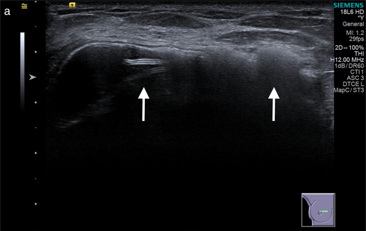
How accurate breast implant rupture ultrasound is
Breast implant rupture ultrasound finds most ruptures, and the recent series find nearly all of them: pooled across 20 studies, 1,987 patients and 3,297 implants checked against surgery, sensitivity is 73.7% and specificity 87.8% (Lacerda Macedo 2021), and recent office and clinic series report 90.9% to 98.3% sensitivity (Glener 2024; Rukanskienė 2021; Spit 2024).
| Estudo | Reader and setting | Frequency and array | Patients and implants | Resultado | Reference standard |
|---|---|---|---|---|---|
| Lacerda Macedo 2021 | Meta-analysis of 20 studies | Pooled across the studies | 1,987 patients, 3,297 implants | Sensitivity 73.7%, specificity 87.8%; AUC 0.7762 | Surgery |
| Glener 2024 | One plastic surgeon, in-office surveillance | High-resolution office ultrasound | 254 patients and 508 implants; 104 implants explored, 20 ruptured | Sensitivity 90.9%, specificity 100%; PPV 100%, NPV 97.6% | Surgery |
| Bengtson and Eaves 2012 | Surgeon and radiologist ultrasound beside MRI | Portable high-resolution linear, 8–16 MHz, 12 MHz best at clinical depth | 15 consecutive patients, 29 breasts | Surgeon ultrasound, radiologist ultrasound and MRI each correct in 29 of 29 | Surgery |
| Rukanskienė 2021 | One breast radiologist, bilateral implants | 12 MHz linear | 76 women, 152 implants | Sensitivity 98.3%, specificity 89.2%; accuracy 94.7% | Surgery |
| Spit 2024 | Clinic cohort | 18 MHz linear | 104 women, 205 implants | Sensitivity 95%, specificity 96%; accuracy 96% | MRI |
| Ferenz 2023 | Surgeons learning office scanning | High-resolution office ultrasound | 480 implants | Above the published sensitivity from the 60th reading on | Surgery |
| Salzman 2022 | Nine practices after one day of training | 7–13 MHz handheld | 584 women, 1,153 implants | Rupture on the scan in 14.0% of women and 8.0% of implants; confirmed in 30 of the 40 women who had surgery (75%) | Surgery in 40 women |
Two things move the number. Extracapsular rupture is the easier read: against MRI, ultrasound found 100% of extracapsular and 63% of intracapsular ruptures (Telegrafo 2015). And readers improve with practice (Ferenz 2023): in a point-of-care series of 5,557 implants scanned at 7 to 18 MHz, the ultrasound call matched surgery in all 89 women who went on to an operation (Kim 2024).
Office screening finds rupture nobody suspected, and patients want the answer: in the nine-practice series, 99.5% of the women wanted to know, and 95.2% wanted a ruptured implant removed (Salzman 2022).
When the scan leaves the shell in doubt, or the patient has symptoms, MRI without contrast is the next study (FDA 2020; ACR 2025). Pooled across 21 studies, MRI ran at 87.0% sensitivity and 89.9% specificity, and samples of symptomatic patients inflated that accuracy (Song 2011). Payers follow the ultrasound: Aetna covers MRI for silent rupture at most once every 2 years and treats MRI to confirm a rupture already shown on ultrasound in a symptomatic patient as experimental, since the ultrasound is enough to proceed to removal (Aetna CPB 0105). Aetna then covers removal of a ruptured silicone implant, intracapsular or extracapsular, after cosmetic augmentation or reconstruction, and treats re-insertion after a cosmetic augmentation as cosmetic (Aetna CPB 0142). On 2011 Medicare amounts, Chung’s decision analysis found ultrasound first with MRI to confirm the optimal strategy for women without symptoms, at $637 per rupture managed, and ultrasound alone optimal for women with symptoms, at $1,622 (Chung 2012).
Extracapsular rupture, siliconoma and silicone in the axillary nodes
Extracapsular rupture puts silicone through both the shell and the capsule into the breast, the muscle or the nodes, and ultrasound shows it as the snowstorm sign: highly echogenic, scattered, reverberating echoes with a sharp front edge and a blurred back edge (Harris 1993). In Kim’s point-of-care series, all 80 capsules that showed a snowstorm held silicone at pathology, and about 90% of those patients had no symptoms (Kim 2024). Free silicone also gathers into a siliconoma, also called a silicone granuloma (Wu 2023): a hyperechoic mass with a snowstorm behind it, or an anechoic to hypoechoic mass of extruded gel (Van Boeckel 2024; Rosculet 1992).
Silicone reaches the axillary nodes with and without rupture. Of 1,217 women with silicone implants in a 10-year single-center cohort, 5.83% had silicone lymphadenopathy, and two-thirds of them, 47 women, had intact implants: gel bleed through an unbroken shell (Berben 2025). After rupture the share rises with the implant’s age: across 299 surgically confirmed ruptured sides with fourth-generation cohesive implants, nodal silicone appeared in 13.6% to 24.4% of them by maker and rupture type, with no significant difference between makers, and the odds rose 13% for each year of implant age (Bresnick 2026). In a 52-patient series ultrasound made the find in 80.7%, 69.3% of patients had no symptoms and every biopsy was benign (Desai 2025).
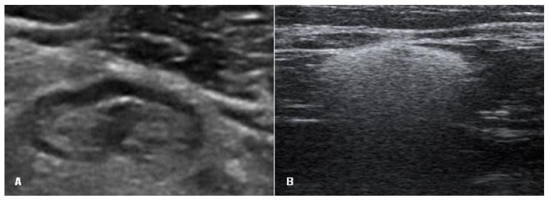
Scan both axillae at every visit (Spit 2024), with the patient turned into the contralateral oblique position and the two sides compared (ACR 2026). Axillary nodes sat a mean 1.6 cm from the skin across 32 nodes measured (Pajcini 2021), the depth of the L20Pro’s 20 and 30 mm presets at 16/20 MHz, with Color and Power Doppler on the hilum. Ultrasound sees more nodal silicone than MRI: in a 104-woman clinic cohort, MRI detected 44% of the nodal silicone that ultrasound found (Spit 2024). Once a rupture is confirmed, record how far the snowstorm reaches and which nodes it involves, so the next study reads against that map (Kim 2024).
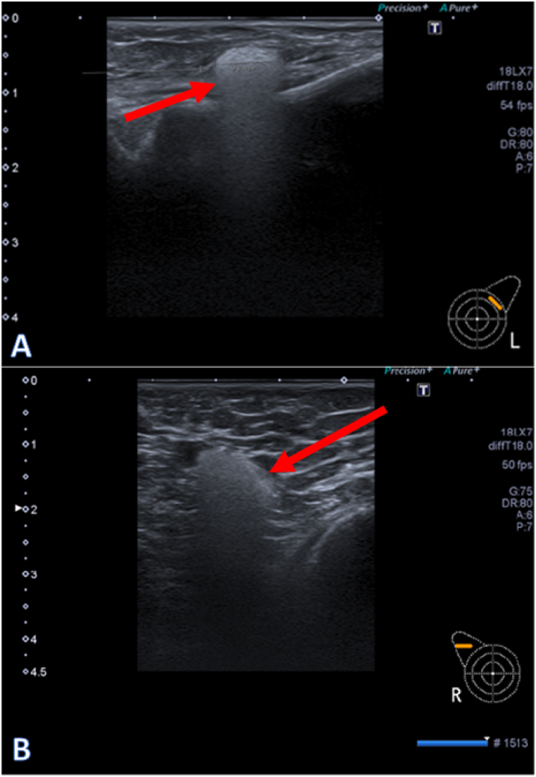
Fluid around an implant: late seroma and guided aspiration
A thin film of fluid around an implant is normal (Kanavou 2023); fluid that appears one year or more after implantation is a late seroma, reported in 0.1% of patients, and its minimum work-up is an ultrasound-guided aspiration with culture and cytology (Bengtson 2011). Ultrasound is the first test: it shows the fluid, any mass in the capsule and the nodes, and it guides the needle (Keane 2023; Longo 2022). In an 852-woman implant clinic, 30 late seromas turned up: 22 on clinical examination and 8 small collections on ultrasound alone (Jaeger 2026).
Every late seroma is sampled because of breast implant-associated anaplastic large cell lymphoma (BIA-ALCL). The FDA’s medical device reports to June 30, 2024 count 1,380 cases: 73% with textured implants, 3% with smooth ones and 24% unspecified. A seroma was the presenting sign in 48%, and the median time from the last implant to diagnosis was 8 years (FDA 2025, MDR report). Most late seromas are not lymphoma: 9% tested positive in the series a 2023 review cites (Vorstenbosch 2023). With smooth implants the risk equals the general population’s risk of ALCL, about 1 in 4 million (Clemens 2024).
The work-up runs in a fixed order. Scan the fluid, the capsule for a mass, the chest wall, the axillary, internal mammary and supraclavicular nodes, and the other implant (Keane 2023). Aspirate under ultrasound guidance and send at least 20 mL of fresh fluid to pathology within 12 hours, with any surplus to microbiology (Longo 2022). The laboratory makes Wright-Giemsa smears and a cell block for immunohistochemistry and flow cytometry, testing for CD30 and ALK (FDA 2019), from fresh, unfixed fluid with the smears made first (Jaffe 2020).
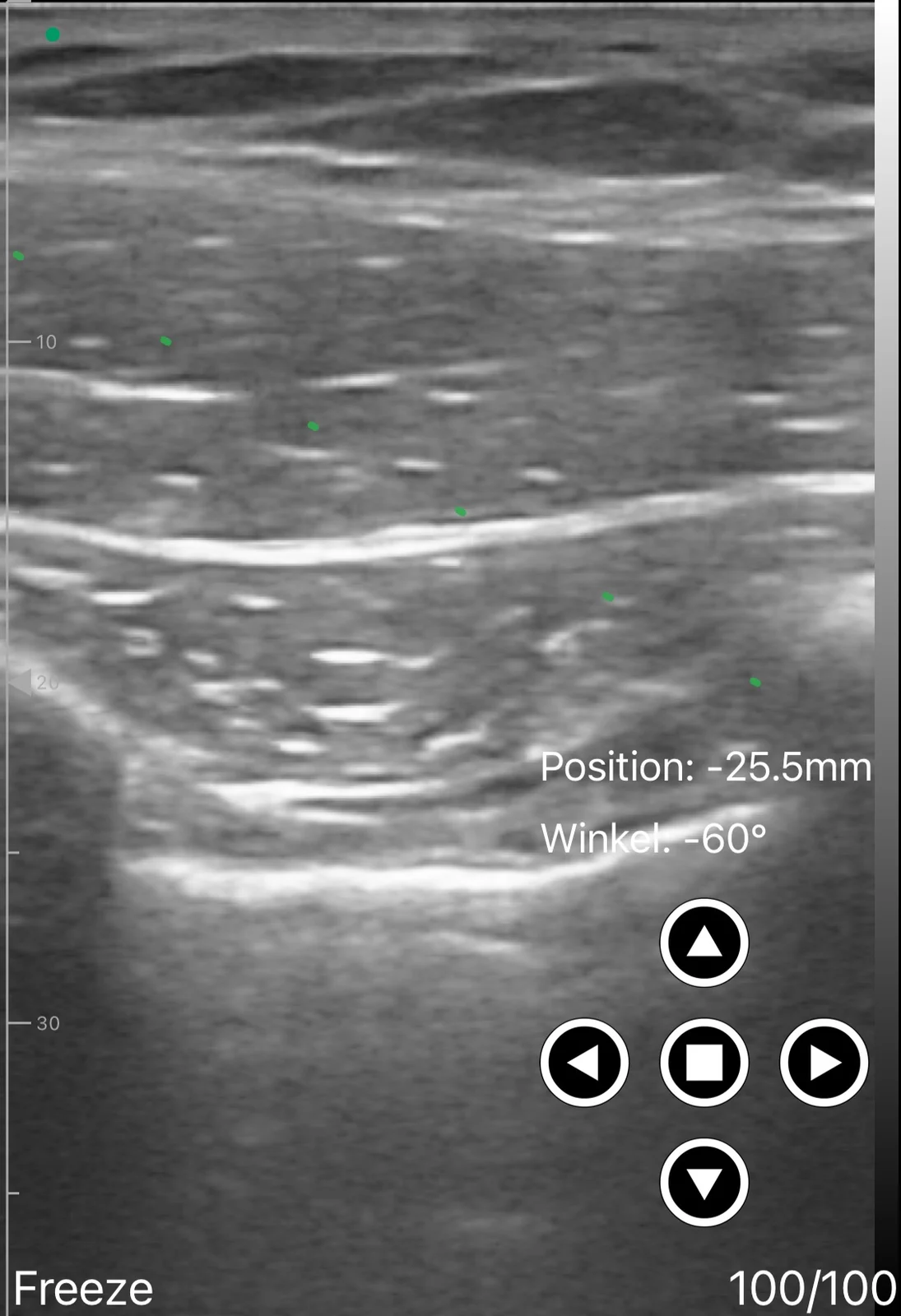
The tap has one rule of its own: keep the shell out of the needle’s path. Stop advancing the moment the tip enters the fluid (Lee 2019), or use a blunt needle: a blunt Veress needle under ultrasound drained 12 patients with no implant damaged (Mazzocchi 2010). The intravenous-cannula variant leaves only the plastic sheath in the cavity; in 71 patients with early seromas after reconstruction it needed fewer taps, 4.48 against 5.80 by needle (Lee 2019). Lay the path in plane and as flat as the collection allows: entering at the end of a 25 mm array, fluid 10 mm deep takes a path of about 39° to mid-image and 22° to the far edge, against 14° and 7° across the L10W’s 80 mm field, so for a broad collection the L10W lays the needle closest to parallel with the shell.
The generic office tap, from skin prep to dressing, is set out in the seroma aspiration steps, and telling fluid from blood in seroma against hematoma on ultrasound. Seromas after body contouring, and the pseudobursa that walls off a chronic one, belong to seroma ultrasound after plastic surgery.
Capsular contracture on ultrasound: capsule thickness and stiffness by Baker grade
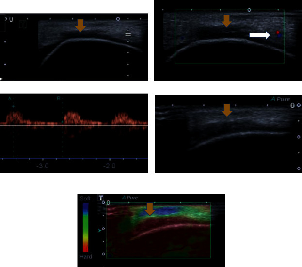
Baker grades contracture by examination: at grade I the breast looks absolutely natural; at II the contracture is minimal, the surgeon can tell surgery was done and the patient has no complaint; at III it is moderate and the patient feels firmness; at IV it is severe and obvious on sight (Mohan 2024). On ultrasound, capsular contracture is a thicker capsule: in 117 patients and 202 breasts, the capsule measured 0.58 mm at Baker II, 1.07 mm at Baker III and 1.89 mm at Baker IV, with cut-offs of 0.8 mm between grades II and III and 1.2 mm between III and IV (Kim 2022). Contracture changes the implant’s shape on the screen too: more radial folds, a rounder, spherical form and a wrinkled shell (Kanavou 2023; Torres-Zuniga 2022). Rupture and contracture travel together: in 105 women with one ruptured and one intact implant, Baker III/IV contracture affected 50% of ruptured sides against 24% of intact ones (odds ratio 4.5) (Bak 2026). Record the thickness in millimeters at the same sites at every visit, so a rise shows against the last study.
Stiffness separates the grades more sharply still. On shear-wave elastography the median capsule read 32.6 kPa in 50 breasts with Baker I–II contracture against 138.0 kPa in 25 with Baker III–IV, and an 82 kPa cut-off found grade III–IV at 100% sensitivity and 98% specificity, where clinicians agreed on the Baker grade at a kappa of 0.55 (Iliescu-Glaja 2026). An inflamed capsule adds pericapsular edema, Doppler flow and a hard capsule on elastography (Fleury 2022). Ten-year Baker III/IV rates in primary augmentation run 18.9% for Natrelle round implants, 12.1% for MemoryGel and 12.9% for Sientra (Spear 2014; Caplin 2021; Sientra label DFU-0002 R7).
The office implant scan, step by step
The office implant scan takes about 20 minutes: with handheld 15 and 20 MHz linears, a dedicated implant exam averaged 19.5 minutes (median 18.64, SD 6.4) in 29 patients with 49 implants (Fleury 2025). Run it the same way every time, so each study reads against the last:
- Probe and preset: the ACR calls for a high-resolution linear array at the highest frequency that still reaches the depth of interest, “typically at least 12 MHz” (ACR 2026). Start at 14 MHz or higher for the shell and capsule, then move to the deep preset that holds the posterior shell and chest wall.
- Position: supine for the medial breast, contralateral oblique for the lateral breast and the axilla (ACR 2026).
- Coverage: both implants and both axillae at every visit; for late fluid or a mass, add the chest wall and the internal mammary and supraclavicular nodes (Spit 2024; Keane 2023).
- Image set: at least six images plus video sweeps per breast: the pocket, the upper, lateral, medial and inferior implant with the space around it, and the shell surface (Kim 2024).
- Labels and planes: right or left, clock face, distance from the nipple in cm and probe orientation on every image; each finding in two orthogonal planes, with and without calipers (ACR 2026).
- Read-out: six items at each visit: the shell, the homogeneity of the gel, the fibrous capsule, the space inside the capsule, the space around it and the axilla (Fleury 2025), plus the capsule thickness in mm (Kim 2022), and the snowstorm extent and node status once rupture is found (Kim 2024).
- Save for the next visit: the same preset and depth, the same views and labels, a cine sweep per view, annotations on each finding and a DICOM copy to the archive.
Medicare’s 2026 national amounts are the billing reference for the scan and for the MRI it replaces or triggers (CMS RVU26D):
| CPT code | Short label | Global | Professional (-26) | Technical (TC) |
|---|---|---|---|---|
| 76641 | Breast ultrasound, unilateral, complete | $100.20 | $33.73 | $66.47 |
| 76642 | Breast ultrasound, unilateral, limited | $83.50 | $31.40 | $52.11 |
| 77046 | Breast MRI without contrast, unilateral | $213.10 | $66.13 | $146.96 |
| 77047 | Breast MRI without contrast, bilateral | $216.44 | $73.15 | $143.29 |
| 77049 | Breast MRI without and with contrast, bilateral | $339.69 | $105.88 | $233.81 |
A bilateral scan bills at $150.30 under CPT 76641 and $125.25 under CPT 76642, and a claim from a facility setting pays the professional component only (CMS RVU26D).
Self-pay practices publish their own prices (practice pages checked November 20, 2026). Watterson Plastic Surgery in Draper, Utah, scans for $100 (page updated June 23, 2026); La Jolla Cosmetic Surgery Centre in San Diego charges $450 for a 20-minute screening (updated September 14, 2026); a 2023 office event at Dr. Kerner’s practice charged $399; and Dan Mills MD and Kirby Plastic Surgery scan the patients whose implants they placed at no cost. Practice pages quote breast MRI at $600–$1,500 (Desert Hills) and about $1,500 (Dan Mills).
What frequency and depth implant ultrasound needs
Implant ultrasound needs two things from one probe: at least 12 MHz for the shell and capsule, the ACR’s “typically at least 12 MHz” (ACR 2026), and a frame deep enough for the posterior shell, which draws 46 to 111 mm down the screen for implants of 2.5 to 6.7 cm projection. The depth comes from the gel: silicone carries sound at 1,060 ± 50.1 m/s when intact and 1,115 m/s when ruptured (Forsberg 1996), against the 1,540 m/s every scanner assumes (Jerban 2023), so the gel draws about 1.45 times its true thickness. The chest wall lies 1 to 4 mm below the posterior shell; the anterior shell sits at 10 to 14 mm, and at 3 to 25 mm under a prepectoral mastectomy flap; the axillary nodes at 14 to 20 mm; and late fluid at about 10 to 17 mm (Hong 2023; Salgarello 2026; Pajcini 2021).
| De mão | Frame | Top MHz | What it reaches | Utilização |
|---|---|---|---|---|
| Suresult L14Pro | Presets of 20, 40, 60 and 80 mm | 14 | The posterior shell and chest wall of implants to about 4.5 cm projection in every plane (smooth moderate-plus sizes to 440 cc, high-profile sizes to 200 cc), and 4.3 cm through a 4 mm gel pad | The whole implant, capsule, nodes and a guided tap with Needle Enhance |
| Suresult L20Pro | Presets of 10, 20, 30 and 40 mm | 20 | The anterior third of the gel: skin, flap or gland, the pectoralis over a subpectoral implant, the anterior shell and capsule, folds, nodes and fluid at 10 to 17 mm | Anterior shell, capsule thickness, the axilla and thin prepectoral flaps |
| Suresult L14 | Presets of 20 to 80 mm | 14 | The same reach as the L14Pro | The whole implant on 192 elements, with in-plane and out-of-plane guide lines |
| Suresult L20 | Presets of 10 to 40 mm | 20 | The same reach as the L20Pro | The anterior implant and nodes, with Needle Enhance |
| Suresult D3Ultra, linear head | Presets of 20, 40, 60 and 100 mm, 40 mm field | 10 | The posterior shell and chest wall to about 5.9 cm projection: every moderate-plus size to 755 cc and high-profile sizes to 490 cc | Large and high-profile implants, and smaller ones under a thick prepectoral flap |
| Suresult D3Ultra, convex head | 90 to 300 mm, 45° field | 5.0 | Projections above about 5.9 cm, including high-profile sizes of 535 to 790 cc | The largest implants and the chest wall behind them |
| Suresult L10W | Presets of 20 to 60 mm, 80 mm field | 10 | The posterior shell to about 3.2 cm projection (moderate-plus 130 cc), and 51% to 96% of an implant’s base width in one frame | Width: folds, contour changes, the spread of fluid along the shell and the flattest in-plane path |
Two Suresult probes split the work by depth: the L14Pro holds the whole implant, pre- or subpectoral, to about 4.5 cm projection, and the L20Pro reads the front of it at 20 MHz, from the skin and flap to the anterior shell, capsule, nodes and fluid. Large and high-profile implants, and small ones under a thick prepectoral flap, go to the D3Ultra’s 100 mm linear preset: a 25 mm flap over a 4.1 cm projection puts the posterior shell near 85 mm. Above about 5.9 cm projection, its convex head covers the rest at 90 to 300 mm. For MHz by depth across the body, see aparelhos de ultrassom de alta frequência, and for linear probes side by side, the best portable linear ultrasound probes.
The 6 best handhelds for breast implant ultrasound, ranked
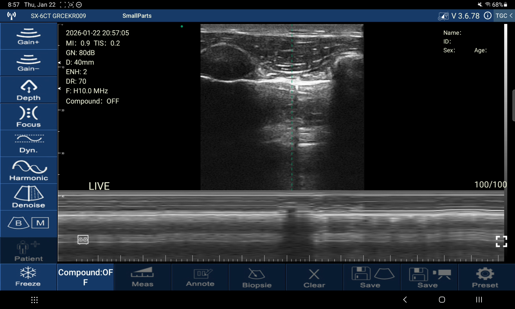
Scored for breast implant ultrasound, the six handhelds finish Suresult L14Pro 100.0, Mindray TE Air e5M 82.4, GE Vscan Air CL 76.9, Clarius L7 HD3 75.0, Butterfly iQ3 73.6 and Philips Lumify L12-4 71.6, on weights that follow the implant visit. The FDA schedule calls for an ultrasound or MRI at 5 to 6 years and every 2 to 3 years after (FDA 2020); the ACR asks for a linear array at the highest frequency that still reaches the depth of interest, typically at least 12 MHz (ACR 2026); the gel draws the posterior shell 46 to 111 mm down the screen; and late fluid is tapped under the probe and sent for cytology (Bengtson 2011). So I weight depth to the posterior shell and chest wall at 20 points and near-field detail for the shell and capsule at 20, Doppler, guided aspiration and measurement at 10 each, records for the next comparison at 15, and the five-year cost of owning every scored tool at 15. Every rival price comes from the maker or seller on November 12, 2026.
Pontuação em resumo
Weights, set before scoring: depth 20 · near-field detail 20 · Doppler 10 · guided aspiration 10 · measurement 10 · records 15 · five-year cost 15. The point rules for each criterion sit under Como atribuí as notas.
| Rank · card | Depth /20 | Near field /20 | Doppler /10 | Aspiration /10 | Measurement /10 | Records /15 | Custo /15 | Total /100 |
|---|---|---|---|---|---|---|---|---|
| 1 · Suresult L14Pro | 20.00, lidera neste critério | 20.00, lidera neste critério | 10.00, lidera neste critério | 10.00, lidera neste critério | 10.00, lidera neste critério | 15.00, lidera neste critério | 15.00, lidera neste critério | 100.0 |
| 2 · Mindray TE Air e5M | 20.00, lidera neste critério | 8.89 | 10.00, lidera neste critério | 10.00, lidera neste critério | 10.00, lidera neste critério | 12.00 | 11.47 | 82.4 |
| 3 · GE Vscan Air CL | 20.00, lidera neste critério | 15.56 | 7.00 | 5.00 | 7.00 | 12.00 | 10.32 | 76.9 |
| 4 · Clarius L7 HD3 | 20.00, lidera neste critério | 17.78 | 8.50 | 7.50 | 7.00 | 6.00 | 8.20 | 75.0 |
| 5 · Butterfly iQ3 | 20.00, lidera neste critério | 15.56 | 5.00 | 7.50 | 10.00, lidera neste critério | 6.00 | 9.56 | 73.6 |
| 6 · Philips Lumify L12-4 | 20.00, lidera neste critério | 15.56 | 7.00 | 5.00 | 7.00 | 12.00 | 5.01 | 71.6 |
- Profundidade: 20 of 20 for all six: Suresult L14Pro, Mindray TE Air e5M, GE Vscan Air CL, Clarius L7 HD3, Butterfly iQ3 and Philips Lumify L12-4, each imaging to 80 mm or deeper.
- Near-field detail: Suresult L14Pro, 20 of 20 at 14 MHz.
- Doppler: Suresult L14Pro and Mindray TE Air e5M, 10 of 10, with Color, Power and PW Doppler in the purchase.
- Guided aspiration: Suresult L14Pro and Mindray TE Air e5M, 10 of 10: Needle Enhance with in-plane and out-of-plane guides on the L14Pro, iNeedle with the VA Grid on the Mindray, all in the purchase.
- Medição: Suresult L14Pro, Mindray TE Air e5M and Butterfly iQ3, 10 of 10, with calipers, volume and annotation.
- Registros: Suresult L14Pro, 15 of 15, with about 165 s of cine, local storage, DICOM export and DICOM send.
- Five-year cost: Suresult L14Pro, 15 of 15, the lowest five-year cost of the six cards.
- Total: Suresult L14Pro, 100.0 of 100.

01 Suresult L14Pro — Best Overall for Breast Implant Ultrasound
10/14 MHz linear on 256 elements and 64 channels · 20, 40, 60 and 80 mm presets with a breast preset · Color, Power and PW Doppler in the price · Needle Enhance with in-plane and out-of-plane guides (Suresult store, checked Nov 20, 2026)
Score: 100.0/100
145 g · 4,400 mAh, about 4 hours of scanning a charge · IP67, drop-tested · its own dual-band Wi-Fi to iOS, Android and Windows (Suresult store, checked Nov 20, 2026)
See the Suresult L14Pro linear probe: $3,293 (Suresult store, checked Nov 20, 2026)
Veredito: Buy the L14Pro if you follow silicone implants on the FDA schedule in your own office and want one probe for every read the visit asks for: 14 MHz for the shell and the capsule, presets to 80 mm for the back of the implant, all three Doppler modes, Needle Enhance for the late-fluid tap, and cine with DICOM for the next comparison, in one price with no subscription (Suresult store, checked Nov 20, 2026).
- Shell and capsule: 14 MHz on the 20 and 40 mm presets for the shell lines, the anterior gel and a capsule that measures 0.58 to 1.89 mm across Baker II to IV (Kim 2022), with a breast preset among the presets (Suresult store, checked Nov 20, 2026)
- Whole implant: the 60 and 80 mm presets frame the posterior shell and chest wall of smooth moderate-plus implants to 440 cc and high-profile implants to 200 cc in every plane, worked from the 1.45 gel factor (Forsberg 1996) and the presets (Suresult store, checked Nov 20, 2026)
- Doppler: Color, Power and PW for flow in an inflamed capsule, a node’s hilum and around a collection before the tap (Suresult store, checked Nov 20, 2026)
- Fluid tap: Needle Enhance, an in-plane needle line with an adjustable angle, an out-of-plane target with a set depth and size, Grid and Flip (Suresult quick user guide, checked Nov 20, 2026)
- Medição: calipers for distance and area, a text comment on any image (Suresult quick user guide, checked Nov 20, 2026), and volume from three diameters
- Registros: about 165 seconds of cine, 1,000 frames, kept on your own device with JPEG, PNG, MP4 and DICOM export (Suresult store, checked Nov 20, 2026); DICOM send is set in the app: “To send studies in DICOM format, set up the connection in Preset › DICOM Settings.” (Suresult quick user guide, checked Nov 20, 2026)
- Preço e plano: $3,293, paid once, with every imaging mode, measurement and export unlocked, free lifetime app updates and no subscription (Suresult store, checked Nov 20, 2026)
- Delivery and cover: door to door with tax and duties paid, No Hidden Fees, a 30-day worry-free trial and a 2-year warranty, extendable to 5 (Suresult store, checked Nov 20, 2026)
Por que ocupa esta posição. The L14Pro takes the maximum on all seven criteria: its 14 MHz top clears the ACR’s “typically at least 12 MHz” (ACR 2026), its 80 mm preset holds the posterior shell and chest wall of implants to about 4.5 cm projection once the gel’s slower sound is counted (Forsberg 1996), and every scored tool comes in the purchase, at the lowest five-year cost of the six cards on November 12, 2026 prices (Suresult store, checked Nov 20, 2026).
Sub-scores: depth 20/20, near-field detail 20/20, Doppler 10/10, guided aspiration 10/10, measurement 10/10, records 15/15, five-year cost 15/15.
The L14Pro and every Suresult product are com liberação 510(k) K211321 (FDA 510(k) K211321).
L14Pro ou L20Pro? Both run 256 elements with all three Doppler modes, Needle Enhance and a 4-hour battery, so depth decides: the L14Pro’s 80 mm preset takes in the whole implant to about 4.5 cm projection, while the Suresult L20Pro high-frequency probe ($3,560) reads its front at 20 MHz on presets to 40 mm (Suresult store, checked Nov 20, 2026). Surveillance on one probe: the L14Pro. Capsule thickness, thin prepectoral flaps and the axilla at the finest detail: add the L20Pro.
Melhor para: plastic surgeons and breast clinics who follow silicone implants on the FDA schedule and read the shell, capsule, nodes and late fluid, then tap that fluid, on one probe in their own office. Choose a sibling if: you want 20 MHz for the capsule and the axilla (the L20Pro), the same band and presets at $2,047 (the L14), or a 100 mm linear preset and a convex head for large implants (the D3Ultra) (Suresult store, checked Nov 20, 2026).
Também nesta família

Suresult L20Pro 16/20 MHz linear probe · $3,560 · 78.9/100: the anterior-implant pick, with 20 MHz on 256 elements for the shell, the capsule and the axilla, presets of 10 to 40 mm, a breast preset and the full Pro kit (Suresult store, checked Nov 20, 2026).

Transdutor de ultrassom sem fio Suresult L14 · $2,047 · 95.0/100: the L14Pro’s band and 20 to 80 mm presets on 192 elements, with all three Doppler modes, both needle guides and a 2-hour battery (Suresult store, checked Nov 20, 2026).

Transdutor de ultrassom linear Suresult L20 · $2,670 · 80.0/100: the L20Pro’s 16/20 MHz band on 192 elements and presets to 40 mm, with Color, Power and PW Doppler, Needle Enhance and a 2-hour battery, for the anterior shell, the capsule and the nodes (Suresult store, checked Nov 20, 2026).

Suresult D3Ultra dual-head ultrasound · $2,848: dual-head 3-in-1, with a 7.5/10 MHz linear head whose 100 mm preset reaches the posterior shell of large and high-profile implants to about 5.9 cm projection, a 3.2/5.0 MHz convex head to 300 mm for the largest, and Color, Power and PW Doppler with Needle Enhance (Suresult store, checked Nov 20, 2026).
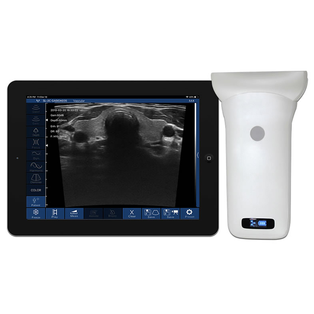
Suresult L10W wide linear ultrasound · $2,136: a 7.5/10 MHz linear with an 80 mm field on 256 elements and presets to 60 mm, which holds 51% to 96% of an implant’s base width in one frame and lays the flattest in-plane path to a broad collection (Suresult store, checked Nov 20, 2026).
Suresult handhelds for plastic surgery · Suresult handhelds for breast care · Suresult FDA clearance and certifications
02 Mindray TE Air e5M — Best for Full Doppler, Needle Aid and Volume in One Purchase
Color, Power and PW Doppler as standard · iNeedle needle enhancement and the VA Grid · one transducer in convex and linear modes · linear B-mode bands to 8.2 MHz within a 2.0–9.0 MHz bandwidth · stated depth to 40 cm (Mindray TE Air e5M datasheet, checked Nov 20, 2026)
Score: 82.4/100
229 ± 3 g · 90 to 120 minutes in B-mode · rated IP68 (Mindray TE Air e5M datasheet, checked Nov 20, 2026) · 3-year warranty direct from Mindray North America (Mindray NA store, checked Nov 20, 2026)
Veredito: Buy the TE Air e5M if the late-fluid tap is part of your implant visits and you want every Doppler mode and a needle aid paid for once: Color, Power and PW Doppler, iNeedle and the VA Grid come standard (Mindray TE Air e5M datasheet, checked Nov 20, 2026), and Mindray North America sells it at one Total Amount with no subscription plan (Mindray NA store, checked Nov 20, 2026).
- Depth and band: linear bands of 2.0–5.2, 4.0–7.4 and 5.0–8.2 MHz on 128 elements, and a stated depth range of 1.5 to 40.0 cm across its linear and convex modes (Mindray TE Air e5M datasheet, checked Nov 20, 2026)
- Doppler: Color, Power and PW Doppler standard, with no subscription plan: “All costs are at the time of purchase” (Mindray TE Air FAQ, checked Nov 20, 2026)
- Fluid tap: iNeedle needle-visualization enhancement and the VA Grid for vascular access (Mindray TE Air e5M datasheet, checked Nov 20, 2026)
- Medição: distance and area in every package, B-mode volume in five exam packages, and comments with text, arrows and 251 body marks (Mindray TE Air e5M datasheet, checked Nov 20, 2026)
- Registros: single frames and cine saved on the device, cine of 125 frames over 2 to 5 seconds, DICOM 3.0 Store, Media Exchange and Worklist, and Tricefy or a shared folder for the archive (Mindray TE Air e5M datasheet, checked Nov 20, 2026)
- Preço e plano: $4,499 as the Total Amount, with a 3-year warranty direct from Mindray North America (Mindray NA store, checked Nov 20, 2026)
- Clearance: FDA 510(k) K240906, cleared July 23, 2024, which added the e5M, and K250024, cleared May 30, 2025
Por que ocupa esta posição. Four full scores, on depth, Doppler, aspiration and measurement, and the lowest rival price, $4,499, put the TE Air e5M second (Mindray NA store, checked Nov 20, 2026); its 9.0 MHz bandwidth top and 2 to 5 second cine cost it points on near-field detail and records (Mindray TE Air e5M datasheet, checked Nov 20, 2026).
Sub-scores: depth 20/20, near-field detail 8.89/20, Doppler 10/10, guided aspiration 10/10, measurement 10/10, records 12/15, five-year cost 11.47/15.
Limitações. The linear B-mode bands end at 8.2 MHz, under the 12 MHz the ACR names for breast ultrasound, and its linear applications are Small Organ, Vascular, Musculo-skeletal and Nerve (Mindray TE Air e5M datasheet, checked Nov 20, 2026).
Melhor para: clinics that tap late fluid and want all three Doppler modes, a needle-enhancement mode, calipers and volume on a transducer that also images deep, in one purchase. Escolha outro se: you want 12 MHz or more on the shell and capsule, or cine long enough to hold a whole sweep (Mindray TE Air e5M datasheet, checked Nov 20, 2026).
Loja Mindray TE Air · TE Air e5M datasheet · TE Air e5M na Mindray North America
03 GE Vscan Air CL — Best for Color and PW on an 8 cm Linear Head With Nothing to Renew
Dual-head probe, scored on its linear head · 3–12 MHz with a 7.7 MHz center frequency · depth up to 8 cm · 40 × 7 mm lens · B, Color, M and PW modes with Harmonics (GE Vscan Air support site, checked Nov 20, 2026)
Score: 76.9/100
205 ± 3 g · 50 minutes of total scan time · IP67 with a MIL-STD-810G drop rating (GE Vscan Air CL datasheet, checked Nov 20, 2026) · standard 3-year warranty (GE HealthCare US shop, checked Nov 20, 2026)
Veredito: Buy the Vscan Air CL if you want Color and PW Doppler, calipers, annotation and DICOM export in one $4,999 purchase with nothing to renew (GE HealthCare US shop, checked Nov 20, 2026), on a linear head that images to 8 cm and a curved head for the rest of the body (GE Vscan Air support site, checked Nov 20, 2026).
- Depth and band: the linear head runs 3–12 MHz to 8 cm, which holds the posterior shell of implants to about 4.5 cm projection; the curved head runs 2–5 MHz to 24 cm (GE Vscan Air support site, checked Nov 20, 2026)
- Doppler: B, Color, B+Color, Harmonics, M and PW on the linear head, every mode in the purchase (GE Vscan Air support site, checked Nov 20, 2026)
- Fluid tap: fluid drainage guidance among the linear head’s indications, and a Centerline Marker for free-hand needle work (GE Vscan Air support site, checked Nov 20, 2026)
- Medição: distance, time, velocity, angle and ellipse measurements (GE Vscan Air support site, checked Nov 20, 2026), with free text and arrows for every preset and mode (GE Vscan Air support site, checked Nov 20, 2026)
- Registros: data for up to 500 exams on the mobile device (GE Vscan Air CL datasheet, checked Nov 20, 2026), and export to a DICOM image server, a network shared folder, MPPS or Tricefy (GE Vscan Air support site, checked Nov 20, 2026)
- Preço e plano: $4,999.00 with a standard 3-year warranty; Innovations, an optional plan at $500 for one year or $1,299 for three, adds no scored tool (GE HealthCare US shop, checked Nov 20, 2026)
- Clearance: FDA 510(k) K250087, cleared May 1, 2025, for the dual-headed probe
Por que ocupa esta posição. Third place rests on the linear head: 8 cm of depth for the full depth credit and a 12 MHz top for most of the near-field points (GE Vscan Air support site, checked Nov 20, 2026), with Color, PW, calipers, annotation and DICOM export all in the $4,999 price (GE HealthCare US shop, checked Nov 20, 2026).
Sub-scores: depth 20/20, near-field detail 15.56/20, Doppler 7/10, guided aspiration 5/10, measurement 7/10, records 12/15, five-year cost 10.32/15.
Limitações. Power Doppler, a needle-enhancement mode and a volume tool for a measured structure sit outside the linear head’s listed features (GE Vscan Air support site, checked Nov 20, 2026); the 8 cm frame holds implants to about 4.5 cm projection, so larger ones go to the 2–5 MHz curved head, and a charge gives 50 minutes of total scan time (GE Vscan Air CL datasheet, checked Nov 20, 2026).
Melhor para: clinics that want Color and PW Doppler, calipers, annotation and DICOM export from a large maker with nothing to renew, on a linear head deep enough for the posterior shell of small and medium implants. Escolha outro se: Power Doppler, needle enhancement or a volume tool has to come in the purchase.
Vscan Air CL na loja GE HealthCare · Vscan Air CL specifications · Vscan Air probes and presets
04 Clarius L7 HD3 — Best for the Maker’s Own Implant Protocol to 15 cm
4–13 MHz linear · max depth 15 cm · 38 mm field of view · Color and Power Doppler with the scanner, PW Doppler with Essentials · Breast (Diagnostic/Interventional) among its applications (Clarius store, checked Nov 20, 2026)
Pontuação: 75,0/100
288 g · more than 60 minutes of scanning · 3-year standard warranty and a 30-day return window (Clarius store, checked Nov 20, 2026) · IP67 with a 1 m drop test (Clarius plastic surgery page, checked Nov 20, 2026)
Veredito: Buy the L7 HD3 if you want the probe Clarius built its own implant teaching on: the Classroom’s “Ultrasound Protocol Breast Implants” with Dr. Marc Salzman lists the L7 HD3 as the product used (Clarius Classroom, checked Nov 20, 2026).
- Depth and band: 4–13 MHz with a max depth of 15 cm, past the posterior shell of every implant size (Clarius store, checked Nov 20, 2026), with imaging optimized to the full 15 cm (Clarius L7 page, checked Nov 20, 2026)
- Doppler: B-mode, M-mode, and Color and Power Doppler included with the scanner; PW Doppler with Essentials (Clarius store, checked Nov 20, 2026)
- Fluid tap: Needle Enhance com Essentials (Clarius Essentials page, checked Nov 20, 2026), and the Center Guide line for injections and other interventions (Clarius Classroom, checked Nov 20, 2026)
- Medição: calipers for distance and area (Clarius beginner’s video guide, checked Nov 20, 2026) and image annotations to mark the area scanned and describe a finding (Clarius Classroom, checked Nov 20, 2026)
- Registros: cine loop playback of 30 seconds; local storage, DICOM and cloud storage with the membership; PDF reports and Procedure Recording Mode in Essentials (Clarius store, checked Nov 20, 2026)
- Breast tools: the Breast Package in the Essentials specialist software (Clarius Essentials page, checked Nov 20, 2026), and the implant protocol and rupture videos in the Classroom (Clarius Classroom, checked Nov 20, 2026)
- Preço e plano: $3,795 for the scanner, with Essentials at $2,500 one-time, $695 a year or $1,785 for three years (Clarius store, checked Nov 20, 2026)
- Clearance: FDA 510(k) K213436, cleared November 15, 2021, which names the L7 HD3 and the L15 HD3
Por que ocupa esta posição. The 15 cm max depth takes the full depth credit and 13 MHz earns 17.78 of 20, the finest near field among the rivals; the tools an implant visit uses sit on Essentials, so the $3,795 scanner plus the $2,500 one-time license, $6,295 over five years, sets its cost score (Clarius store, checked Nov 20, 2026).
Sub-scores: depth 20/20, near-field detail 17.78/20, Doppler 8.5/10, guided aspiration 7.5/10, measurement 7/10, records 6/15, five-year cost 8.20/15.
Limitações. Essentials is required for PW Doppler, Needle Enhance, local storage, DICOM, cloud storage and PDF reports, and cine plays back 30 seconds (Clarius store, checked Nov 20, 2026).
Melhor para: surgeons who want the maker’s own implant protocol, taught on the same probe, with the finest near field of the rival cards and depth to spare for the largest implants. Escolha outro se: you would rather own PW Doppler, needle enhancement, storage and DICOM outright than hold a membership.
Também nesta família
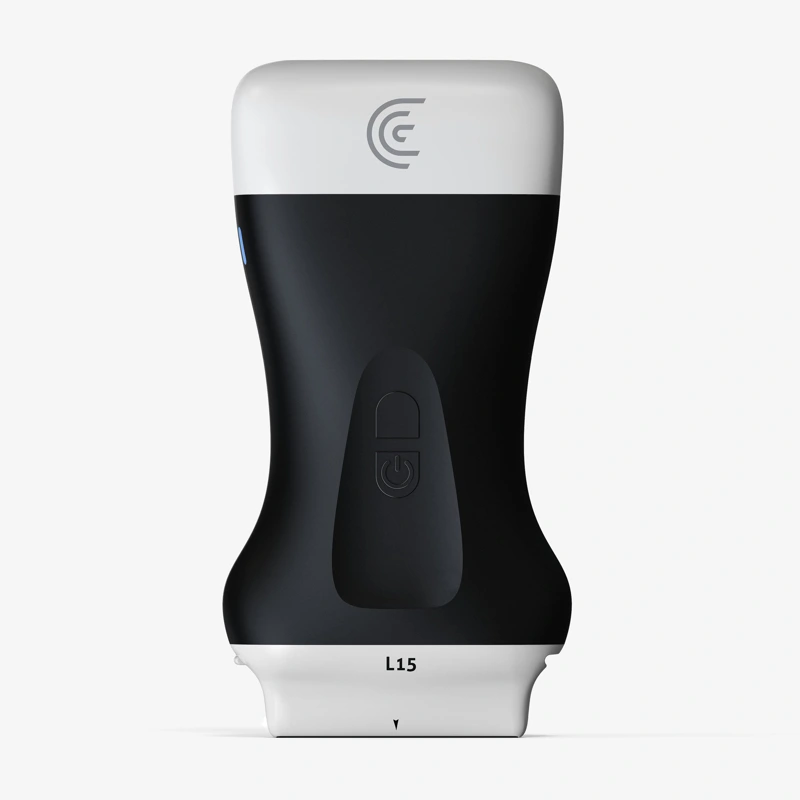
Clarius L15 HD3 · $3,795 plus Essentials · 77.2/100 (Clarius store, checked Nov 20, 2026). A 5–15 MHz linear with a 50 mm field and a max depth of 10 cm, which holds the posterior shell of implants to about 5.9 cm projection and takes the full depth and near-field credit (Clarius store, checked Nov 20, 2026). It is the scanner in Clarius’s “Breast Implant Surveillance with Ultrasound” demonstration (Clarius Classroom, checked Nov 20, 2026).
Clarius plastic surgery · Clarius breast implant protocol · Preços da Clarius e custos do Essentials em 2026
05 Butterfly iQ3 — Best for Calipers, Volume and Biplane on One Whole-Body Probe
Single-chip probe in linear, curved and phased formats · 1–12 MHz (Butterfly iQ3 specs page, checked Nov 20, 2026) · depth 1 to 30 cm, MSK-Soft Tissue preset to 12 cm (Butterfly iQ3 manual, checked Nov 20, 2026) · Color, Power, PW, Needle Viz and Biplane with Advanced (Butterfly pricing page, checked Nov 20, 2026)
Score: 73.6/100
300 g · 2,220 mAh, up to 2 hours a charge · IPX7 and IP6X, 4-ft drop, MIL-STD-810G · 3-year warranty with an optional 2 more (Butterfly iQ3 specs page, checked Nov 20, 2026)
Veredito: Buy the iQ3 if you want linear and elliptical calipers, a volume calculation, biplane needle views and every Doppler mode on one whole-body probe, bought with Advanced in a single payment (Butterfly US store, checked Nov 20, 2026). Its manual names ultrasound rupture screening for patients without symptoms and MRI for symptomatic or equivocal cases, the same split as the FDA schedule (Butterfly iQ3 manual, checked Nov 20, 2026).
- Depth and band: 1–12 MHz on one chip, with the MSK-Soft Tissue preset to 12 cm and the Small Organ preset, whose indications include the breast, to 6 cm (Butterfly iQ3 manual, checked Nov 20, 2026)
- Doppler: Color, Power and PW Doppler with Advanced (Butterfly pricing page, checked Nov 20, 2026), at $420 a year or $1,500 for five years of access (Butterfly pricing page, checked Nov 12, 2026)
- Fluid tap: Needle Viz, Biplane and the out-of-plane needle view with Advanced (Butterfly pricing page, checked Nov 20, 2026), plus a midline marker (Butterfly iQ3 specs page, checked Nov 20, 2026)
- Medição: up to 4 linear calipers and 1 elliptical caliper, a generic volume calculation and on-screen annotation (Butterfly iQ3 specs page, checked Nov 20, 2026), with a manual volume of 0.52 × D1 × D2 × D3 (Butterfly iQ3 manual, checked Nov 20, 2026)
- Registros: start-stop clips, unlimited cloud storage with Advanced, and Basic DICOM for PACS and worklist sold as an add-on (Butterfly pricing page, checked Nov 20, 2026)
- Preço e plano: $5,399 in one payment with no subscription (Butterfly US store, checked Nov 20, 2026): the $3,899 probe with $1,500 for five years of Advanced (Butterfly pricing page, checked Nov 12, 2026)
- Clearance: FDA 510(k) K232808, cleared January 4, 2024
Por que ocupa esta posição. A depth range to 30 cm takes the full depth credit (Butterfly iQ3 manual, checked Nov 20, 2026), and calipers, the volume calculation and annotation take the full 10 on measurement; Doppler, Needle Viz, cloud storage and DICOM sit on paid tiers and score as paid (Butterfly pricing page, checked Nov 20, 2026), and the $5,399 five-year total earns 9.56 of 15 on cost (Butterfly US store, checked Nov 20, 2026).
Sub-scores: depth 20/20, near-field detail 15.56/20, Doppler 5/10, guided aspiration 7.5/10, measurement 10/10, records 6/15, five-year cost 9.56/15.
Limitações. Color, Power and PW Doppler, Needle Viz and Biplane need Advanced; Basic DICOM is a separate purchase (Butterfly pricing page, checked Nov 20, 2026); the battery runs up to 2 hours (Butterfly iQ3 specs page, checked Nov 20, 2026).
Melhor para: clinics that measure every finding, with linear and elliptical calipers, a volume calculation and annotation, and want biplane needle views on one whole-body probe that reaches the deepest implant with room to spare. Escolha outro se: you want Doppler and DICOM in the purchase without a software tier.
iQ3 one-time purchase at the Butterfly store · Butterfly membership pricing · Especificações do Butterfly iQ3
06 Philips Lumify L12-4 — Best for a Battery-Free Linear Bought Outright
12–4 MHz linear · scan depth up to 12 cm · 34 mm footprint, 34.5 mm field of view · 2D, PW and steerable color Doppler and M-mode (Philips L12-4 product page, checked Nov 20, 2026)
Score: 71.6/100
Under 136 g with a battery-free transducer · up to 7.5 hours of continuous scanning (Philips L12-4 product page, checked Nov 20, 2026)
Veredito: Buy the Lumify L12-4 if you already run Lumify, or want a light, battery-free linear bought outright with every software feature (Philips Lumify page, checked Nov 20, 2026), PW and steerable color Doppler (Philips L12-4 product page, checked Nov 20, 2026) and DICOM to PACS (Philips Lumify FAQ, checked Nov 20, 2026).
- Depth and band: 12–4 MHz with scan depth up to 12 cm, deep enough for the posterior shell of the largest implants (Philips L12-4 product page, checked Nov 20, 2026)
- Doppler: 2D, PW and steerable color Doppler and M-mode, all in the outright purchase (Philips L12-4 product page, checked Nov 20, 2026)
- Fluid tap: a center line marker for needle work (Philips Lumify anesthesiology page, checked Nov 20, 2026)
- Medição: distance and ellipse area and circumference, with text annotation on Android devices (Philips Lumify resources guide, checked Nov 20, 2026)
- Registros: loops of 1 to 10 seconds, hundreds of exams on the device, and four export routes: email, PACS over DICOM, a shared network and a local directory (Philips Lumify FAQ, checked Nov 20, 2026)
- Preço e plano: $10,307.95 new and outright at PartsSource with a 90-day warranty (PartsSource listing, checked Nov 20, 2026); the Philips Lumify page lists a 3–5-year warranty (Philips Lumify page, checked Nov 20, 2026)
- Clearance: FDA 510(k) K252558, cleared May 5, 2026, which names the L12-4
Por que ocupa esta posição. Scan depth to 12 cm takes the full depth credit and a 12 MHz top earns 15.56 of 20 on near-field detail (Philips L12-4 product page, checked Nov 20, 2026), and PW and color Doppler, distance and ellipse measurements and DICOM to PACS come in the outright purchase; the $10,307.95 price, the highest five-year cost in the ranking, places it sixth (PartsSource listing, checked Nov 20, 2026).
Sub-scores: depth 20/20, near-field detail 15.56/20, Doppler 7/10, guided aspiration 5/10, measurement 7/10, records 12/15, five-year cost 5.01/15.
Limitações. Power Doppler and a needle-enhancement mode sit outside its listed features (Philips L12-4 product page, checked Nov 20, 2026); loops run 1 to 10 seconds (Philips Lumify FAQ, checked Nov 20, 2026), text annotation works on Android devices only (Philips Lumify resources guide, checked Nov 20, 2026), and the PartsSource listing carries a 90-day warranty (PartsSource listing, checked Nov 20, 2026).
Melhor para: clinics already on Lumify, or anyone who wants a battery-free linear with PW and color Doppler and DICOM to PACS bought outright, deep enough for the posterior shell of the largest implants. Escolha outro se: you want Power Doppler, needle enhancement or the lowest price in this ranking.
Lumify L12-4 na PartsSource · Página do produto Philips Lumify L12-4 · Ultrassom portátil Philips Lumify
Five-year cost: what each handheld costs with the tiers its scored tools need
Over five years the Suresult L14Pro costs $3,293, paid once: every tool I scored comes with the probe, and its imaging app carries no subscription. On prices read November 20, 2026, the rivals run from $4,499 for the Mindray TE Air e5M and $4,999 for the GE Vscan Air CL to $5,399 for the Butterfly iQ3 with five years of Advanced, $6,295 for the Clarius L7 HD3 with its one-time Essentials license, and $10,307.95 for the Philips Lumify L12-4 bought outright from PartsSource.
| De mão | Preço de compra | Requisitos dos recursos avaliados | Total em cinco anos, com pagamento único do software | Five-year total, software paid yearly |
|---|---|---|---|---|
| Suresult L14Pro |
$3,293 | Nothing beyond the probe | $3,293, one-time | $3,293; no subscription to renew |
| Mindray TE Air e5M |
$4,499 | All standard | $4,499 | $4,499; no subscription plan |
| GE Vscan Air CL |
$4,999 | All in the purchase | $4,999 | $4,999; the optional Innovations plan ($500 a year) adds no scored tool |
| Clarius L7 HD3 |
$3,795 | Essentials for PW, Needle Enhance, storage, DICOM and PDF reports: $2,500 one-time or $695 a year | $6,295 | $7,270, with $695 in each of the five years |
| Butterfly iQ3 |
$3,899 | Advanced for Color, Power and PW, Needle Viz, Biplane and cloud storage; Basic DICOM sold separately | $5,399, five years of Advanced in one payment | $5,999, with Advanced at $420 a year |
| Philips Lumify L12-4 |
$10,307.95 | Every Lumify software feature, with the probe bought outright | $10,307.95 | $10,307.95; nothing to renew |
Two guides take the cost question further: the best handheld ultrasound with no subscription ranks the probes that need no plan at all, and the Clarius price and subscription guide sets out every Clarius membership and license.
Como atribuí as notas
I scored every card out of 100 on seven criteria, with weights declared on November 12, 2026, before any rival carried a score, each tied to a step of implant surveillance:
- Depth to the posterior shell and chest wall (20): 20 × (the deepest image depth in mm − 40) ÷ 40, held between 0 and 20, so a frame of 80 mm or more earns 20 and 40 mm earns 0.
- Near-field detail for the shell, folds and capsule (20): 20 × (top frequency in MHz − 5) ÷ 9, held between 0 and 20, so 14 MHz and up earns 20, 13 MHz 17.78, 12 MHz 15.56 and 9 MHz 8.89.
- Doppler for flow in the capsule and the nodes (10): Color 4, Power 3 and PW 3.
- Guided aspiration of peri-implant fluid (10): needle enhancement 5, and 5 for in-plane or out-of-plane guides or biplane; I count a centerline, midline or access-grid aid the maker names for needle work as a guide.
- Measurement (10): calipers in mm 4, volume from three diameters or an ellipsoid tool 3, and annotation or a body marker 3.
- Records for serial comparison (15): cine of a stated 60 seconds or more 3, exam storage on the device or in the app 4, DICOM export 4, and an archive or report route (PACS, cloud or PDF report) 4.
- Five-year cost (15): the cheapest card lead’s five-year total divided by this card’s, times 15. A Suresult total is the probe’s price, with no license; a rival total adds every tier its scored tools need over five years. At the November 12, 2026 prices, the L14Pro’s total was the lowest and set the scale.
A tool in the purchase earns its full points, a tool behind a paid tier half, and a tool missing from the maker’s listing nothing; a tool the maker sells without naming a tier counts as included, and ties break on the lower five-year cost. Each rival is scored on the probe its maker uses for breast work: GE on its linear head, Mindray on the stated maxima of its one transducer.
| De mão | Depth and near field | Doppler | Guided aspiration | Medição | Records | Custo em cinco anos |
|---|---|---|---|---|---|---|
| Suresult L14Pro | 80 mm: 20 · 14 MHz: 20 | Color 4 · Power 3 · PW 3 | Needle Enhance 5 · in-plane and out-of-plane guides 5 | Calipers 4 · volume 3 · annotation 3 | Cine of about 165 s: 3 · storage 4 · DICOM export 4 · DICOM send 4 | $3,293: 15 |
| Mindray TE Air e5M | 40 cm: 20 · 9.0 MHz: 8.89 | Color 4 · Power 3 · PW 3 | iNeedle 5 · VA Grid 5 | Calipers 4 · B-mode volume 3 · Comments and body marks 3 | Cine of 2–5 s 0 · storage 4 · DICOM 3.0 Store 4 · Tricefy and shared folder 4 | $4,499: 11.47 |
| GE Vscan Air CL | 8 cm: 20 · 12 MHz: 15.56 | Color 4 · Power 0 · PW 3 | Enhancement 0 · Centerline Marker 5 | Calipers 4 · volume 0 · free text and arrows 3 | Cine 0 · 500 exams 4 · DICOM image server 4 · shared folder and Tricefy 4 | $4,999: 10.32 |
| Clarius L7 HD3 | 15 cm: 20 · 13 MHz: 17.78 | Color 4 · Power 3 · PW 1.5 on Essentials | Needle Enhance 2.5 on Essentials · Center Guide 5 | Calipers 4 · volume 0 · Annotations 3 | 30 s cine 0 · storage 2 · DICOM 2 · cloud and PDF reports 2, all on Essentials | $6,295: 8.20 |
| Butterfly iQ3 | 30 cm: 20 · 12 MHz: 15.56 | Color 2, Power 1.5 and PW 1.5 on Advanced | Needle Viz 2.5 on Advanced · midline marker 5 | Calipers 4 · Generic Volume Calculation 3 · annotation 3 | Cine 0 · storage 2, DICOM 2 and cloud archive 2, on Advanced or the Basic DICOM add-on | $5,399: 9.56 |
| Philips Lumify L12-4 | 12 cm: 20 · 12 MHz: 15.56 | Color 4 · Power 0 · PW 3 | Enhancement 0 · center line marker 5 | Calipers 4 · volume 0 · text annotation 3 | Loops of 1–10 s 0 · hundreds of exams 4 · DICOM to PACS 4 · email and shared network 4 | $10,307.95: 5.01 |
Siblings are scored on the same criteria and cost scale inside their maker’s card: the Suresult L14 95.0, L20 80.0 and L20Pro 78.9, and the Clarius L15 HD3 77.2. The L14Pro, the Suresult lead declared before scoring, outscores each: its 80 mm presets earn the depth credit that the 40 mm L20Pro and L20 score 0 on, and Needle Enhance adds the 5 aspiration points the L14 lacks. The L7 HD3 leads the Clarius card as the probe Clarius uses in its implant protocol video; the L15 HD3 pairs 15 MHz with a 10 cm frame and scores 77.2 to the L7 HD3’s 75.0.
I reran the order six ways, as declared, and under sixteen alternative readings of the sources: the L14Pro stays first in five of the six reruns and fourteen of the sixteen readings. In S4, which scores the Suresult card on the 40 mm L20Pro, the Mindray TE Air e5M leads, 83.29 to 80.00. S7, declared for makers that only quote, had no case to run: every rival publishes a price. The two readings least favorable to the L14Pro are RC14, which stacks S3 and S5, leaves DICOM send uncredited, counts Clarius Procedure Recording as cine and credits GE’s Auto Bladder Volume, putting the Clarius L7 HD3 first at 87.98; and RC15, which also withholds credit from the centerline and midline markers, the VA Grid and Mindray’s convex-mode figures, putting the Butterfly iQ3 first at 87.12. In both, the L14Pro holds 86.00.
| Cenário | Ficha da Suresult | Mindray TE Air e5M | GE Vscan Air CL | Clarius L7 HD3 | Butterfly iQ3 | Philips Lumify L12-4 | Order, first to sixth |
|---|---|---|---|---|---|---|---|
| S0, cenário declarado | 100.00 | 82.36 | 76.88 | 74.98 | 73.62 | 71.56 | L14Pro, Mindray, GE, Clarius, Butterfly, Philips |
| S1, critério de custo retirado | 85.00 | 70.89 | 66.56 | 66.78 | 64.06 | 66.56 | L14Pro, Mindray, Clarius, GE, Philips, Butterfly |
| S2, critério de profundidade retirado | 80.00 | 62.36 | 56.88 | 54.98 | 53.62 | 51.56 | L14Pro, Mindray, GE, Clarius, Butterfly, Philips |
| S3, full depth credit moved to 100 mm, counted from 60 mm | 90.00 | 82.36 | 66.88 | 74.98 | 73.62 | 71.56 | L14Pro, Mindray, Clarius, Butterfly, Philips, GE |
| S4, Suresult card scored on the L20Pro | 80.00 | 83.29 | 77.72 | 75.64 | 74.39 | 71.97 | Mindray, L20Pro, GE, Clarius, Butterfly, Philips |
| S5, rival paid features counted as included | 100.00 | 82.36 | 76.88 | 84.98 | 87.12 | 71.56 | L14Pro, Butterfly, Clarius, Mindray, GE, Philips |
| S6, records weighted 5 and near-field detail 30 | 100.00 | 78.81 | 76.66 | 79.87 | 77.39 | 71.34 | L14Pro, Clarius, Mindray, Butterfly, GE, Philips |
| RC14, S3 and S5 with three rival-favoring readings | 86.00 | 82.36 | 66.63 | 87.98 | 87.12 | 71.56 | Clarius, Butterfly, L14Pro, Mindray, Philips, GE |
| RC15, RC14 with three credits withheld | 86.00 | 72.58 | 61.63 | 82.98 | 87.12 | 66.56 | Butterfly, L14Pro, Clarius, Mindray, Philips, GE |
Breast implant ultrasound questions: schedule, rupture, nodes, fluid and cost
How often should silicone breast implants be checked with ultrasound?
First at 5 to 6 years after the operation, then every 2 to 3 years, for silicone implants without symptoms, by ultrasound or MRI (FDA 2020; ACR 2025). Symptoms, or an ultrasound that leaves the shell in doubt, go to MRI without contrast.
Can ultrasound detect an extracapsular breast implant rupture?
Yes, and it is the plainest rupture sign on the screen: free silicone draws a snowstorm of bright, reverberating echoes with a sharp front edge and a blurred back edge (Harris 1993). Against MRI, ultrasound found every extracapsular rupture in one series (Telegrafo 2015).
Can an ultrasound detect capsular contracture?
Yes: the capsule thickens from 0.58 mm at Baker II to 1.07 mm at III and 1.89 mm at IV (Kim 2022), and on shear-wave elastography an 82 kPa cut-off separated grades III–IV from I–II at 100% sensitivity and 98% specificity (Iliescu-Glaja 2026).
What percentage of breast implants rupture?
Under 10% of primary augmentation patients over 10 years in the core studies behind today’s silicone implants: 9.3% for Natrelle round, 9.81% for MemoryGel confirmed at explant (24.2% counting suspected ruptures) and 8.5% for Sientra, with 5.7% across the MRI cohort of the Natrelle 410 study (Natrelle label L034rev15; Caplin 2021; Sientra label DFU-0002 R7; Maxwell 2015).
How many years do breast implants last?
Most last well past a decade: on repeat MRI, modern implants still intact at 3 years had an estimated rupture-free survival of 98% at 5 years and 83% to 85% at 10 (Hölmich 2003), and older-generation gel had a median life span of 16.4 years (Goodman 1998).
Is breast implant rupture ultrasound as accurate as MRI?
In trained hands, yes: in 15 consecutive patients, surgeon ultrasound, radiologist ultrasound and MRI each called 29 of 29 breasts correctly against surgery (Bengtson and Eaves 2012), and recent office and clinic series run at 90.9% to 98.3% sensitivity (Glener 2024; Rukanskienė 2021; Spit 2024).
What does the stepladder sign mean on ultrasound?
Intracapsular rupture: the collapsed shell lies folded inside the capsule, drawn as stacked horizontal echogenic lines parallel to the probe in the anterior gel, the ultrasound counterpart of the MRI linguine sign (Seiler 2017). In cohesive gel, read it together with the shell line and the gel (Rochira 2016).
How do radial folds differ from rupture on ultrasound?
A radial fold runs in from the periphery, perpendicular to the capsule, and stays joined to the shell; rupture lines lie parallel to the probe in the anterior gel (Jaeger 2026; Kanavou 2023). A fold becomes a rupture sign when gel fills its loop, the keyhole or teardrop (Seiler 2017).
What does fluid around a breast implant years after surgery mean?
A late seroma: fluid one year or more after implantation, reported in 0.1% of patients, worked up by ultrasound-guided aspiration with culture and cytology (Bengtson 2011). The fluid is sampled because of BIA-ALCL, which presented as a seroma in 48% of the FDA’s reported cases (FDA 2025, MDR report).
Can ultrasound detect BIA-ALCL?
Ultrasound finds the effusion or the capsule mass of BIA-ALCL and guides the needle that samples it (Keane 2023; Longo 2022); the diagnosis comes from the aspirate, by cytology and a cell block tested for CD30 and ALK (FDA 2019; Jaffe 2020).
What does silicone in the axillary lymph nodes look like on ultrasound?
A snowstorm inside the node: the node keeps a sharp upper border and sides and loses its lower border in bright, reverberating echoes, where a normal node is oval with a fatty hilum (Rukanskienė 2021). Two-thirds of the women with silicone lymphadenopathy in one cohort had intact implants (Berben 2025).
Do saline implants need ultrasound screening?
No: the FDA schedule covers silicone gel implants only, and the ACR rates no imaging as needed for a saline implant without symptoms (FDA 2020; ACR 2025). A saline rupture shows itself as a deflation the patient sees (Krisnan 2021).
What does an in-office breast implant ultrasound cost?
Self-pay practices publish $100 to $450 (practice pages checked November 20, 2026). Medicare’s 2026 national amount for a complete unilateral breast ultrasound, CPT 76641, is $100.20, or $150.30 for both sides, and bilateral breast MRI without contrast pays $216.44 (CMS RVU26D).
Which handheld ultrasound is best for breast implant surveillance?
I pick the Suresult L14Pro at $3,293, 100.0/100 in my ranking: 14 MHz for the shell and capsule, an 80 mm preset for the posterior shell of implants to about 4.5 cm projection, all three Doppler modes, Needle Enhance, volume, cine and DICOM in one price. For larger implants, the D3Ultra’s 100 mm linear preset reaches the posterior shell to about 5.9 cm projection.
Fontes
- U.S. Food and Drug Administration. Breast implants: certain labeling recommendations to improve patient communication. Guidance for industry and Food and Drug Administration staff, September 29, 2020. — fda.gov, November 20, 2026
- U.S. Food and Drug Administration. FDA update on the safety of silicone gel-filled breast implants, June 2011. — fda.gov, November 20, 2026
- Allergan. Natrelle silicone-filled breast implants, directions for use L034 Rev.12, September 2017. — rxabbvie.com, November 20, 2026
- U.S. Food and Drug Administration. Labeling for approved breast implants, content current as of February 6, 2025. — fda.gov, November 20, 2026
- Allergan (AbbVie). Natrelle silicone-filled breast implants, directions for use L034rev15, November 2024. — rxabbvie.com, November 20, 2026
- U.S. Food and Drug Administration. Things to consider before getting breast implants, content current as of March 8, 2023. — fda.gov, November 20, 2026
- Sientra (Tiger Aesthetics Medical). Silicone gel breast implants, directions for use DFU-0002 R7, October 19, 2026. — tiger-aesthetics.com, November 20, 2026
- Carr LW, Roberts J, Mericli AF, et al. Breast implant imaging surveillance among U.S. plastic surgeons: U.S. Food and Drug Administration recommendations versus clinical reality. Plast Reconstr Surg. 2020;145(6):1381–1387. — doi.org, November 20, 2026
- Moosavi A, Ha J, Papoutsis B, et al. Breast implant imaging surveillance practice: survey of breast imaging radiologists in the Society of Breast Imaging. J Breast Imaging. 2024;6(3):271–276. — doi.org, November 20, 2026
- Caplin DA, Calobrace MB, Wixtrom RN, et al. MemoryGel breast implants: final safety and efficacy results after 10 years of follow-up. Plast Reconstr Surg. 2021;147(3):556–566. — doi.org, November 20, 2026
- Hölmich LR, Fryzek JP, Kjøller K, et al. The diagnosis of silicone breast-implant rupture: clinical findings compared with findings at magnetic resonance imaging. Ann Plast Surg. 2005;54(6):583–589. — doi.org, November 20, 2026
- Spear SL, Murphy DK; Allergan Silicone Breast Implant U.S. Core Clinical Study Group. Natrelle round silicone breast implants: core study results at 10 years. Plast Reconstr Surg. 2014;133(6):1354–1361. — doi.org, November 20, 2026
- Maxwell GP, Van Natta BW, Bengtson BP, Murphy DK. Ten-year results from the Natrelle 410 anatomical form-stable silicone breast implant core study. Aesthet Surg J. 2015;35(2):145–155. — doi.org, November 20, 2026
- Hölmich LR, Friis S, Fryzek JP, et al. Incidence of silicone breast implant rupture. Arch Surg. 2003;138(7):801–806. — doi.org, November 20, 2026
- Goodman CM, Cohen V, Thornby J, Netscher D. The life span of silicone gel breast implants and a comparison of mammography, ultrasonography, and magnetic resonance imaging in detecting implant rupture: a meta-analysis. Ann Plast Surg. 1998;41(6):577–585. — doi.org, November 20, 2026
- Vernice NA, Boyd CJ, Hemal K, et al. Management of asymptomatic breast implant rupture in older adult patients: a scoping review, discussion, and recommendations. Plast Reconstr Surg Glob Open. 2026;14(6):e7821. — doi.org, November 20, 2026
- Kanavou T, Mastorakos DP, Mastorakos PD, et al. Imaging of the reconstructed breast. Diagnostics (Basel). 2023;13(20):3186. — doi.org, November 20, 2026
- Rukanskienė D, Bytautaitė G, Česnauskaitė A, et al. The value of ultrasound in the evaluation of the integrity of silicone breast implants. Medicina (Kaunas). 2021;57(5):440. — doi.org, November 20, 2026
- Torres-Zuniga S, Ziade NV, Zambrano JC. Ultrasound criteria and Baker scale for breast implant capsular contracture diagnosis. Plast Reconstr Surg Glob Open. 2022;10(10):e4582. — pmc.ncbi.nlm.nih.gov, November 20, 2026
- Krisnan RNK, Chotai N. Imaging spectrum of augmented breast and post-mastectomy reconstructed breast with common complications: a pictorial essay. Korean J Radiol. 2021;22(7):1005–1020. — doi.org, November 20, 2026
- Palmon LU, Foshager MC, Parantainen H, et al. Ruptured or intact: what can linear echoes within silicone breast implants tell us? AJR Am J Roentgenol. 1997;168(6):1595–1598. — doi.org, November 20, 2026
- Jaeger M, Randquist C, Gahm J. Outcome analysis of 852 breast implant check assessments: the added value of clinician-operated ultrasound. Plast Reconstr Surg Glob Open. 2026;14(3):e7513. — doi.org, November 20, 2026
- Shah AT, Jankharia BB. Imaging of common breast implants and implant-related complications: a pictorial essay. Indian J Radiol Imaging. 2016;26(2):216–225. — doi.org, November 20, 2026
- Hong P, Kang JK, Hwang SH, Lee KA. Feasibility of imaging modalities combined with a silicone gel-filled breast implant in Korean women. Gels. 2023;9(3):232. — doi.org, November 20, 2026
- Salgarello M, Domar NL, Luca R, et al. Are thin flaps still a contraindication? Early outcomes of prepectoral DTI reconstruction in a large single-center cohort. Front Oncol. 2026;16:1779735. — doi.org, November 20, 2026
- De Faria Castro Fleury E, Junqueira MRDS, Teixeira TSE, et al. Dedicated ultrasonography versus magnetic resonance imaging for breast implant evaluation, initial study. Med Devices (Auckl). 2025;18:177–189. — doi.org, November 20, 2026
- Bresnick SD. Silicone axillary lymphadenopathy associated with rupture of fourth-generation cohesive silicone breast implants following breast augmentation. Aesthet Surg J. 2026;46(9):1007–1011. — doi.org, November 20, 2026
- DeBruhl ND, Gorczyca DP, Ahn CY, et al. Silicone breast implants: US evaluation. Radiology. 1993;189(1):95–98. — doi.org, November 20, 2026
- Seiler SJ, Sharma PB, Hayes JC, et al. Multimodality imaging-based evaluation of single-lumen silicone breast implants for rupture. Radiographics. 2017;37(2):366–382. — doi.org, November 20, 2026
- Rosculet KA, Ikeda DM, Forrest ME, et al. Ruptured gel-filled silicone breast implants: sonographic findings in 19 cases. AJR Am J Roentgenol. 1992;159(4):711–716. — doi.org, November 20, 2026
- Rochira D, Cavalcanti P, Ottaviani A, Tambasco D. Longitudinal ultrasound study of breast implant rupture over a six-year interval. Ann Plast Surg. 2016;76(2):150–154. — doi.org, November 20, 2026
- Bak EEF, Gudjonsdottir LR, Weltz TK, et al. Breast implant rupture is strongly associated with capsular contracture: an intrapatient study. Plast Reconstr Surg. 2026;157(4):504e–513e. — doi.org, November 20, 2026
- Wu HH, Weng YT, Chou YY, Wang CH. Rupture of 40-year-old silicone gel breast implants: a case report. BMC Geriatr. 2023;23(1):589. — doi.org, November 20, 2026
- Lacerda Macedo AC, Carvalho G, Uggioni MLR, et al. Accuracy of ultrasonography in breast implant rupture diagnosis: systematic review and meta-analysis. Plast Reconstr Surg. 2021;148(5):939–947. — doi.org, November 20, 2026
- Glener AD, Sergesketter AR, Adams WP Jr. Outcomes of in-office, high resolution ultrasound silicone breast implant surveillance by plastic surgeons. Aesthet Surg J. 2024;45(1):48–55. — doi.org, November 20, 2026
- Spit KA, Azahaf S, de Blok CJM, et al. Ultrasound versus MRI for evaluation of silicone leakage from silicone breast implants. Heliyon. 2024;10(12):e33325. — doi.org, November 20, 2026
- Bengtson BP, Eaves FF 3rd. High-resolution ultrasound in the detection of silicone gel breast implant shell failure: background, in vitro studies, and early clinical results. Aesthet Surg J. 2012;32(2):157–174. — doi.org, November 20, 2026
- Ferenz S, McGuire P, Glicksman C. The efficacy and associated learning curve of office-based high-resolution ultrasound to detect shell failure in breast implants. Aesthet Surg J. 2023;43(6):657–661. — doi.org, November 20, 2026
- Salzman MJ. Silent rupture of silicone gel breast implants: high-resolution ultrasound scans and surveys of 584 women. Plast Reconstr Surg. 2022;149(1):7–14. — doi.org, November 20, 2026
- Telegrafo M, Moschetta M. Role of US in evaluating breast implant integrity. J Ultrasound. 2015;18(4):329–333. — doi.org, November 20, 2026
- Kim JH, Kim YG, Song KY, et al. Exploration of point-of-care ultrasonography for silicone breast implant rupture detection and classification. Medicina (Kaunas). 2024;60(2):306. — doi.org, November 20, 2026
- Expert Panel on Breast Imaging; Bennett DL, Brown A, Freer PE, et al. ACR Appropriateness Criteria® breast implant evaluation: update 2025. J Am Coll Radiol. 2026;23(4):658–680. — doi.org, November 20, 2026
- Song JW, Kim HM, Bellfi LT, Chung KC. The effect of study design biases on the diagnostic accuracy of magnetic resonance imaging for detecting silicone breast implant ruptures: a meta-analysis. Plast Reconstr Surg. 2011;127(3):1029–1044. — doi.org, November 20, 2026
- Aetna. Clinical Policy Bulletin 0105: magnetic resonance imaging of the breast, last reviewed August 6, 2026. — aetna.com, November 20, 2026
- Aetna. Clinical Policy Bulletin 0142: breast implant removal, last reviewed March 19, 2026. — aetna.com, November 20, 2026
- Chung KC, Malay S, Shauver MJ, Kim HM. Economic analysis of screening strategies for rupture of silicone gel breast implants. Plast Reconstr Surg. 2012;130(1):225–237. — doi.org, November 20, 2026
- Harris KM, Ganott MA, Shestak KC, et al. Silicone implant rupture: detection with US. Radiology. 1993;187(3):761–768. — doi.org, November 20, 2026
- Van Boeckel V, Danthine D, Nizet JL. A case of a giant siliconoma mimicking localized breast cancer. J Belg Soc Radiol. 2024;108(1):74. — doi.org, November 20, 2026
- Berben JA, Heuts EM, van Nijnatten TJA, van der Hulst RRWJ. Prevalence of silicone lymphadenopathy in women with breast implants: a single-center retrospective study. JPRAS Open. 2025;44:1–10. — doi.org, November 20, 2026
- Desai A, Smartz T, Tabibi O, et al. Presentation and management of silicone lymphadenopathy: a single institutional retrospective cohort study. Aesthetic Plast Surg. 2025;49(12):3417–3424. — doi.org, November 20, 2026
- American College of Radiology. ACR practice parameter for the performance of a diagnostic breast ultrasound examination, revised 2026. — gravitas.acr.org, November 20, 2026
- Pajcini M, Wapnir I, Tsai J, et al. Influence of imaging features and technique on US-guided tattoo ink marking of axillary lymph nodes removed at sentinel lymph node biopsy in women with breast cancer. J Breast Imaging. 2021;3(5):583–590. — doi.org, November 20, 2026
- Bengtson B, Brody GS, Brown MH, et al. Managing late periprosthetic fluid collections (seroma) in patients with breast implants: a consensus panel recommendation and review of the literature. Plast Reconstr Surg. 2011;128(1):1–7. — doi.org, November 20, 2026
- Keane GC, Keane AM, Diederich R, et al. The evaluation of the delayed swollen breast in patients with a history of breast implants. Front Oncol. 2023;13:1174173. — doi.org, November 20, 2026
- Longo B, Di Napoli A, Curigliano G, et al. Clinical recommendations for diagnosis and treatment according to current updated knowledge on BIA-ALCL. Breast. 2022;66:332–341. — doi.org, November 20, 2026
- U.S. Food and Drug Administration. Medical device reports of breast implant-associated anaplastic large cell lymphoma, content current as of February 6, 2025. — fda.gov, November 20, 2026
- Vorstenbosch J, Chu JJ, Ariyan CE, et al. Clinical implications and management of non-BIA-ALCL breast implant capsular pathology. Plast Reconstr Surg. 2023;151(1):20e–30e. — doi.org, November 20, 2026
- Clemens MW, Myckatyn TM, Di Napoli A, et al. American Association of Plastic Surgeons consensus on breast implant-associated anaplastic large-cell lymphoma. Plast Reconstr Surg. 2024;154(3):473–483. — doi.org, November 20, 2026
- U.S. Food and Drug Administration. Questions and answers about breast implant-associated anaplastic large cell lymphoma (BIA-ALCL), content current as of October 23, 2019. — fda.gov, November 20, 2026
- Jaffe ES, Ashar BS, Clemens MW, et al. Best practices guideline for the pathologic diagnosis of breast implant-associated anaplastic large-cell lymphoma. J Clin Oncol. 2020;38(10):1102–1111. — doi.org, November 20, 2026
- Al Shaibani N, Kaazan Moussallem G, Simon N, et al. J Surg Case Rep. 2026;2026(10):rjag917. — doi.org, November 20, 2026
- Lee J, Park HY, Kim WW, et al. Comparison between ultrasound-guided aspiration performed using an intravenous cannula or a conventional needle in patients with peri-prosthetic seroma. Medicine (Baltimore). 2019;98(51):e18511. — doi.org, November 20, 2026
- Mazzocchi M, Dessy LA, Marchetti F, et al. The use of the Veress needle to drain mammary periprosthetic fluid. In Vivo. 2010;24(2):219–222. — pubmed.ncbi.nlm.nih.gov, November 20, 2026
- Mohan AS, Sullivan J, Tenenbaum MM, et al. Toward a consensus approach for assessing capsular contracture severity and progression: a systematic review. Plast Reconstr Surg. 2024;153(1):7–22. — doi.org, November 20, 2026
- Kim JH, Nam SE, Sung JY, et al. The value of capsule thickness on breast ultrasound as an indicator of the severity of capsular contracture and its correlation with the Baker classification. Aesthetic Plast Surg. 2022;46(2):621–629. — doi.org, November 20, 2026
- Iliescu-Glaja M, Simion F, Stoian D, et al. Shear-wave elastography as an objective diagnostic tool for capsular contracture after breast implant surgery: a comparative study. Diagnostics (Basel). 2026;16(11):1601. — doi.org, November 20, 2026
- de Faria Castro Fleury E, Brawer AE. Fundamentals of breast implant illness and device imaging. Int J Inflam. 2022;2022:4155530. — doi.org, November 20, 2026
- Centers for Medicare & Medicaid Services. Physician fee schedule relative value file RVU26D, October 2026 release. — cms.gov, November 20, 2026
- Watterson Plastic Surgery. Why ultrasound screenings matter for breast implants, modified June 23, 2026. — wattersonplasticsurgery.com, November 20, 2026
- La Jolla Cosmetic Surgery Centre. San Diego breast implant ultrasound, modified September 14, 2026. — ljcsc.com, November 20, 2026
- Dr. Kerner. Schedule an ultrasound to screen for a “silent rupture” of breast implants, May 2, 2023. — drkerner.com, November 20, 2026
- Dan Mills MD. Breast implant rupture and ultrasound detection, Orange County, CA. — danmillsmd.com, November 20, 2026
- Kirby Plastic Surgery. Breast implant ultrasound, Fort Worth. — kirbyplasticsurgery.com, November 20, 2026
- Desert Hills Plastic Surgery. MRIs for silicone breast implant rupture in asymptomatic women. — deserthillsplasticsurgery.com, November 20, 2026
- Forsberg F, Conant EF, Russell KM, Moore JH Jr. Quantitative ultrasonic diagnosis of silicone breast implant rupture: an in vitro feasibility study. Ultrasound Med Biol. 1996;22(1):53–60. — doi.org, November 20, 2026
- Jerban S, Barrère V, Andre M, et al. Quantitative ultrasound techniques used for peripheral nerve assessment. Diagnostics (Basel). 2023;13(5):956. — doi.org, November 20, 2026
- Loja Mindray North America. TE Air e5M. — mindrayna.my.site.com, November 20, 2026
- Mindray. TE Air e5M datasheet, Rev 2.0. — mindray.com, November 20, 2026
- Mindray. TE Air frequently asked questions. — mindray.com, November 20, 2026
- Mindray North America. TE Air e5M handheld ultrasound. — mindray.com, November 20, 2026
- U.S. Food and Drug Administration. 510(k) K240906, TE Air Diagnostic Ultrasound System (Shenzhen Mindray Bio-Medical Electronics Co., Ltd.), 23 de julho de 2024. — accessdata.fda.gov, November 20, 2026
- U.S. Food and Drug Administration. 510(k) K250024, TE Air Diagnostic Ultrasound System (Shenzhen Mindray Bio-Medical Electronics Co., Ltd.), May 30, 2025. — accessdata.fda.gov, November 20, 2026
- GE HealthCare store. Vscan Air CL. — gehealthcare.com, November 20, 2026
- GE HealthCare. Suporte do Vscan Air: especificações. — vscanair-support.gehealthcare.com, November 20, 2026
- GE HealthCare. Suporte do Vscan Air: sobre o Vscan Air. — vscanair-support.gehealthcare.com, November 20, 2026
- GE HealthCare. Suporte do Vscan Air: medidas básicas. — vscanair-support.gehealthcare.com, November 20, 2026
- GE HealthCare. Suporte do Vscan Air: configuração. — vscanair-support.gehealthcare.com, November 20, 2026
- GE HealthCare. Vscan Air support: setting up the device for a procedure. — vscanair-support.gehealthcare.com, November 20, 2026
- GE HealthCare. Vscan Air CL data sheet. — gehealthcare.com, November 20, 2026
- GE HealthCare. Vscan Air support: annotating an image. — vscanair-support.gehealthcare.com, November 20, 2026
- GE HealthCare. Vscan Air support: probes and presets. — vscanair-support.gehealthcare.com, November 20, 2026
- GE HealthCare. Vscan Air support: export data to the DICOM image server. — vscanair-support.gehealthcare.com, November 20, 2026
- U.S. Food and Drug Administration. 510(k) K250087, Vscan Air (GE Medical Systems Ultrasound and Primary Care Diagnostics), May 1, 2025. — accessdata.fda.gov, November 20, 2026
- Clarius Mobile Health. Clarius L7 HD3 handheld ultrasound scanner, store page with Essentials and Clarius Care plans. — store.clarius.com, November 20, 2026
- Clarius Mobile Health. Aparelho de ultrassom portátil Clarius L15 HD3, página da loja. — store.clarius.com, November 20, 2026
- Clarius Mobile Health. Essentials information. — store.clarius.com, November 20, 2026
- Clarius Mobile Health. Clarius for plastic surgery. — clarius.com, November 20, 2026
- Clarius Mobile Health. Ultrasound protocol: breast implants. — clarius.com, November 20, 2026
- Clarius Mobile Health. Breast implant surveillance with ultrasound. — clarius.com, November 20, 2026
- Clarius Mobile Health. Implant rupture. — clarius.com, November 20, 2026
- Clarius Mobile Health. Annotations. — clarius.com, November 20, 2026
- Clarius Mobile Health. Center Guide. — clarius.com, November 20, 2026
- Clarius Mobile Health. Guia em vídeo para iniciantes sobre o ultrassom portátil Clarius. — clarius.com, November 20, 2026
- Clarius Mobile Health. Clarius L7 scanner. — clarius.com, November 20, 2026
- U.S. Food and Drug Administration. 510(k) K213436, Clarius Ultrasound Scanner (Clarius Mobile Health Corp.), November 15, 2021. — accessdata.fda.gov, November 20, 2026
- Butterfly Network. Especificações do Butterfly iQ3. — butterflynetwork.com, November 20, 2026
- Butterfly Network. Manual do usuário do Butterfly iQ3, revisão I. — manual.butterflynetwork.com, November 20, 2026
- Butterfly Network. Preços e planos de assinatura. — butterflynetwork.com, November 20, 2026
- Butterfly Network store. Butterfly iQ3, one-time payment. — store.butterflynetwork.com, November 20, 2026
- U.S. Food and Drug Administration. 510(k) K232808, Butterfly iQ3 Ultrasound System (Butterfly Network, Inc.), January 4, 2024. — accessdata.fda.gov, November 20, 2026
- Philips. Transdutor de matriz linear Lumify L12-4. — usa.philips.com, November 20, 2026
- Philips. Ultrassom portátil Lumify. — usa.philips.com, November 20, 2026
- Philips. Oferta de produtos Lumify, perguntas frequentes. — usa.philips.com, November 20, 2026
- Philips. Lumify para anestesiologia. — usa.philips.com, November 20, 2026
- Philips. Lumify global resources guide. — philips.com, November 20, 2026
- PartsSource. Transdutor Philips Lumify L12-4, peça 453561734017. — partssource.com, November 20, 2026
- U.S. Food and Drug Administration. 510(k) K252558, Lumify Diagnostic Ultrasound System (Philips Ultrasound, LLC), May 5, 2026. — accessdata.fda.gov, November 20, 2026
- Suresult L14Pro. — suresultmed.com, November 20, 2026
- Suresult L20Pro. — suresultmed.com, November 20, 2026
- Suresult L14. — suresultmed.com, November 20, 2026
- Suresult L20. — suresultmed.com, November 20, 2026
- Suresult D3Ultra. — suresultmed.com, November 20, 2026
- Suresult L10W. — suresultmed.com, November 20, 2026
- Suresult quick user guide. — suresultmed.com, November 20, 2026
- U.S. Food and Drug Administration. 510(k) K211321, Wireless Probe Type Ultrasound Scanner, October 13, 2022. — accessdata.fda.gov, November 20, 2026
- Certificações e conformidade da Suresult. — suresultmed.com, November 20, 2026
Which Suresult probe to order for implant surveillance
Order the L14Pro at $3,293 if one probe has to cover the whole implant: it reads the shell and capsule at 14 MHz and holds the posterior shell and chest wall of implants to about 4.5 cm projection, and it is the probe I would buy for the surveillance schedule. Choose the L20Pro at $3,560 when the work is the anterior shell, capsule thickness, the axilla or a thin prepectoral flap, where 20 MHz draws the finest lines. For large and high-profile implants, the D3Ultra at $2,848 reaches the posterior shell to about 5.9 cm projection on its 100 mm linear preset, and its convex head goes deeper still. Every order travels door to door with tax and duties paid, comes with a 30-day worry-free trial and a 2-year warranty that extends to 5 years, and costs what checkout shows: No Hidden Fees.