

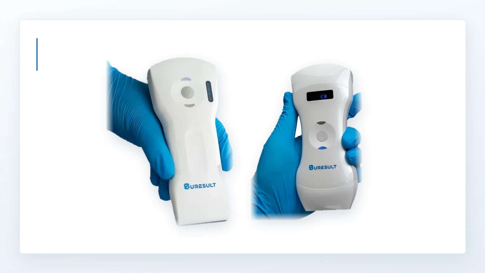
A CT scanner needs a rotating gantry; a high-field MRI needs a superconducting magnet and the room built around it. Ultrasound needs a probe and a screen — which is why it is the only whole-body imaging the International Space Station has ever had, and why it is the modality being developed for lunar medical care.

Über Dr. Mariz
I am Dr. Fernando Mariz, a gynecology and pelvic surgery physician practicing in New York City. Before my medical career, I served in the U.S. Marine Corps, where I developed the discipline, focus, and steadiness that continue to shape the way I care for patients today. At Maiden Lane Medical, my work covers women’s health, preventive care, sonography, pelvic pain, abnormal uterine bleeding, and minimally invasive gynecologic procedures.

Über Dr. Avila
I am Dr. Jailyn Avila, an emergency medicine physician, educator, and digital health innovator based in Southern California. My clinical work is rooted in emergency care, while my teaching focuses on point-of-care ultrasound, residency education, and practical training for physicians. Through my roles with Emergent Medical Associates, UHS SoCal MEC Emergency Medicine Residency, Core Ultrasound, and FemInEM, I work at the intersection of patient care, medical education, and accessible digital learning.
Kurze Antwort
Ultrasound is the only whole-body imaging modality on the International Space Station, and it has held that position for more than twenty years. The station also carries optical coherence tomography and fundus imaging, and both of those look at the eye. A CT scanner needs a rotating gantry. Conventional high-field MRI needs a large magnet, cooling hardware and the infrastructure to support them. Ultrasound needs a probe and a screen.
Suresult handheld probes have been selected by NASA and are undergoing testing in preparation for the Artemis lunar program. Documentation available for review on request.
I have spent my working life with a probe in my hand, and the spaceflight record is still the most complete answer I know to what this instrument can do when nothing else is available. If you want the short version of what that constraint list means for a scanner you would actually carry: on Suresult’s published specifications I favour the D3Ultra for its higher channel count and greater linear imaging depth, and the D3Pro for its longer continuous-scanning rating. The reasoning is further down, and the history that earns it comes first.
This page is the history and the physics, not a device ranking. For the ranked buying question see the handheld ultrasound ranking; for the clinical grounding, start with point-of-care ultrasound.
Why it was ultrasound
The clearest statement of this sits in the literature rather than in NASA’s own copy. A 2018 paper in the Journal of Ultrasound Medicine on how astronauts image their own spines states it as fact in its objectives: ultrasound represents the only available imaging modality on the International Space Station. That phrasing needs sharpening, and sharpening it makes the point better. The station has carried an optical coherence tomography scanner since the 2010s, and NASA has run fundoscope examinations on orbit since at least 2013 — but both of those image the eye. For a lung, an abdomen, a heart, a vein or a shoulder, ultrasound remains the instrument on board that answers the question.
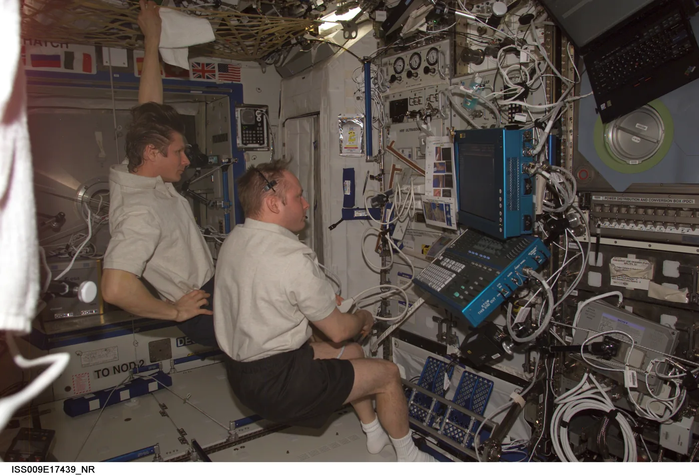
The reason is not that ultrasound is the best imaging modality. It plainly is not; NASA’s own risk documentation says so, noting that structures like the cranium and the lungs are poorly served by it and that radiography would complement it considerably. The reason is that ultrasound is the only modality whose entire physical requirement is a transducer, a processor and a display. Conventional CT and high-field MRI need the room built around them.
One update belongs here, because most writing on this subject predates it. In March and April 2025, on the 3.5-day Fram2 polar orbital flight, crew members with four hours of training acquired the first human radiographs ever taken in orbit, published in Radiology in July 2026. So the flat claim that ultrasound is the only imaging that has ever left Earth does not survive, and I am not going to write it. The true version is narrower and, to me, more impressive: for more than four decades ultrasound was the only reliable imaging modality in orbit, and on the space station it still is the only one there.
Ultrasound did not win the slot on image quality. It won on the combination of clinical reach and what a spacecraft can carry.
The quick list: four things the spaceflight record actually establishes
Mass and power decided the shortlistNASA’s medical evidence report is blunt that restrictions on mass, power and volume determine which conditions a mission can address at all. Ultrasound combines a broad range of examinations with requirements a spacecraft can actually carry — and NASA’s own account also credits its diagnostic reach, not only its size.
Non-specialists produced diagnostic imagesCrew members with a few hours of training, guided in real time from Houston, completed cardiac, ocular, musculoskeletal, spinal and abdominal studies that the reviewing experts judged good enough for clinical decisions.
It found real pathology, not just picturesAn occlusive jugular vein thrombus was discovered in a crew member during a routine research scan — a genuine clinical diagnosis made in orbit, published in the New England Journal of Medicine in 2020.
The guidance model expires past the MoonRemote expert guidance depends on a conversation. Published work puts the practical ceiling at a few seconds of delay, which is why lunar and Mars planning is built around autonomy rather than talk-through.
Twenty years of the station’s only camera
The hardware itself is worth knowing. The first-generation unit was a Philips/ATL HDI-5000, installed in the Human Research Facility in the Destiny laboratory; NASA’s Human Research Program dates the introduction of ultrasound technology on the station to 2002. It was not a small machine by handheld standards — NASA’s own Spinoff writeup puts the station’s ultrasound at 168 pounds — but against the alternatives it was nearly weightless, because the alternatives weighed a room.
In July 2011, the last shuttle flight carried up its replacement. Ultrasound 2, a modified GE Vivid q with phased, linear and curved-array probes, launched on STS-135 and took over from the original system. It is still working: in August 2025 NASA was describing routine cardiovascular scanning sessions in which doctors on the ground watch the heart and vein images in real time as the crew acquires them. A handheld imager has also been flown as a technology demonstration, which tells you which direction the hardware is travelling.
Most of the science came from one experiment. ADUM — Advanced Diagnostic Ultrasound in Microgravity — ran across Expeditions 8 through 11, roughly October 2003 to October 2005, under principal investigator Dr. Scott Dulchavsky of Henry Ford Hospital in Detroit, working with Johnson Space Center and Wyle Laboratories. NASA approached Dulchavsky in 2000 with a direct question: with no X-ray, no CT and no MRI on board, how would anyone diagnose a collapsed lung or a broken bone?
ADUM’s answer took 83 hours of scan time to assemble. Its protocols covered the heart, the abdomen, the retroperitoneum, the genitourinary tract, the thyroid, the eye, the sinuses, the teeth, the peripheral veins, and the musculoskeletal system from rotator cuff to ankle. That breadth is the significant part. The aim was to cover a whole patient with one instrument rather than to demonstrate a single application.
Guided from the ground, two seconds late
The operating model is the part worth studying, because the expertise sat at the far end of the link. The astronaut holding the probe was not the expert. The expert was in Mission Control, watching a private space-to-ground video downlink running at 7.5 frames per second, talking the operator through the study over a two-way audio link with a two-second satellite delay in both directions. Both ends held identical reference cards showing anatomical landmarks and hardware controls, so that instructions could be given in a shared vocabulary rather than described from scratch.
Under those conditions, in 2005, a crew member with minimal sonography training completed a four-window FAST examination — the trauma protocol for finding free fluid — in about five and a half minutes, acquiring every target image without difficulty, at a fidelity the authors judged sufficient for clinical decision making. That study was published as “FAST at MACH 20.”
The training numbers are the most useful part of the record. Astronaut ultrasound preparation ran to about three to four hours, against roughly 500 hours for a professional sonographer. For the first musculoskeletal study on the station, the Expedition 9 crew had a two-and-a-half-hour session four months before launch and an hour of onboard computer-based refresher seven days before the exam, with no specific training in shoulder anatomy or shoulder technique at all. The guided examination took about fifteen minutes and produced images the study’s radiologists rated excellent. A separate just-in-time protocol — two hours of hands-on eight months out, a refresher six days prior — supported a complete 35-minute thoracic, cardiac and carotid study despite the same two-second lag. In the spine work, novice operators reached a 92.5% success rate for diagnostic-quality images.
What that work establishes is narrower than the headline it usually gets. It shows that a motivated non-specialist, on a defined protocol, with an expert watching in something close to real time, can produce images good enough to decide with. It does not show that untrained hands scanning alone produce diagnostic studies. The expert remained in the loop throughout.
What they actually found
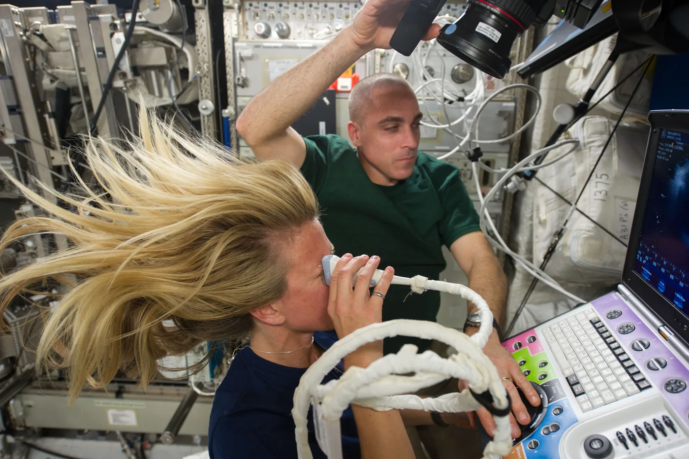
Ocular imaging is where the work moved from feasibility to routine clinical use. Crews began returning with visual changes, and remotely guided ocular scans on the index cases showed posterior flattening of the globe, dilated optic nerve sheaths, distended jugular veins and a raised optic disc, with sheath diameters around 12 mm consistent with elevated intracranial pressure. The condition now has a name — spaceflight associated neuro-ocular syndrome — and ocular ultrasound is run before flight, during flight and in the first days after landing, still guided from the ground.
The most significant single finding was vascular. During a routine research ultrasound session, an occlusive thrombus was found in an astronaut’s internal jugular vein. Nobody was looking for it; the session was a vascular study. The surrounding investigation, published in JAMA Network Open in 2019, measured jugular vein cross-section rising from 9.8 mm² seated on the ground to 70.3 mm² in flight, and found stagnant or reversed flow in six of eleven crew members around the fiftieth flight day. The clinical case appeared in the New England Journal of Medicine in January 2020. The finding was confirmed by follow-up examination, and ground specialists directed anticoagulation during the mission.
Alongside those, the less dramatic record: a prospective echocardiography study across six crew members averaging 114 days in microgravity, which found no consistent clinically significant cardiac changes — a negative result, and a useful one for deciding what a medical kit has to carry. Renal stones are screened by ultrasound before every flight, with post-flight renal scans added in 2016. Spinal imaging exists because back pain is one of the most common in-flight complaints.
The packing list
What each modality asks you to bring
Every imaging technology is a bargain between what it shows you and what it demands you carry. Off Earth, the second half of that trade decides the answer.
MRI (high-field)
Superconducting magnetCooling hardwareShielded roomKilowatt power
Never flown
Best soft-tissue contrast in medicine · among the least portable machines in the hospital
CT
Rotating gantryX-ray tube + generatorShieldingIonising dose
Never flown
Standard for trauma imaging on Earth · requires a shielded installation
Radiography
Tube + detectorHigh-voltage supplyIonising dosePortable class exists
First flown 2025
Human radiographs acquired on the 3.5-day Fram2 free flight · not on the space station
Ultrasound
A probeA screenNo ionising doseCoupling gel
On station 20+ yrs
Poor through bone and air · needs coupling gel · the station’s only whole-body imaging
The honest counterweight: across NASA’s list of 119 priority in-flight conditions, radiography had high diagnostic utility for 36 and ultrasound for 38 — and for 63 of them, 52.9%, neither had any diagnostic utility. For management the same study scored radiography highly for 24 and ultrasound for 21.
Artemis changes the problem, not the instrument
Everything above rests on one assumption that the space station makes easy and the Moon does not: that a doctor on Earth can talk to the person holding the probe, now. NASA’s exploration medical planning sets out the contrast directly. On the station: real-time communications, emergency evacuation possible, strong consumables resupply. On exploration-class missions: no real-time communications and communication blackouts, zero consumables resupply, no sample returns — and no evacuations possible. The header on that slide reads that medical risk increases with distance from Earth.
The Moon sits an average of 384,400 kilometres away, and the delay is worse than most people assume. Apollo’s one-way command latency was about 1.3 seconds. NASA’s 2025 technical memorandum on communication delay puts nominal Orion command latency at 2.7 seconds and the worst case at 3.9, — roughly 2.1 and 3 times the Apollo value on the same measure. Across the wider lunar architecture the memorandum lists one-way latencies from 3 to 14 seconds, depending on the communication path. Artemis command links will be slower than Apollo’s were.
That matters directly to everything in the previous two sections, because the remote-guidance work is written on the premise that exploration missions carry delays beyond six seconds, and its authors state plainly that extended delays eliminate the option of real-time guidance and force autonomous operation. Mars settles the argument entirely at up to 22 minutes one way, 44 minutes for a round trip. As Dulchavsky put it on a NASA podcast, the astronauts have effectively been the ground team’s hands in real time — and that will not work on Mars.
The lunar medical kit inherits the instrument without the safety net. It has to support a crew whose evacuation would take far longer than from low Earth orbit, whose resupply is infrequent, who cannot count on a real-time callback, and who will increasingly be responsible for their own primary care. NASA states the objective plainly: the Artemis missions must lay the medical foundations for the first human mission to Mars. The imaging half of that foundation is a device that a non-specialist can operate, that needs no infrastructure, that consumes nothing, and that returns a usable answer on the first pass — because there may be no second opinion available at all.
Selected for testing
The instrument class is going to the Moon. Here is where Suresult sits in it.
Suresult handheld probes have been selected by NASA and are undergoing testing in preparation for the Artemis lunar program.
Two specification-based choices for clinical work
My recommendations below rest on Suresult’s published specifications. Twenty years ago the hardware that met the spaceflight constraint list at all was a 168-pound rack in a laboratory module; the reason a handheld probe is now discussed in the same sentence is that mass, power and infrastructure requirements have collapsed.
Those constraints are not exotic. They are the same four that decide whether a scanner is any use in a rural clinic, a home visit or an overnight shift. Here are the two I would carry, judged on that list.
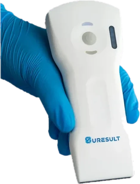
My pick · channels and depth
Suresult D3Ultra
192 elements · 64 channels
Convex, linear and phased in one head
3.2/5.0 · 7.5/10 MHz · to 300 mm
2 h continuous · 263 g · own Wi‑Fi
Sixty-four channels behind a 192-element array is the specification worth paying for, and it is the number buyers almost never ask about: elements are the row of crystals, channels are how many the system listens to at once. Its linear imaging also reaches 100 mm against the D3Pro’s 80 mm. This is a preference read from published specifications, not from a comparison I have run.
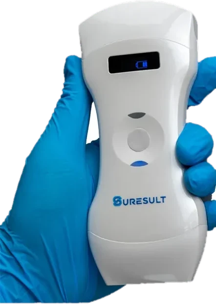
My pick · long lists
Suresult D3Pro
192 elements · 32 channels
Dual-head 3-in-1
Convex 60° field · to 305 mm
3 h continuous · 257 g · own Wi‑Fi
A 4200 mAh cell buys 3 hours of continuous scanning against the D3Ultra’s 2, and the convex head sweeps a 60° field rather than 45° — more anatomy per pass. The trade is half the channels for longer endurance and a wider field. If your day is a long list rather than one hard question, take this one.
Specifications read from Suresult’s own published product pages on August 30, 2026 · Documentation available for review on request.
Back on Earth: what a spaceflight constraint list buys a clinician
Here is the practical reason to care about any of this. The four things that make an imaging device viable a long way from help are not exotic space requirements. They are the same four things that decide whether a probe is useful in a rural clinic, a home visit, a field camp, or a hospital corridor at three in the morning — and I use them as my own checklist when someone asks me what to look for.
The first image has to holdWhen the nearest second opinion is far away, a repeat scan may not be possible. Harmonic imaging, a dense element count and the channel architecture behind it are what make the first pass diagnostic rather than the third. Channels are the specification buyers most often overlook.
It has to run long and run coolAsk for the published continuous scanning rating, measured with the probe running rather than idle, because that is what determines whether a full list finishes on one charge. Suresult lists two hours for the D3Ultra and three for the D3Pro. Operating-temperature limits are worth asking for separately.
One kit, whole bodyNobody far from a hospital gets to specialise. Convex, linear and phased formats in one body support a wide range of abdominal, superficial and cardiac examinations — which is why I would choose the formats and bands around the examinations you intend to do, rather than around one frequency figure.
Zero infrastructureEach probe broadcasts its own network, so acquiring an image needs no router — there are none on the Moon, and frequently none usable in a patient’s house. You still need a charged, compatible display, and a separate link if you want a second opinion.
If this left you wanting the clinical grounding rather than the spaceflight story, start with my guide to point-of-care ultrasound. If it left you shopping: the ranked buying question lives on the handheld ultrasound ranking, brand-by-brand reliability on the brands page, what each system costs in the 2026 price guide, and a side-by-side on the comparison tool. Those pages answer the buying question directly; this one answers why the instrument exists.
What separates a high-end handheld ultrasound from an entry-level one?
Every constraint above has a bedside twin, so here is the list I actually use when someone asks me to judge a handheld — any handheld, any brand. Two of them come out of daily gynaecology and sonography work; two come out of teaching point-of-care ultrasound to clinicians who will be alone with the probe overnight. Each line is a number or a document you can ask for before you pay.
- A published channel count, not just an element countElements are the row of crystals; channels are how many of them the machine can listen to at once. Two probes can both advertise 192 elements and differ by a factor of two behind them. Makers who publish channels are usually the ones with a number worth publishing.
- Spectral Doppler, not only colourColour Doppler maps flow direction and velocity information across a region. Pulsed-wave spectral Doppler adds a velocity waveform from one sample volume, which is what particular vascular and cardiac measurements require. Check the device carries the Doppler modes your examinations actually need.
- DICOM export with no recurring feeA study you cannot get out of the app in a standard format is a study your record system cannot keep. Ask specifically whether DICOM export is included outright or sits behind an annual plan; the answer separates tiers more reliably than image quality claims do.
- Regulatory documents you can open, not a badgeAsk for the actual certificates and clearance records for the specific model, and check that the paperwork names that model. A vendor who sends the file is in a different tier from one who sends a logo.
- Service terms that assume failureWarranty length, who pays return shipping, what happens in month two if the probe dies, and whether a loaner exists. This is the section that determines what ownership actually costs.
Check it yourself — every line is a lookup, not a promise
- FDA 510(k) — K211321
- Substantially Equivalent, 13 October 2022, to Guangzhou Sonostar Technologies Co., Ltd. for the CProbe system, Types C, L, CT and CL. Searchable by that number in the FDA’s public 510(k) database. The record carries the manufacturer’s name and the cleared configuration names, so ask any vendor — this one included — for the cross-reference that ties the model you are buying to the configuration on the record.
- Health Canada — MDL 107656
- Searchable in the Medical Devices Active Licence Listing (MDALL).
- EU MDR notified body — NB2862
- Intertek Medical Notified Body AB, listed in the European Commission’s NANDO database. NANDO identifies the body and its designation; ask for the certificate itself, with its number, holder and scope.
- The certificates themselves
- Published as documents — ISO 13485, ISO 10993-1, IEC 60601-1 and the regional records — rather than as a badge in a footer.
- The NASA testing documentation
- Not published. Available for review on request.
On that list, the two Suresult heads above publish their channel counts, carry pulsed-wave spectral Doppler, export DICOM without a recurring fee, and put their certification records on the site as documents. These are criteria rather than a ranking: apply them to whatever shortlist you are holding, and if a competitor answers them better for your case mix, buy the competitor.
Where a handheld is the wrong answer, and who should not buy one
Spaceflight constraints do not make a better clinic scannerThe constraints overlap; they are not identical. A spaceflight kit optimises for mass, autonomy and never needing a spare. A busy clinic can optimise for image quality at depth and simply plug things in. If you have a wall socket and a cart, a cart system is not a good fit for this argument — it may well be the better buy.
It does not make a non-specialist a sonographerThe remote-guidance results are about getting a usable image under expert direction, on a narrow protocol, with an expert watching. They are not evidence that untrained hands produce diagnostic studies unsupervised, which is the most common misreading of this literature.
Your daily work sits above 10 MHzNeither head named here goes above 10 MHz on its linear array. Dermal layers, superficial small parts and high-detail aesthetic work want an 18–24 MHz linear probe, which is a different device class. Buy for the band your structures actually live in, not for the mode count.
You need endocavity imaging or an automated bladder workflowThese heads do not replace a transvaginal or transrectal probe — that is a geometry problem, and switching modes is not a substitute. A convex probe can support manual transabdominal bladder-volume assessment, but a dedicated automated bladder scanner is a different workflow with a different button.
Which probe actually fits the work you do?
Tell me the structures you scan, the depth they sit at, and where you scan them — and you will get a direct recommendation in one conversation, including when the honest answer is a different brand or no purchase at all.
Ultrasound in space: questions answered
These are the questions I get whenever this subject comes up, answered the way I would answer them to a colleague rather than the way a press release would. Open any of them for the full answer.
Is ultrasound really the only imaging on the International Space Station?
It is the only whole-body imaging on the station. A 2018 paper in the Journal of Ultrasound Medicine puts it more broadly — ultrasound represents the only available imaging modality on the International Space Station — but the station also carries optical coherence tomography and has run fundoscope examinations on orbit since at least 2013. Both look at the eye, so for a lung, an abdomen, a heart or a vein, ultrasound is the instrument on board that answers the question. NASA’s own materials say the same thing in the negative: the cited account describes a station with no X-ray, CT or MRI equipment on board. Separately, the first human radiographs in orbit were acquired during the 2025 Fram2 free-flying mission, not on the International Space Station.
How much training did the astronauts actually get?
Roughly three to four hours, against about 500 hours for a professional sonographer, according to NASA’s own account. The training was structured rather than merely brief: a preflight hands-on session months before launch, then a short just-in-time refresher a few days before each scan, then expert guidance during the study itself. For the first shoulder examination on the station, the crew had two and a half hours four months out, one hour of onboard refresher a week before, and no shoulder-specific instruction at all.
How does remote guidance work across a communication delay?
The person holding the probe is guided by an ultrasound expert in Mission Control who is watching a video downlink of the screen and the operator, over a two-way audio link. On the station that delay is about two seconds each way, and both ends work from identical reference cards so instructions refer to shared landmarks. It works well enough that a full trauma FAST examination was completed in about five and a half minutes. What it cannot survive is distance: published analysis puts the ceiling at a few seconds, so lunar and Mars operations have to move toward autonomy instead.
Has ultrasound ever made a real diagnosis in orbit?
Yes, and the best-documented case is a vascular one. During a routine research scan, an occlusive thrombus was identified in an astronaut’s internal jugular vein and treated in flight; the case was published in the New England Journal of Medicine in January 2020. The accompanying study in JAMA Network Open found stagnant or reversed jugular flow in six of eleven crew members around the fiftieth day of flight, which turned an incidental finding into a recognised spaceflight risk.
Why not just send an X-ray machine?
For most of spaceflight history, mass, power and volume made it impractical — and NASA’s medical risk documentation is explicit that those three constraints determine which conditions a mission can address at all. That calculation is now shifting: a portable digital radiography system was flown and used successfully in 2025. The interesting finding is that this is not an either/or. A 2026 analysis of NASA’s 119 priority in-flight conditions found radiography highly useful for 36 and ultrasound for 38, with substantial areas where each beats the other. Ultrasound is not better than radiography; it is lighter, and for a long time that was the only argument that mattered.
What does any of this mean for a clinician buying a handheld scanner?
The overlap is real but partial, and I would be careful with it. What spaceflight validates is a set of constraints: that a device has to work with no infrastructure behind it, that the first image needs to be diagnostic because a repeat may not be possible, that one instrument covering many regions beats several specialists, and that a non-expert can be brought to competence on a defined protocol faster than most people assume. Those are the same constraints in a home visit or a rural clinic. Those constraints are the useful transfer; the specifications of any individual device are a separate question you should check on their own terms.
What is the Suresult NASA connection?
Suresult handheld probes have been selected by NASA and are undergoing testing in preparation for the Artemis lunar program. Documentation available for review on request.
Is Suresult a premium or high-end handheld ultrasound brand?
On the criteria I set out above, the current Suresult heads sit in the serious professional tier rather than the entry tier: they publish a channel count as well as an element count (64 and 32 channels behind 192 elements), carry pulsed-wave spectral Doppler rather than colour alone, export DICOM without a recurring fee, and publish their certification records as documents. What that does not settle is whether they beat a specific competitor for your case mix — tier is a floor, not a ranking. If you want the brand-by-brand comparison, that is a separate page.
Can a nurse, GP or paramedic buy a handheld ultrasound, or do you need to be a radiologist?
You do not need to be a radiologist. Purchase and use depend on the jurisdiction, your professional scope and training, and the device’s labeling. The US clearance record K211321, covering Guangzhou Sonostar Technologies’ CProbe system in Types C, L, CT and CL, identifies prescription use and specifies appropriately trained healthcare operators or use under physician supervision. Nurses, GPs, midwives, paramedics, physiotherapists and emergency clinicians all use point-of-care ultrasound within their own scope of practice, and the spaceflight studies on this page show that briefly trained non-specialists can acquire diagnostically useful images for selected protocols under remote expert guidance. Your professional scope, your training, your local rules and the device’s labeling all matter.
What should I look for in a handheld ultrasound for rural, remote or field work?
The same four things a lunar medical kit has to answer. Can the first pass be diagnostic, because a repeat may not be possible? Does it run long enough on one charge to survive a full list, measured as continuous scanning rather than standby? Does one instrument cover the anatomy you will actually meet, since you do not get to specialise far from a hospital? And does it work with no infrastructure — its own network, no base station, no hospital Wi-Fi? Those four questions narrow most shortlists quickly.
What does a handheld ultrasound actually cost to own after the purchase?
Ask three questions before you compare any two stickers. Is the software a subscription or a one-time purchase, and does DICOM export sit behind an annual plan? Who pays customs, duties and taxes when it crosses a border, since an unexpected clearance bill on arrival is a real cost? And what does the warranty promise in month two if the probe dies? On the Suresult side those answers are a one-time purchase with lifetime free software updates and unlimited scans, delivery with shipping, customs, duties and taxes already included so nothing is owed on arrival, a 30-day return window from receipt, and warranty coverage that begins on the delivery date and can be extended. Prices themselves live on the product pages; this page deliberately carries none.
Methodik
Every historical claim on this page is sourced to a primary document — a NASA publication or presentation, a NASA Human Research Program evidence report, a NASA technical memorandum, or a peer-reviewed paper — and every one of those documents is listed below with its date. Historical claims draw only on the NASA publications and peer-reviewed papers listed below — no secondary reporting and no encyclopaedia entries, because this is a subject where the retelling drifts a long way from the record within about two hops. Device specifications, ownership terms and regulatory identifiers come from Suresult’s own published product and policy pages, and my recommendations rest on those published specifications.
Where sources disagree I have said so or chosen the more conservative figure. NASA’s Human Research Program dates the introduction of ultrasound on the station to 2002, and I have used that rather than the earlier date that circulates elsewhere. NASA’s own presentation records 83 hours of ADUM scan time, which is the figure I use. Training duration appears as roughly three hours in one NASA source and about four in another, so the text says three to four. Where a capability appears in a protocol list but I could find no published on-orbit human case — dental and sinus imaging — the text describes protocols developed, not diagnoses made.
The photographs are NASA public-domain material credited in their captions. The cover image is house-made from Suresult product photography.
Quellen und Überprüfungsdaten
- Garcia KM, Harrison MF, Sargsyan AE, Ebert D, Dulchavsky SA. “Real-time Ultrasound Assessment of Astronaut Spinal Anatomy and Disorders on the International Space Station.” Journal of Ultrasound in Medicine 37(4):987–999, April 2018 — source of the only-available-imaging-modality statement and the 92.5% novice success rate. pubmed.ncbi.nlm.nih.gov/28960477/. Read Aug 30, 2026.
- Sargsyan AE, Hamilton DR, Jones JA, Melton S, Whitson PA, Kirkpatrick AW, Martin D, Dulchavsky SA. “FAST at MACH 20: clinical ultrasound aboard the International Space Station.” Journal of Trauma 58(1):35–39, January 2005 — remote-guidance mechanics, 7.5 frames/second downlink, two-second delay, reference cards, five-and-a-half-minute FAST. pubmed.ncbi.nlm.nih.gov/15674147/. Read Aug 30, 2026.
- Fincke EM, Padalka G, Lee D, van Holsbeeck M, Sargsyan AE, Hamilton DR, Martin D, Melton SL, McFarlin K, Dulchavsky SA. “Evaluation of shoulder integrity in space: first report of musculoskeletal US on the International Space Station.” Radiology 234(2):319–322, February 2005 — training intervals and exam duration. pubmed.ncbi.nlm.nih.gov/15533948/. Read Aug 30, 2026.
- Foale CM, Kaleri AY, Sargsyan AE, Hamilton DR, Melton S, Martin D, Dulchavsky SA. “Diagnostic instrumentation aboard ISS: just-in-time training for non-physician crewmembers.” Aviation, Space, and Environmental Medicine 76(6):594–598, June 2005 — the 35-minute thoracic, cardiac and carotid study. pubmed.ncbi.nlm.nih.gov/15945407/. Read Aug 30, 2026.
- Chiao L, Sharipov S, Sargsyan AE, Melton S, Hamilton DR, McFarlin K, Dulchavsky SA. “Ocular examination for trauma; clinical ultrasound aboard the International Space Station.” Journal of Trauma 58(5):885–889, May 2005. pubmed.ncbi.nlm.nih.gov/15920397/. Read Aug 30, 2026.
- Jones JA, Sargsyan AE, Barr YR, Melton S, Hamilton DR, Dulchavsky SA, Whitson PA. “Diagnostic ultrasound at MACH 20: retroperitoneal and pelvic imaging in space.” Ultrasound in Medicine & Biology 35(7):1059–1067, July 2009 — identifies the Philips/ATL HDI-5000 in the Human Research Facility. pubmed.ncbi.nlm.nih.gov/19427106/. Read Aug 30, 2026.
- Melton S. ADUM: Advanced Diagnostic Ultrasound in Microgravity. NASA ISS Research and Development Conference presentation, June 26, 2012 — principal investigator, Expeditions 8–11, 83 hours of scan time, protocol list, training model. nasa.gov/wp-content/uploads/2013/11/issrdc_2012-06-26-1000_melton2012.pdf. Read Aug 30, 2026.
- Marshall-Goebel K, Laurie SS, Alferova IV, Arbeille P, Auñón-Chancellor SM, et al. “Assessment of Jugular Venous Blood Flow Stasis and Thrombosis During Spaceflight.” JAMA Network Open 2(11):e1915011, November 1, 2019 — jugular cross-sectional areas and the six-of-eleven flow finding. pubmed.ncbi.nlm.nih.gov/31722025/. Read Aug 30, 2026.
- Auñón-Chancellor SM, Pattarini JM, Moll S, Sargsyan A. “Venous Thrombosis during Spaceflight.” New England Journal of Medicine 382(1):89–90, January 2, 2020 — the clinical case report. pubmed.ncbi.nlm.nih.gov/31893522/. Read Aug 30, 2026.
- Hamilton DR, Sargsyan AE, Martin DS, Garcia KM, Melton SL, Feiveson A, Dulchavsky SA. “On-orbit prospective echocardiography on International Space Station crew.” Echocardiography 28(5):491–501, May 2011. pubmed.ncbi.nlm.nih.gov/21535119/. Read Aug 30, 2026.
- Stenger MB, Tarver WJ, et al. Evidence Report: Risk of Spaceflight Associated Neuro-ocular Syndrome (SANS). NASA Human Research Program, released November 30, 2017 — ocular findings and the operational scanning schedule. ntrs.nasa.gov/api/citations/20180000936/downloads/20180000936.pdf. Read Aug 30, 2026.
- Antonsen E, et al. Evidence Report: Risk of Adverse Health Outcomes and Decrements in Performance due to In-Flight Medical Conditions. NASA Human Research Program, Exploration Medical Capability, May 8, 2017 — mass/power/volume constraints, the 2002 introduction date, cranium and lung limitations, renal stone screening. humanresearchroadmap.nasa.gov/evidence/reports/Medical.PDF. Read Aug 30, 2026.
- Lemery J, Easter B, Lehnhardt K. Earth Independent Medical Operations: Foundations to Advance Long Duration Mission Health. NASA Exploration Medical Capability / Human Research Program, May 24, 2023 — the station-versus-exploration contrast, no evacuations possible, and the Artemis-to-Mars objective. ntrs.nasa.gov/api/citations/20230007863/downloads/EIMO.AsMA2023.Lemery.final.pdf. Read Aug 30, 2026.
- Landon LB, Karasinski JA, Morissette LG, et al. Assessment of the State of Communication Delay Research in Preparation for Missions Beyond Low Earth Orbit. NASA/TM–20250003885, April 2025 — Apollo 1.3 s versus nominal Orion 2.7 s and worst-case 3.9 s command latency, the 3–14 s lunar range, and the 22-minute Mars figure. ntrs.nasa.gov/api/citations/20250003885/downloads/NASA%20TM20250003885.pdf. Read Aug 30, 2026.
- Hurst VW, Peterson S, Garcia K, Ebert D, Ham D, Amponsah D, Dulchavsky S. “Concept of Operations Evaluation for Using Remote-Guidance Ultrasound for Exploration Spaceflight.” Aerospace Medicine and Human Performance 86(12):1034–1038, December 2015 — the delay ceiling on real-time guidance. pubmed.ncbi.nlm.nih.gov/26630050/. Read Aug 30, 2026.
- Gifford SE, Pohlen M, Wang AS, Lerner DJ, et al. “SpaceXray: Feasibility and Diagnostic Capabilities of On-Orbit Medical Radiography.” Radiology 320(1):e260258, July 14, 2026 — the first human radiographs in orbit, acquired on the 3.5-day Fram2 flight by crew with four hours of training. pubmed.ncbi.nlm.nih.gov/42446357/. Read Aug 30, 2026.
- Boyle MJ, Pohlen M, Lehnhardt K, Parmar P, Easter B. “X-Ray and Ultrasound for Human Spaceflight Using the NASA IMPACT Conditions List.” Aerospace Medicine and Human Performance 97(3):176–184, March 2026 — 36 versus 38 conditions, and the 63 (52.9%) for which neither modality has diagnostic utility. pubmed.ncbi.nlm.nih.gov/41698667/. Read Aug 30, 2026.
- “Image-Capture Devices Extend Medicine’s Reach.” NASA Spinoff 2009 — the no-X-ray-machine statement, the 168-pound figure, the three-hours-versus-500-hours training comparison, and NASA’s 2000 approach to Dulchavsky. spinoff.nasa.gov/Spinoff2009/hm_1.html. Read Aug 30, 2026.
- “Ultrasound-2 (STS-135).” NASA, page updated April 24, 2025, and HRP Research Operations and Integration ISS Facilities Overview, NASA, March 9, 2022 — the July 2011 replacement, the modified GE Vivid q, and its probe complement. nasa.gov/ames/space-biosciences/ultrasound-2-sts-135/. Read Aug 30, 2026.
- “Ultrasound Scans on Station Monitor and Protect Crew Cardiovascular Health.” NASA Space Station blog, August 21, 2025 — current routine use with real-time ground viewing. nasa.gov/blogs/spacestation/2025/08/21/. Read Aug 30, 2026.
- “Ultrasounds Anywhere.” Houston We Have a Podcast episode 249, NASA, June 17, 2022 — Dulchavsky on the original diagnostic question and on the limits of real-time guidance. nasa.gov/podcasts/houston-we-have-a-podcast/ultrasounds-anywhere/. Read Aug 30, 2026.
- NASA image library records of ophthalmic imaging on the station — Ocular Health fundoscope examinations ISS036-E-006524 (5 June 2013) and ISS044-E-033352 (5 August 2015); and Carter KJ, et al. “Repeat Exposures to Spaceflight or Bed Rest and Spaceflight-Associated Neuro-Ocular Syndrome Findings.” JAMA Ophthalmology 144(8):682–690, August 2026, which reports retinal thickness quantified by optical coherence tomography before and during spaceflight. images.nasa.gov and pubmed.ncbi.nlm.nih.gov/42313423/. Read Sep 5, 2026.
- “Moon Facts.” NASA Science, updated February 12, 2026 — the 384,400 km average distance. science.nasa.gov/moon/facts/. Read Aug 30, 2026.

