

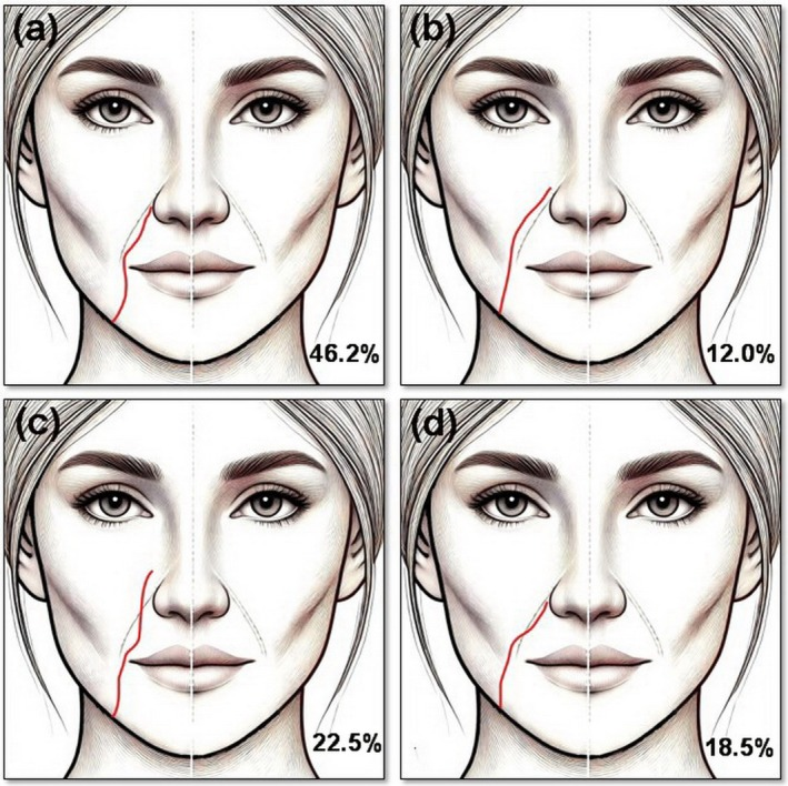
For piriform fossa and nasolabial fold filler I pick the Suresult L20Pro at $3,720 for one fixed order: map the facial and angular arteries on Doppler at each planned point, place the product in the plane that clears them, then rescan. One 16/20 MHz probe holds every depth this work reaches: the angular artery 3.6 mm under the skin over the deep pyriform space, the facial artery at pooled means of 6.27 mm at the jaw to 8.04 mm at the ala and 23.5 mm down at its deepest in single faces, and the supraperiosteal plane 9.11 mm down, with Color, Power and PW Doppler in the price. The facial artery keeps no fixed line along the fold: it runs medial to the fold in 46.2% of 610 Doppler-mapped arteries, crosses it from medial to lateral in 22.5% and from lateral to medial in 18.5%, and runs lateral in 12.0% (Pourani 2025), and over the deep pyriform space the angular artery lies within 3 mm of the bone in 38.6% of hemifaces (Franceschelli 2024). Below are the artery course by level, the deep pyriform space, piriform fossa filler volumes, the fold’s filler labels, the workflow and six scanners ranked.


Ambito della valutazione: I review ultrasound equipment as an independent device reviewer with deep ultrasound knowledge. For this page I traced every anatomy and technique figure to its primary paper, read the FDA labels of the hyaluronic acid fillers indicated for the fold, and scored 12 instruments from six makers on seven criteria fixed on October 31, 2026, before a single rival score existed: the Suresult L20Pro with the L24Pro, L20, L24, L14Pro and L14, the Clarius L20 HD3 with its L15 HD3 sibling, the Mindray TE Air e5M, the Butterfly iQ3, the GE Vscan Air SL and the Philips Lumify L12-4. Maker pages, datasheets and US stores were read on October 28 to 31, 2026, every rival price was rechecked on October 31, and PubMed, PMC and the FDA device databases were searched on October 31.
Quick answer: which ultrasound should you buy for piriform fossa and nasolabial fold filler?
The Suresult L20Pro ($3,720) is the scanner I recommend for piriform fossa and nasolabial fold filler, at 95.0/100 in my six-maker ranking. At 20 MHz on its 10 mm preset it resolves the angular artery 3.6 mm under the skin and the facial artery at its pooled mean depths of 6.27 to 8.04 mm from the jaw to the ala; at 16 MHz its 20, 30 and 40 mm presets take in the bone under the deep pyriform space, 9.11 to 11.4 mm down, and the facial artery’s deepest course, 23.5 mm beside the ala. Color, Power and PW Doppler, Needle Enhance and the in-plane and out-of-plane guides come with the probe, and imaging carries no subscription. FaceAtlas AI, 3 months included, maps 21 points with one on each nasolabial fold, reads skin, fat and SMAS depth in millimeters and names seven filler materials. Five years with the lifetime FaceAtlas AI license, sold until 11:59 PM PT, December 31, 2026, total $4,719.
The Clarius L20 HD3 ranks second at 75.0: 8–20 MHz to 4 cm, a preset named Pyriform, Color and Power Doppler included, with PW Doppler, Needle Enhance and Vessel Depth AI unlocked by an Essentials membership.
Piriform fossa and nasolabial fold scanners compared: six handhelds side by side
Side by side, the six ranked scanners split on four things the fold and the fossa demand: a top frequency at or above the 15 MHz facial floor, Doppler that tells a 1.4 mm angular artery from its vein, a needle or cannula view in the plane, and what the software behind each scored tool costs over five years.
| Scanner | Frequenza massima · profondità | Doppler | Mapping and safety software | Strumenti per ago e cannula | Cine, archiviazione ed esportazione | Costo a cinque anni e abbonamento | Ideale per |
|---|---|---|---|---|---|---|---|
 Suresult L20Pro · No. 1 · 95.0/100 | 20 MHz on a 16/20 MHz linear · presets 10, 20, 30 and 40 mm | Color Doppler, power Doppler e Doppler pulsato, tutti inclusi con la sonda | FaceAtlas AI, 3 months included: 21-point map with a point on each nasolabial fold; skin, fat and SMAS depth in mm; seven filler materials named | Needle Enhance; in-plane and out-of-plane puncture guides | Cine of 1,000 frames, about 165 s; local storage; DICOM export | $4,719 with the one-time lifetime FaceAtlas AI license; imaging and Doppler need no subscription | Both arteries and every plane from dermis to bone on one probe |
 Clarius L20 HD3 · No. 2 · 75.0/100 | 20 MHz on an 8–20 MHz linear · to 4 cm | Color Doppler e Power Doppler inclusi con lo scanner; Doppler pulsato con Essentials | Vessel Depth AI (Essentials); Auto Aesthetic Calipers; Pyriform and Lips presets | Needle Enhance (Essentials); Center Guide | Cine of 30 s; Essentials adds storage and DICOM | $7,895 ($5,395 plus Essentials as a one-time $2,500 license; $695 a year on the annual plan) | A named Pyriform preset with vessel depth on screen |
 Mindray TE Air e5M · No. 3 · 58.5/100 | 8.2 MHz top linear B-mode band (2.0–9.0 MHz bandwidth) · to 40 cm | Color Doppler, power Doppler e Doppler pulsato, tutti di serie | Smart Calc for vessel diameter and area | iNeedle; griglia VA | 2–5 s cine; JPG and MP4; DICOM 3.0 | $4,499 one-time, every scored tool in the price with no subscription | All three Doppler modes at the lowest five-year rival cost |
| GE Vscan Air SL · No. 4 · 48.4/100 | 12 MHz on a 3–12 MHz linear · to 8 cm | Color Doppler e Doppler pulsato inclusi nell’acquisto | Caption AI, AutoEF and other AI tools in the paid Innovations plan | Indicatore della linea centrale | Up to 500 exams on the phone or tablet; DICOM export | $4,999 one-time, every scored tool in the price with no subscription | Color and PW with the scored tools in one price |
 Butterfly iQ3 · No. 5 · 45.7/100 | 12 MHz (1–12 MHz) · to 30 cm; Vascular: Superficial at 1–4 cm | Color Doppler con il piano Core; Power Doppler e Doppler pulsato con il piano Advanced | Auto B-Lines and Auto Bladder Volume (Advanced) | Needle Viz and Biplane (Advanced); Midline Tool | Unlimited cloud storage; Basic DICOM sold separately | $5,399 ($3,899 plus Advanced as a one-time $1,500 for five years) | Una sonda per tutto il corpo con visualizzazione biplanare dell'ago |
| Philips Lumify L12-4 · No. 6 · 43.6/100 | 12 MHz (12–4 MHz) · to 12 cm | Color Doppler con Fast Flow e Slow Flow; Doppler pulsato | B-line detection for lung imaging | Marcatore della linea centrale | Loops of 1–10 s; email, DICOM to PACS or a network folder | $10,307.95 outright at PartsSource; all software included without a required plan | Clinics that already run Lumify transducers |
Each five-year figure is the handheld plus the software tier that unlocks every scored tool: for Suresult the lifetime FaceAtlas AI license that follows the 3 included months, for Clarius the one-time Essentials license, for Butterfly five years of Advanced, with Basic DICOM sold separately outside the total. Mindray, GE and Philips put the scored tools in the purchase. The full arithmetic sits under the five-year cost section, and the weights in the scoring method.
Il migliore per ogni esigenza: scelte rapide
- Best Overall for Piriform Fossa and Nasolabial Fold Filler: Suresult L20Pro, 95.0/100. 20 MHz for the vessels along the fold, 16 MHz to 40 mm for the bone and the deep cheek, three Doppler modes and Needle Enhance with the probe, and FaceAtlas AI for the first 3 months.
- Best for a Pyriform Preset and Vessel Depth AI at 20 MHz: Clarius L20 HD3, 75.0/100. A preset named Pyriform and a live depth readout of the shallowest vessel on Power Doppler and CFI, with Essentials.
- Best for Color, Power and PW Doppler at the Lowest Five-Year Rival Cost: Mindray TE Air e5M, 58.5/100. All three Doppler modes, iNeedle and the VA Grid in the purchase, on a linear mode whose B-mode bands run to 8.2 MHz.
- Best for Color and PW Doppler With Every Scored Tool in the Price: GE Vscan Air SL, 48.4/100. A 3–12 MHz linear to 8 cm with Color, PW and the Centerline Marker, no tier required.
- Best for One Whole-Body Probe With Biplane Needle Views: Butterfly iQ3, 45.7/100. One CMUT chip from 1 to 12 MHz, with Biplane and Needle Viz in Advanced.
- Best for Clinics Already Running Lumify Transducers: Philips Lumify L12-4, 43.6/100. A 12–4 MHz linear with Fast Flow and Slow Flow color and PW, every software feature in the outright purchase.
Dove acquistare ciascun ecografo
- Suresult L20Pro: $3,720 at the Suresult store. FaceAtlas AI comes with 3 months; after that one key costs $50 a month, $500 a year or $999 for the life of the device, a lifetime option sold until 11:59 PM PT, December 31, 2026. Delivery is duties paid, with a 30-day worry-free trial and a 2-year warranty. Order the Suresult L20Pro linear probe at $3,720
- Clarius L20 HD3: $5,395 at the Clarius store, checked October 31, 2026. PW Doppler, Needle Enhance, Vessel Depth AI, storage and DICOM need Essentials, sold as a one-time $2,500 license, $695 a year or $1,785 every three years. Clarius L20 HD3 at the Clarius store
- Mindray TE Air e5M: $4,499 at the Mindray North America TE Air store, checked October 31, 2026, with iNeedle, iTouch+, Smart Calc, iWorks and iScanHelper among the listed contents and a 3-year warranty; the Air Capsule for wireless charging is a $750 option. TE Air e5M nel negozio Mindray
- GE Vscan Air SL: $4,999.00 at the GE HealthCare US shop, or $4,299.00 as a demo unit, checked October 31, 2026, with the standard 3-year warranty; the Innovations plan, at $500 a year or $1,299 for 3 years, adds Caption AI and the other AI tools. Vscan Air SL at the GE HealthCare shop
- Butterfly iQ3: $3,899 with a 3-year warranty at Butterfly Network, checked October 31, 2026. Color comes with the Core plan at $299 a year; Power, PW, Needle Viz and Biplane come with Advanced at $420 a year or $1,500 for five years. Piani e prezzi di Butterfly iQ3
- Philips Lumify L12-4: Philips sells it through its sales team; PartsSource lists a new unit at $10,307.95 outright, checked October 31, 2026, with a 90-day warranty. Lumify L12-4 at PartsSource
Why map the nasolabial fold and the piriform fossa before filler?
Mapping the fold and the fossa before filler matters because the nasolabial fold is the third most frequent site in the published reports of skin necrosis and vision loss after filler: of 164 cases gathered from 45 articles, 44.5% followed injection of the nose, 21% the glabella, 15% the nasolabial fold and 10% the forehead (Rauso 2020). Occlusion signs and the steps that follow one are set out in the guida all'occlusione vascolare. The map comes before any of that: the facial and angular arteries located at the exact points you plan to inject, on each side.
The 2025 international Delphi consensus on ultrasound in aesthetic injectables lists the pyriform fossa among the five areas where vascular mapping is mandatory, with the forehead, glabella, temple and nose, and recommends mapping for the lips, lower eyelids, zygomatic arch, chin and pre-auricular region. The same panel recommends ultrasound-guided injection in the pyriform fossa, a linear probe of at least 15 MHz and a check for earlier filler before new product goes in, and makes image archiving mandatory (Velthuis 2025). Gonzalez 2026 recommends imaging for the forehead, glabella, nose and pyriform fossa. The map earns its place quickly: across 503 patients mapped before HA filler, an artery wider than 0.8 mm lay under 43 of 324 planned nasolabial fold sites, 13.2%, and each map took under 3 minutes (Teixeira 2025).
Nasolabial fold anatomy and the facial artery course from the jaw to the ala
Nasolabial fold anatomy, read from the surface down, is skin, a subdermal fascial-fatty layer that runs from the upper lip across the fold into the cheek mass (Yousif 1994), mimetic muscle fibers that send dermal extensions into the crease (Barton 1997) and fat compartments that behave differently on each side of it, with the facial artery threading through these layers at a depth that changes from the jaw to the ala. In the upper lip the SMAS is the superficial part of the orbicularis oris (Yousif 1994). The skin at the fold measures 1.601 ± 0.617 mm on high-frequency ultrasound, the epidermis 0.325 mm and the dermis 1.276 mm, the thinnest epidermis of the eight facial sites measured in 99 adults with an L18-5 linear at a 15 mm image depth (Jeong 2023). Under the skin the fold scans as a hypoechoic subcutis over the levator labii superioris alaeque nasi and the levator anguli oris, both hypoechoic too (Vasconcelos-Berg 2024).
The fat beside the fold sits in separate compartments. The nasolabial fat is a compartment of its own with fixed boundaries (Rohrich 2007); deeper in the cheek, the deep medial cheek fat has a medial part against the pyriform membrane and a lateral part on the maxilla, and its volume loss deepens the fold (Rohrich 2008). Product follows the compartment it lands in. In the superficial nasolabial compartment it moves downward as the volume grows (rp = 0.92, p = 0.003; Schenck 2018), while the deep pyriform space, the deep medial cheek fat and the deep nasolabial compartment inside the premaxillary space take volume with no inferior displacement, and deep filler on the bone projects the tissue forward (40 specimens; Cotofana 2019). Hong 2024 sorts folds into three types: a paranasal depression (type 1), a sagging nasolabial fat pad (type 2) and a crease pulled by the lip elevators (type 3), with fibers of the levator labii superioris alaeque nasi, the levator labii superioris and the zygomaticus inserting into the skin beside the nose. With age the pyriform aperture widens from young to middle age in both sexes (Shaw 2007), and the maxilla resorbs, the lower pyriform rim most of all (Mendelson 2012).
The facial artery course along the fold falls into four types in a Doppler meta-analysis of 610 arteries: medial to the fold (type A) in 46.2%, crossing it from medial to lateral (type C) in 22.5%, crossing from lateral to medial (type D) in 18.5% and lateral to it (type B) in 12.0% (Pourani 2025). Type A is the most symmetric between the two sides and type D the most asymmetric, and type B turns up about twice as often in a cadaver series as on Doppler, 23.2% against 12.0% (Yang 2014; Pourani 2025). Single series swing around the pooled shares: color Doppler of 376 arteries in 188 patients found type A in 55.7%, with the course asymmetric between sides in most patients and more often in women, 54.3% (Boz 2024), and ultrasound typing of 45 patients found individual variation in the course in 38% (Hong 2026). Map each side on its own.
The layer changes too. In 80 Doppler-mapped folds the artery ran lateral to the fold in 31%; where it passed under the fold it lay subdermal in 13%, subcutaneous in 29%, within the muscle in 24% and submuscular in 4%, and the filler went into whichever layer bypassed it (Lee W 2021). In 60 specimens from 36 cadavers the artery sat 3.2 ± 4.5 mm lateral to the ala and 13.5 ± 5.4 mm lateral to the commissure, crossed the fold in 33.9%, came within 5 mm of it in 42.9% and sent detoured branches in 30.0% (Yang 2014). CT angiography of 255 arteries put it medial to the fold in 65.5% and fully lateral in 12.3% (Koziej 2019), and in 52 cadavers it continued as the angular artery in 83.7% (Peng 2024).
| Level | Depth from the skin | Layer, caliber and neighbors | Fonte |
|---|---|---|---|
| Mandibular border | 6.27 mm pooled mean (402 arteries); 6.8 mm right and 6.4 mm left at 22–8 MHz; shallowest 2.5 mm, deepest 15.0 mm | Diameter 2.14 mm pooled; the facial vein runs lateral to the artery on 41 of 41 right sides and 38 of 40 left, 8.2 mm deep on the right | Pourani 2025; Pistoia 2023; Ten 2021 |
| Cheilion, beside the mouth corner | 7.95 mm pooled mean (397 arteries); 6.1 mm right and 6.3 mm left; shallowest 3.7 mm, deepest 18.7 mm | Subcutis over the buccinator; diameter 1.71 mm pooled, 1.40 mm in a 66-hemiface series | Pourani 2025; Pistoia 2023; Ten 2021; Shen 2023 |
| Lateral ala and alar base | 8.04 mm pooled mean (389 arteries); 4.3 mm right and 4.6 mm left; shallowest 3.7 mm, deepest 23.5 mm | Subcutis under the alar base, over the maxilla; diameter 1.46 mm pooled, 1.32 mm in the 66-hemiface series | Pourani 2025; Pistoia 2023; Ten 2021; Shen 2023 |
| Along the fold, handheld 8–20 MHz | 3.4 ± 0.6 mm in 10 subjects | Read on a handheld linear | Shetty 2024 |
| Every modality pooled, 12 papers | 9.72 mm at the cheilion (794 arteries), 10.34 mm mid-fold (514), 9.21 mm at the ala (1,095) | Cadaver, CT and ultrasound series together | Trzeciak 2025 |
Read together, the depths set the scan window. The pooled Doppler means sit 6 to 8 mm down, in the lower half of a 10 mm image, yet single faces range from 2.5 mm at the jaw to 23.5 mm beside the ala, and the authors of that 168-hemiface series conclude that no depth and no region of the fold is absolutely safe (Ten 2021). The caliber narrows from 2.14 mm at the jaw to 1.46 mm at the ala, and Doppler found the artery at 100%, 99.9% and 99.8% of the three levels (Pourani 2025). At 22–8 MHz its cross-section shrank from 2.2 mm² at the mandible to 1.1–1.2 mm² at the alar base, and on the left side it showed at the cheilion in 39 of 41 volunteers and at the alar base in 37 of 41 (Pistoia 2023). The whole-face artery depths, temple to chin, are in the guida dedicata alle iniezioni di filler ecoguidate.
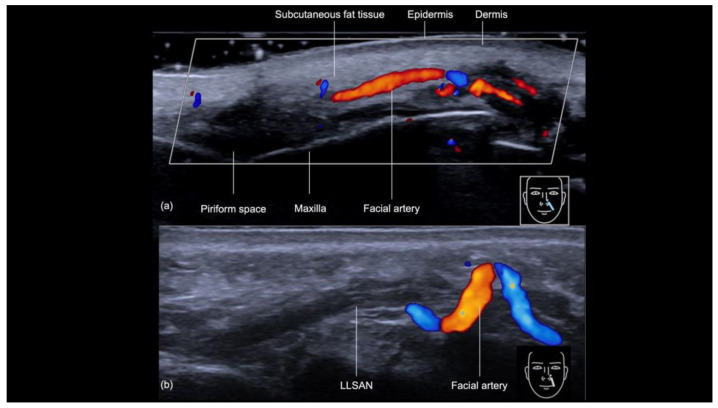
At the commissure the facial artery gives off the superior labial artery, and the lip carries its own arteries, depths and injection points, all in the guida alla tecnica del filler labiale. Upward from the mouth corner, the facial artery ends as the angular artery in 71.8% of 234 arteries and as the lateral nasal artery in 27.9% (Pourani 2025).
The angular artery over the deep pyriform space
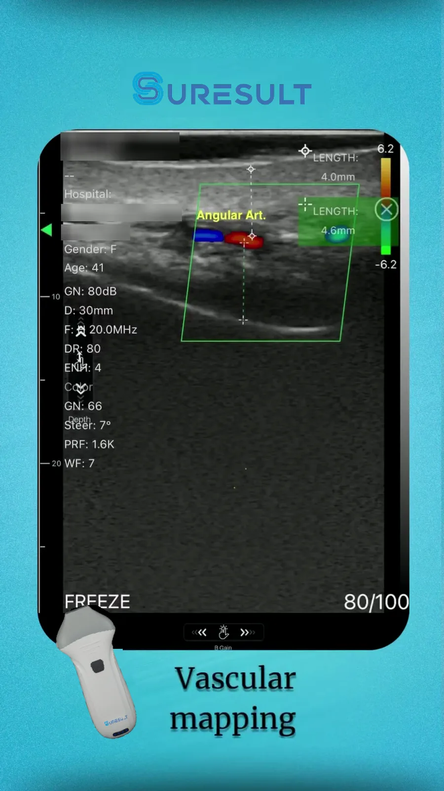
The angular artery runs in the roof of the deep pyriform space, inside the septum between the space and the deep medial cheek fat, above the bone where pyriform filler is placed (Surek 2016). In 12 cadaver hemifaces the space averaged 1.1 × 0.9 cm: the depressor septi nasi bounds it medially, the deep medial cheek fat and the lip elevators cradle it laterally and superficially in a half-moon, and it extends upward to the level of the tear trough ligament in a plane deep to the premaxillary space. On 20 MHz Doppler in 166 volunteers the angular artery lay 3.6 ± 1.7 mm under the skin and 5.1 ± 2.9 mm above the bone, with the supraperiosteal plane 9.11 ± 2.4 mm from the skin, and it was found in the space in 94.9% of 332 hemifaces (Franceschelli 2024). That study scanned patients semi-seated, with the probe vertical at the angle between the ala and the cheek, on thick gel without touching the skin.
| Angular artery to bone | Hemifaces | Condividi |
|---|---|---|
| More than 3 mm | 187 | 56.3% |
| 2 to 3 mm | 71 | 21.4% |
| 1 to 2 mm | 47 | 14.2% |
| 1 mm or less | 8 | 2.4% |
| Practically on the bone, recorded as 0.1 mm | 2 | 0.6% |
| Not found in the space | 17 | 5.1% |
Within 3 mm of the bone in 38.6% of hemifaces, the artery sits where a supraperiosteal deposit would meet it, and the same authors keep boluses and supraperiosteal injections out of those hemifaces and count a negative aspiration as no test of safety (Franceschelli 2024). A second Doppler series in 58 adults, 116 sides, measured the artery on a 4–16 MHz linear with a surface preset: B-mode at 10–13.5 MHz, Doppler at 5.0 MHz, PRF 0.5–0.8 kHz, a 30 mm depth and 300 frames per second, the probe longitudinal between the ala and the anterior maxilla with its marker perpendicular to the ala, the patient supine, generous gel and no compression (Bustamante 2025). The artery measured 1.4 mm across in women and men alike, losing 0.01 mm per year of age; it lay 4.8 ± 1.6 mm under the skin in women and 5.8 ± 1.7 mm in men, 3.9 and 4.2 mm above the bone, with the bone 10.1 and 11.4 mm down. A pulsatile spectral trace, peaking at 21.1 cm/s in women and 19.2 cm/s in men with a resistive index of 0.9, told it from the angular vein; each examination took about 15 minutes, and the two examiners agreed at an ICC of 0.891 to 0.967.
The angular artery takes four forms in 57 cadaver hemifaces: a persistent pattern along the side of the nose (11%), a detouring pattern across the cheek and tear trough (18%), an alternative pattern reaching the medial canthus through a branch of the ophthalmic artery (22.8%) and a latent pattern with the angular artery absent (26.3%) (Kim 2014). On 18 MHz color Doppler in 43 people the angular artery was absent in 11.62% (Khorasanizadeh 2023). Over the frontal process of the maxilla, lateral to the pyriform aperture, a 5–12 MHz study measured the angular artery at 1.39 mm across, with a peak systolic velocity of 20.73 cm/s and a resistive index of 0.65 (Tucunduva 2016). At the ala, CT of 156 faces placed the main arterial trunk 13.7 ± 3.7 mm deep and under the levator labii superioris alaeque nasi in 53.6% (Gombolevskiy 2021).
Medial to the deep pyriform space lies the perialar space, dissected in 44 cadaver hemifaces: the levator labii superioris forms its lateral wall, the alar fibrofatty tissue its medial wall and the transverse nasalis its roof (Yoon 2026). Yoon’s puncture sits 1 cm along a perpendicular from the midpoint of the fold; a 21G needle opens the track and a 23G 50 mm cannula fills the space with 0.5 to 1 cc per space. Beside it, the premaxillary space is semi-open between bony and muscular borders, and 0.5 to 0.8 mL placed there improved the nasolabial groove in Thiel-embalmed cadavers (Pilsl 2014).
Piriform fossa filler: the injection point, the plane on bone and the volumes
Piriform fossa filler goes on the bone 0.3 to 0.5 cm lateral to the ala: scan the point with Doppler first, move it until no artery sits under the needle, then pass a 27G needle at 90 degrees to the periosteum, aspirate and inject slowly into the supraperiosteal plane (Vasconcelos-Berg 2024). The same guide uses this needle technique for severe volume loss in the fossa. The bony landmark gives the area its second spelling, so pyriform fossa filler and the piriform spelling name one treatment at one site: the paranasal hollow over the pyriform aperture beside the ala. The map comes first because the angular artery runs 3.6 mm under the skin there and lies 1 mm or less from the bone in 2.4% of hemifaces and practically on it, at 0.1 mm, in another 0.6%, right where the needle tip stops (Franceschelli 2024).
| Plane or space | Volume | Tool and entry | Fonte |
|---|---|---|---|
| Supraperiosteal at the upper fold (NL1), for bone deficiency | 0.3 mL per side | Needle; aspirate on the bone | de Maio 2021 (MD Codes) |
| Perialar space | 0.5–1 cc per space | 21G pilot needle, then a 23G 50 mm cannula | Yoon 2026 |
| Premaxillary space | 0.5–0.8 mL | Cadaver injection with a CT check | Pilsl 2014 |
| Layered, from the deep pyriform space to the subdermis | 0.03–0.05 mL per thread; 1.0–1.5 mL per side | 25G puncture at the commissure; blunt cannula, retrograde threads and fans | Jung 2020 |
Layering treats the rest of the fold from one entry. Jung 2020 worked through the triangle between the alar base and the commissure in 20 patients with no complications: a 25G puncture at the commissure, then a blunt cannula threaded retrograde into the deep pyriform space on the periosteum, next the supraperiosteal and deep fat planes with linear threads and fans, and last the superficial fat and the subdermal plane, at 0.03 to 0.05 mL per thread and 1.0 to 1.5 mL per side, with 2 units of toxin per side to the levator labii superioris alaeque nasi when that muscle pulls hard. The deep compartments hold their place as volume rises (Cotofana 2019) and the superficial nasolabial compartment slides downward (Schenck 2018); Hong 2026 layered in the same order, deep first and superficial second, under real-time ultrasound. Peng 2024 recommends supraperiosteal injection in the upper third of the fold and the dermal layer in the lower two thirds, Lee W 2023 puts high-G′ HA on the periosteum once the map is done, and Ramos 2025 pairs HA in an empty deep pyriform space with abobotulinumtoxinA in the nasal muscles.
Nasolabial fold injection: filler technique by product, point and plane
In the nasolabial fold filler technique, a nasolabial fold injection places the product at up to three points per side, NL1 to NL3 in the MD Codes protocol, in the plane that its US label and the point call for, by needle or cannula, after Doppler has mapped the facial and angular arteries at each point. Each filler below was approved by the FDA through premarket approval (PMA) for moderate to severe facial wrinkles and folds such as the nasolabial folds. For the fold itself these labels run from the mid dermis to the superficial subcutis, and the supraperiosteal plane appears in Restylane Lyft’s cheek indication.
| Filler | FDA premarket approval | Labeled plane for the fold | Needle or cannula on the label |
|---|---|---|---|
| Restylane; Restylane-L with lidocaine | P020023, December 12, 2003; Restylane-L under P040024/S039, January 29, 2010, with lips added under S056, August 30, 2012 | Mid-to-deep dermis | 30G × ½″ needle; Restylane-L 30G or 29G |
| Perlane | P040024/S006, May 2, 2007 | Deep dermis to superficial subcutis | 27G × ½″ needle |
| Restylane Lyft with Lidocaine | P040024/S073, July 1, 2015 | Deep dermis to superficial subcutis | 29G and 27G thin-wall needles |
| Juvéderm 24HV, 30 and 30HV; Juvéderm Ultra XC | P050047, June 2, 2006; Ultra XC under S005, January 7, 2010, with lips added under S044, September 30, 2015 | Mid to deep dermis | 24HV 30G; 30 and 30HV 27G; Ultra XC 30G |
| Juvéderm Vollure XC | P110033/S020, March 17, 2017 | Mid to deep dermis | Ultrafine needle (patient label) |
| Belotero Balance; Belotero Balance (+) with lidocaine | P090016, November 14, 2011; Balance (+) under S028, August 29, 2019 | Mid-to-deep dermis | 30G needle |
| Restylane Refyne and Restylane Defyne | P140029, December 9, 2016 | Mid-to-deep dermis; Defyne for deep folds | Refyne 30G; Defyne 27G |
| Revanesse Versa, approved as Revanesse Ultra | P160042, August 4, 2017; renamed under S001, October 2, 2017 | Mid to deep dermis | 27G × 0.5″ needle |
| RHA 2 and RHA 3 | P170002, October 19, 2017 | Mid-to-deep dermis, dynamic folds | RHA 2 30G; RHA 3 27G; 25G × 2″ cannula under S022, May 12, 2023 |
| RHA 4 | P170002, October 19, 2017 | Deep dermis to superficial subcutaneous tissue, dynamic folds | 27G needle; 25G × 2″ cannula under S022, May 12, 2023 |
| RHA Kiss Volume, formerly RHA 3 Mepi | P170002/S026, October 13, 2023; renamed under S055, July 27, 2026 | Mid-to-deep dermis, dynamic folds | 27G or 30G needle for the lips and the fold under S055 |
| Evolysse Smooth and Evolysse Form | P240022, February 13, 2025 | Dermal and subdermal, dynamic folds | Smooth 30G; Form 30G or 27G |
| saypha MagIQ | P240008, September 8, 2025 | Mid to deep dermis | 27G × ½″ needle |
MD Codes set three points per side along the fold: NL1 at the upper fold beside the nasal flare, where the protocol flags the facial artery and its branches to the flare, NL2 at the center and NL3 at the lower fold (de Maio 2021). For bone deficiency at the upper fold it places 0.3 mL per side on the bone at NL1 with a needle, aspirating on the bone. Mild to moderate folds take 0.3 mL at NL1 and 0.2 mL at NL2 in the subcutaneous plane by cannula; severe folds take 0.5, 0.3 and 0.2 mL at NL1, NL2 and NL3, 1.0 mL per side. The cannula serves dynamic folds and a deep-dermal needle serves fine lines, with the volumes drawn from Vycross products.
The cannula route for the fold enters 1 to 1.5 cm lateral to the commissure along the natural wrinkle line, uses a blunt cannula of 25G or larger in the subcutaneous plane and runs retrograde from the nasal end to the labial end, with the probe laid along the cannula in-plane and scanning while the product goes in (Vasconcelos-Berg 2024). The artery averages 13.5 ± 5.4 mm lateral to the commissure in cadavers (Yang 2014), right beside that entry, so the map at the entry and along the track comes before the puncture. In Lee W 2026 a finger-held 15 MHz probe ran Doppler through each cannula injection from an entry lateral to the fold: the artery lay in the subcutaneous fat above the fold, the left side took a mean of 0.98 mL (0.5–2 mL) and the right 0.95 mL (0.5–1.5 mL), and none of the 33 patients had a vascular complication.
Gauge, entry and tool choice for each point are compared in needle or cannula for the fold and the fossa.
Radiesse, a calcium hydroxylapatite filler, holds its own nasolabial fold indication under FDA premarket approval P050052 of December 22, 2006, for subdermal implantation with a 25G to 27G needle.
Map, inject, rescan: the ultrasound workflow for the fold and the fossa
The ultrasound workflow for the fold and the fossa runs in five steps, map, plan, inject under guidance, confirm and rescan, built on Rocha’s three-step Doppler method of arterial mapping, real-time guided filling and a perfusion check, used in 480 patients at 18 MHz (Rocha 2022). Teixeira 2025 times the method’s guided filling at 5 to 20 minutes and its perfusion check at 1 to 3 minutes, and the arterial map alone at 1 to 3 minutes in a series of 503 patients.
- Map and mark, face at rest. Scan with the face relaxed: a smile pressed the angular vein against the maxilla and cut its flow from 6.12 to 0.52 cm/s, to zero in 80.8% of observations after 3 seconds (Cotofana 2020). Scan each side separately, hold the probe perpendicular to the vessel, mark its cross-section on the skin point by point and join the points into a line (Sigrist 2024). Look for earlier filler before any new product goes in (Velthuis 2025).
- Plan the plane at each point. Where the artery passes under the fold, pick the layer that bypasses it, as Lee W 2021 did in 80 folds. Where it runs within 3 mm of the bone at the fossa, keep the deposit off the bone at that point (Franceschelli 2024). The labels hold the fold’s product in the dermis or the superficial subcutis, and MD Codes put NL1 on the bone for bone deficiency (de Maio 2021).
- Inietti sotto guida ecografica. At the fold, lay the probe along the cannula and scan while injecting, confirming the tip in its layer and clear of the facial artery and vein branches; at the fossa, clear the point on Doppler, then set the 27G needle on the bone at 90 degrees (Vasconcelos-Berg 2024). Lee W 2026 kept a finger-held 15 MHz probe on Doppler through every cannula pass.
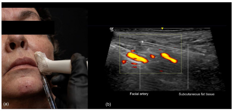
- Confirm the placement. Under ultrasound, cannula filler held its intended plane in about 90% of 100 injections at day 0, day 14 and day 30, while the product area went from 22.51 to 12.9 mm² (Calomeni 2023). Layering the volume-deficient folds of 20 patients deep then superficial with real-time depth confirmation, Hong 2026 reported no vascular compromise or malposition, a median GAIS of 2 at 12 weeks and rescans showing even spread in the target planes.
- Ripeta la scansione. Bring the probe back after each pass to confirm the cannula sits outside the vessels and in the planned plane (Sigrist 2024), then close with the perfusion check of Rocha’s third step (Rocha 2022).
Fresh hyaluronic acid has a recognizable look at each frequency. At 70 MHz, HA in the fold showed as several anechoic oval areas in the dermis, which thickened from 2.04 to 2.28 mm, 11%, and still measured 2.19 mm at 24 weeks (Salvia 2023). At 20 MHz it formed well-demarcated hypoechoic or anechoic areas in the subcutis, one product spreading as many small bubbles and the other as larger clumps (Qiao 2019). At 15 MHz it read anechoic, with a distinct boundary, good through-transmission and no flow (Jiang 2022, as summarized in Salvia 2023). Earlier filler belongs on the pre-map as well: in 94 patients scanned at the nasolabial groove the material was HA in 35, polyacrylamide in 34, fat in 10, silicone oil in 7, calcium hydroxylapatite in 6 and a silicone implant in 2, told apart by echo intensity, texture, boundary, fluidity and flow (Jiang 2022), and a blinded sonographer found the filler at the injection site in all 36 subjects after lip or fold filler (Grippaudo 2010). HA sitting where new product is planned can be dissolved first, as set out in the guide to dissolving HA filler under ultrasound.
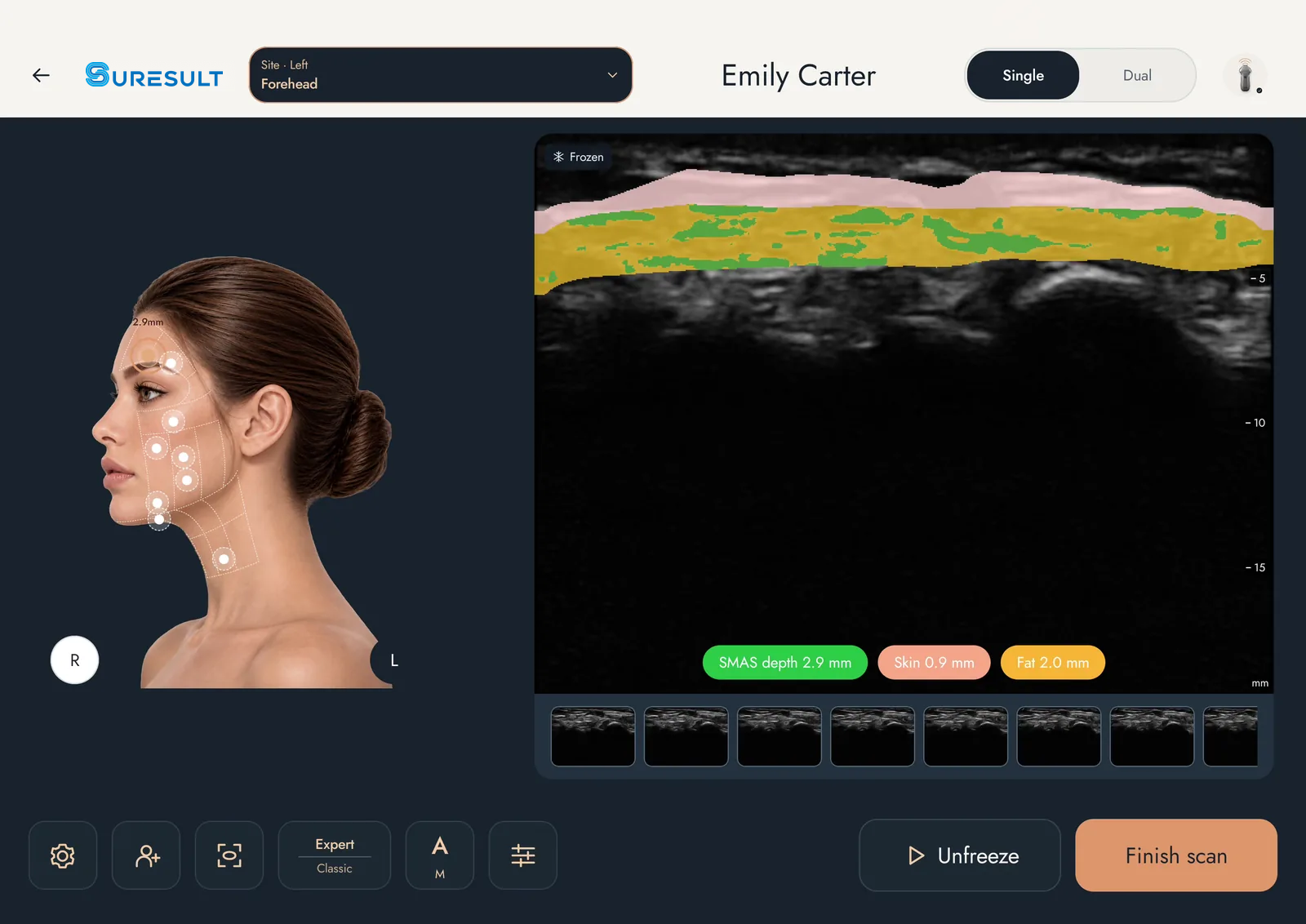
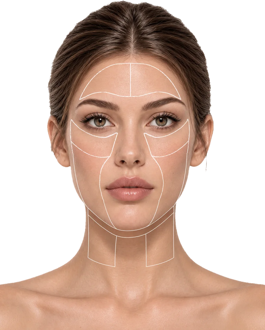
FaceAtlas AI turns the map into a record on all six Suresult linears. Color Power Doppler (CPD) shows blood-flow signals on the live scan before the needle goes in; a freeze gives skin thickness, subcutaneous fat thickness and depth to the SMAS in millimeters, and a 5-second clip keeps the motion. Filler Scan names the material already in the tissue and measures its depth and size, for example HA at 2.6 mm deep over 20.7 mm², across seven materials: HA, collagen, PLLA, PCL, CaHA, PMMA and PVA, and the filler assessment adds abnormality screening and complication evaluation. The 21-point map runs from forehead to jawline with two nasolabial fold points and two perioral points, one of each per side, so every follow-up rescans the same spot. Dual View sets the raw scan beside the AI layers while the filler goes in, one click puts this visit beside the last, and each visit exports as a US Letter PDF.
Doppler settings for the facial and angular arteries
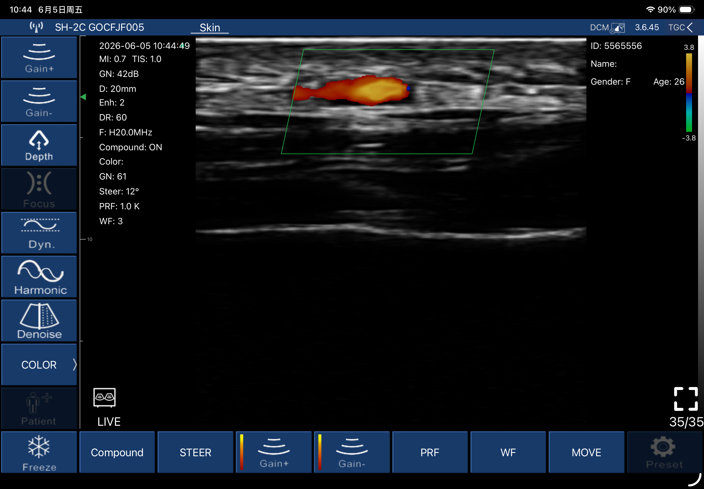
Small facial arteries need slow-flow settings: a velocity scale near 3 cm/s with the PRF and the wall filter brought down to match, plenty of gel and almost no probe pressure (Sigrist 2026). Conventional wall filters hide slow flow in small vessels (Pistoia 2023), which is why the filter comes down with the scale. The table gives each step for the fold and the fossa; what each Doppler mode shows is explained in the portable Doppler ultrasound guide.
| Step or setting | Value at the fold and the fossa | Fonte |
|---|---|---|
| Face position | At rest; a smile stopped angular vein flow within 3 seconds in 80.8% of observations | Cotofana 2020 |
| Accoppiamento | Abundant gel and no compression | Pistoia 2023; Bustamante 2025; Sigrist 2026 |
| Modalità | Color Doppler in every study; pulsed (spectral) Doppler for mapping | Chammas 2025 |
| Fold sweep | Oblique plane along the fold, from the ala to the mouth corner | Chammas 2025 |
| Fossa sweep | Longitudinal between the ala and the anterior maxilla, marker perpendicular to the ala; or vertical at the angle between the ala and the cheek | Bustamante 2025; Franceschelli 2024 |
| Probe to vessel | Perpendicular; each cross-section marked on the skin and the marks joined into a line | Sigrist 2024 |
| Box colore | Steered along the vessel | Sigrist 2024 |
| Scala delle velocità | About 3 cm/s for small arteries, with PRF and wall filter matched | Sigrist 2026 |
| PRF at the fossa | 0.5–0.8 kHz, Doppler at 5.0 MHz | Bustamante 2025 |
| Wall filter | Matched to the low scale; conventional filters hide slow flow in small vessels | Sigrist 2026; Pistoia 2023 |
| Arteria o vena | A pulsatile spectral trace marks the artery; flow that stops completely when the probe presses on the vessel is a vein | Bustamante 2025; Lee W 2023 |
Every Suresult linear carries these controls on its own Doppler: PRF, wall filter, steer, color gain and Move & Zoom in Color and PDI, and PW gain, steer, sample volume and angle in PW, with peak systolic velocity, end-diastolic velocity, resistive index and vessel diameter measured on the trace. For coupling, the Cuscinetti in gel per ecografia estetica Suresult are clear, sterile hydrogel pads of Ø35 × 4 mm, each in its own sealed pack, latex-free and ISO 10993 biocompatibility-tested, with a 5-year shelf life. One pad stays on from the Doppler map to the last pass, the needle goes straight through it, it repositions on the skin, and it fits all six Suresult linears and other brands’ linear probes, at $140 for 50 with shipping and tax included. The 4 mm stand-off uses 4 mm of display depth, so the L20Pro’s 10 mm preset shows about 6 mm of tissue under a pad. Sterile coupling for the in-plane probe is set out in the sterile coupling section of the filler injections guide.
What frequency and depth the fold and the fossa need
The fold and the fossa need a linear probe of at least 15 MHz, the floor in the 2025 Delphi consensus and in the WFUMB position paper, which puts axial resolution at 100 µm at 15 MHz (Velthuis 2025; Chammas 2025). Above the floor, frequency trades depth for detail: 20 MHz reaches about 10 mm and 10 MHz about 35 mm (Mlosek 2020), 18–22 MHz shows vessels of about 0.1 mm (Jaguś 2020), and Veronese 2024 names 24 MHz the method of choice for reading the facial dermis and hypodermis in vivo. The table matches each target to the band and the preset that hold it, using the deepest reading of each target as its limit.
| Struttura di interesse | Depth from the skin | Banda | La specifica decisiva | Fonte |
|---|---|---|---|---|
| Skin over the fold | 1.6 mm; dermis 1.3 mm | 24 or 20 MHz | 10 mm preset puts the skin in the top sixth of the image; 24 MHz on the L24Pro gives the finest near field | Jeong 2023 |
| Angular artery over the fossa | 3.6 mm mean; 4.8–5.8 mm in the second series | 20 or 24 MHz | 10 mm preset holds both series on gel alone; with a 4 mm gel pad the deepest reading sits at 9.8 mm, inside the L24Pro’s 15 mm or the L20Pro’s 20 mm preset; Power and PW tell it from the vein | Franceschelli 2024; Bustamante 2025 |
| Facial artery, alar base to commissure | 7.95–8.04 mm pooled means; 4.3–6.3 mm in the 22–8 MHz series | 20 MHz | 10 mm preset holds every mean on gel alone, 12.0 mm with the pad on, inside the L24Pro’s 15 mm or the L20Pro’s 20 mm preset; Color, Power and PW on the same probe | Pistoia 2023; Pourani 2025 |
| Facial artery at the mandibular border | 6.27 mm pooled mean; 6.4–6.8 mm in the 22–8 MHz series | 20 or 24 MHz | 10 mm preset; 10.8 mm with the pad on, held by the L24Pro’s 15 mm or the L20Pro’s 20 mm preset | Pourani 2025; Pistoia 2023 |
| Bone under the deep pyriform space | 9.11–11.4 mm means | 16 to 20 MHz | L20Pro 20 mm preset, 15.4 mm with the pad on; L24Pro 15 mm preset without the pad, 20 mm with it | Franceschelli 2024; Bustamante 2025 |
| Deepest facial artery course | 18.7 mm at the cheilion; 23.5 mm at the ala | 16 MHz | L20Pro 20 mm and 30 mm presets | Ten 2021 |
That makes the L20Pro the one probe for the whole fold: 20 MHz at 10 mm for the dermis and both arteries, and 16 MHz from 20 to 40 mm for the bone, the deepest arterial course and the deep cheek, with Color, Power and PW Doppler, Needle Enhance, in-plane and out-of-plane guides and a 25 mm field. The L24Pro adds the finest near field in the line, 24 MHz on 256 elements, for the dermis over the fold and the angular artery at 3.6 mm, and images to 25 mm. The L14Pro and L14 run at 10/14 MHz with presets to 80 mm for body work; for the fold and the fossa, the 15 MHz floor points to the 16/20 and 18/24 MHz bands. The 15, 20 and 24 MHz trade-offs are compared in the guida agli ecografi ad alta frequenza, and the three Suresult linear bands head to head in L14 vs L20 vs L24 for aesthetic ultrasound.
The 6 best scanners for piriform fossa and nasolabial fold filler, ranked
For piriform fossa and nasolabial fold filler the Suresult L20Pro ranks first at 95.0/100, ahead of the Clarius L20 HD3 at 75.0, the Mindray TE Air e5M at 58.5, the GE Vscan Air SL at 48.4, the Butterfly iQ3 at 45.7 and the Philips Lumify L12-4 at 43.6. I scored each card on the seven criteria set out under How I scored, with the rival prices read on October 31, 2026.

01 Suresult L20Pro — Best Overall for Piriform Fossa and Nasolabial Fold Filler
16/20 MHz linear on 256 elements · 10/20/30/40 mm presets · Color, Power and PW Doppler in the price · Needle Enhance plus in-plane and out-of-plane guides · $3,720
Punteggio: 95,0/100
4,400 mAh, up to 4 hours a charge · 145 g · IP67, drop-tested · no subscription for imaging or Doppler · $3,720
See the Suresult L20Pro high-frequency probe: $3,720
Verdetto: Buy the L20Pro if you fill both the fold and the fossa. I rank it first because one probe maps the angular artery and the facial artery along the whole fold at 20 MHz, switches to 16 MHz for the bone under the deep pyriform space, carries all three Doppler modes and Needle Enhance in its price, and keeps each map for the rescan.
- Both arteries: 20 MHz on the 10 mm preset holds the angular artery at 3.6 mm and the facial artery’s pooled mean depths of 6.27 to 8.04 mm; a PW trace tells the 1.4 mm angular artery from its vein
- Depth range: 16 MHz on the 20, 30 and 40 mm presets reaches the bone at 9.11 to 11.4 mm, the facial artery’s deepest course at 23.5 mm and the deep cheek
- Needle and cannula: Needle Enhance, Flip, Grid and the in-plane and out-of-plane puncture guides across a 25 mm linear face
- Documentazione: cine of up to 1,000 frames, about 165 seconds; local storage; JPEG, PNG, MP4 and DICOM export; apps for iOS, Android and Windows
- Mapping software: FaceAtlas AI carries two nasolabial fold points and two perioral points in its 21-point map, reads skin, fat and SMAS depth in millimeters, shows blood flow on Color Power Doppler before the needle goes in, and names HA among seven filler materials with its depth and area

The FaceAtlas AI facial map with its zones outlined from forehead to jawline. - FaceAtlas AI license: the L14Pro, L20Pro and L24Pro each include 3 months, counted from the day you press Start; the L14, L20 and L24 run it on a key at $50 per month, $500 per year or $999 for the life of the device, and the lifetime key is sold until 11:59 PM PT, December 31, 2026
Perché occupa questa posizione. The L20Pro takes the maximum on six of the seven criteria: 20 MHz for resolution, the 40 mm preset for depth, all three Doppler modes in the purchase, FaceAtlas AI mapping in the 3 included months, Needle Enhance with the guides, and 1,000 frames of cine with DICOM. Its five-year total, $4,719 with the lifetime key, sold until 11:59 PM PT, December 31, 2026, scores 9.975 of 15 against the lowest total I scored, the Suresult L14 at $3,138.
Sub-scores: resolution 20/20, depth 10/10, Doppler 25/25, mapping software 15/15, needle and cannula 10/10, records 5/5, five-year cost 9.975/15.
The L20Pro and its five Suresult siblings are la clearance 510(k) K211321.
L20Pro, L24Pro o L14Pro? All three Pros run on 256 elements with Color, Power and PW Doppler, Needle Enhance, a 4-hour battery and 3 months of FaceAtlas AI, so the band decides. The L20Pro covers the fold and the fossa from the dermis to the bone. The Suresult L24Pro handheld ultrasound ($4,185) adds 24 MHz, the finest near field in the line, for the 1.3 mm dermis over the fold and the angular artery at 3.6 mm, with presets to 25 mm. The Sonda lineare Suresult L14Pro ($3,441) runs 10/14 MHz to 80 mm for the body, and the 15 MHz Delphi minimum hands the face to the other two.
Ideale per: injectors who map the facial and angular arteries, fill the fold and the fossa and rescan on one probe from the dermis to the bone. Scelga un modello della famiglia se: you want the 24 MHz near field on 192 elements at $3,441 (the L24), or the 16/20 MHz band at $3,000 with FaceAtlas AI on a key (the L20). Confronti con attenzione se: you want a preset named Pyriform and an on-screen depth of the shallowest vessel on a membership (the Clarius L20 HD3).
Altri modelli della famiglia
Suresult L24Pro 18/24 MHz linear ultrasound · $4,185 · 92.4/100. The superficial pick: 24 MHz on 256 elements for the dermis over the fold and the angular artery 3.6 mm down, presets from 10 to 25 mm, the full Pro kit and 3 months of FaceAtlas AI.
Sonda ecografica lineare Suresult L20 · $3,000 · 89.3/100. Same 16/20 MHz band and 10–40 mm presets as the L20Pro, on 192 elements, with Color, Power and PW Doppler, Needle Enhance, a 2-hour battery and FaceAtlas AI on a key.
Suresult L24 high-frequency probe · $3,441 · 86.4/100. 18/24 MHz to 25 mm on 192 elements with all three Doppler modes and Needle Enhance, a 2-hour battery and FaceAtlas AI on a key.
Suresult L14Pro linear ultrasound · $3,441 · 83.6/100. Scored for the record: 10/14 MHz with presets to 80 mm and the full Pro kit for body work, while the fold and the fossa take the 16/20 or 18/24 MHz band.
Suresult L14 wireless ultrasound probe · $2,139 · 75.5/100. Scored for the record and as the cost reference: $3,138 over five years with a lifetime key, sold until 11:59 PM PT, December 31, 2026. It reaches 80 mm at 10/14 MHz with all three Doppler modes and the puncture guides.
Suresult Ø35 × 4 mm gel pads for aesthetic scans · FaceAtlas AI · Ecografi palmari Suresult per la medicina estetica
02 Clarius L20 HD3 — Best for a Pyriform Preset and Vessel Depth AI at 20 MHz
8–20 MHz linear · 4 cm image depth · 25 mm field of view · Pyriform, Lips and Cheek presets · Color and Power Doppler included, PW with Essentials · $5,395 plus Essentials
Score: 75.0/100
290 g · about 60 minutes of scanning and 90 minutes to charge · 3-year standard warranty · $5,395
Verdetto: Buy the L20 HD3 if you want a 20 MHz wireless linear whose maker has built its aesthetics tools around the midface: a preset named Pyriform, Vessel Depth AI that prints the depth of the shallowest vessel on Power Doppler and CFI, and classroom videos on pyriform fossa mapping, with an Essentials membership covering PW Doppler, Needle Enhance, local storage and DICOM.
- Presets: Aesthetics, Cheek, Chin/Jaw, Dermatology, Lips, Pyriform and Vascular, on the scanner
- Vessel tools: Color and Power Doppler with the scanner; PW Doppler and Vessel Depth AI, which shows vessel depth in real time on Power Doppler and CFI, with Essentials
- Measurement and AI: Auto Aesthetic Calipers for skin, fat and tissue thickness; T-Mode AI and the Aesthetics Protocol on the membership
- Needle tools and records: Needle Enhance with Essentials; Center Guide for injections; 30-second cine; local storage and DICOM on the membership, with MP4 export
- Prezzo e piano: $5,395, checked October 31, 2026; Essentials yearly at $695, per 3 years at $1,785 or once at $2,500; Clarius Care at $265, $530 or $795 for 1, 2 or 3 years
Perché occupa questa posizione. 20 MHz takes the full resolution credit and the 4 cm image the full depth credit. Color and Power come with the scanner and PW with Essentials, for 21 of the 25 Doppler points; Vessel Depth AI earns half the map credit on the membership and the calipers the full layer credit, with no tool that names the material; Needle Enhance on Essentials plus the Center Guide earn 7.5 of 10; and $7,895 over five years with one-time Essentials is the second-highest total in the ranking.
Sub-scores: resolution 20/20, depth 10/10, Doppler 21/25, mapping software 7.5/15, needle and cannula 7.5/10, records 3/5, five-year cost 5.962/15.
Limiti. PW Doppler, Vessel Depth AI, Needle Enhance, local storage and DICOM all sit in Essentials; cine runs 30 seconds; no tool names the filler already in the tissue.
Ideale per: injectors who want a Pyriform preset, a live depth readout of the shallowest vessel and the maker’s own pyriform and angular-artery teaching videos, and who will hold Essentials. Scelga altro se: you want PW Doppler, needle enhancement and DICOM in the purchase price, or software that names the filler already in the fold.
Altri modelli della famiglia
Clarius L15 HD3 · $3,795 plus Essentials, checked October 31, 2026 · 64.0/100. A 5–15 MHz linear with a 50 mm field and images to 7 cm on the same membership tiers; five years with one-time Essentials come to $6,295.
Clarius aesthetics · L20 HD3 at the Clarius store · Clarius prices and Essentials costs in 2026
03 Mindray TE Air e5M — Best for Color, Power and PW Doppler at the Lowest Five-Year Rival Cost
2-in-1 convex and linear transducer · linear B-mode bands to 8.2 MHz in a 2.0–9.0 MHz bandwidth · images to 40 cm · Color, Power and PW Doppler standard · iNeedle and VA Grid · $4,499
Score: 58.5/100
229 g · 90 to 120 minutes of B-mode scanning · IP68 · 3-year warranty · $4,499
Verdetto: Buy the TE Air e5M if you want Color, Power and PW Doppler, a needle-enhancement mode and a vascular access grid in one purchase with nothing to renew, on a 2-in-1 transducer that adds a convex mode for whole-body imaging. Its linear B-mode bands top out at 8.2 MHz, under the 15 MHz minimum the 2025 Delphi panel sets for facial injectables.
- Doppler: Color, Power and PW standard, Color and PW at 4.0, 4.4 and 5.0 MHz on the linear; Color to 146 cm/s, Power on 30 scale levels, a PW scale from 1.0 cm/s and a 0.5–20 mm sample volume
- Strumenti per l’ago: iNeedle needle-visualization enhancement, left or right, and the VA Grid for vascular access, both specific to the e5M
- Software: Smart Calc for automatic vessel diameter and area, iTouch+ image optimization, iWorks protocols and iScanHelper tutorials; Small Organ, Vascular, Musculo-skeletal and Nerve applications on the linear, among 23 exam modes
- Documentazione: cine up to 125 frames, preset from 2 to 5 seconds; JPG and MP4 export; DICOM 3.0; patient data kept on board, with no cloud and no subscription
- Prezzo e clearance: $4,499 at the Mindray North America store, checked October 31, 2026, with no subscription plan; the Air Capsule for wireless charging costs $750; the FDA cleared the e5M transducer with iNeedle, Smart Calc, iWorks and iScanHelper under 510(k) K240906 on July 23, 2024
Perché occupa questa posizione. All three Doppler modes in the purchase take the full 25 points, iNeedle with the VA Grid the full needle credit, DICOM with onboard storage 3 records points, and $4,499, the lowest five-year total among the rivals, 10.462 of 15. The resolution rule starts counting at 10 MHz, so linear bands that end at 8.2 MHz score 0, and Smart Calc measures vessel diameter and area, which the software criterion does not credit.
Sub-scores: resolution 0/20, depth 10/10, Doppler 25/25, mapping software 0/15, needle and cannula 10/10, records 3/5, five-year cost 10.462/15.
Limiti. The linear B-mode bands run from 2.0 to 8.2 MHz, under the 15 MHz floor for facial work; cine holds 2 to 5 seconds; Mindray names no layer-depth, injection-point or filler-material tool.
Ideale per: whole-body practices that want all three Doppler modes and the needle tools paid once, with no plan to renew. Scelga altro se: you image the dermis over the fold or the 3.6 mm angular artery at 15 MHz or more, or want layer depths and filler mapping in the software.
TE Air e5M sul sito Mindray North America · Negozio Mindray TE Air
04 GE Vscan Air SL — Best for Color and PW Doppler With Every Scored Tool in the Price
3–12 MHz linear centered at 7.7 MHz · images to 8 cm · 40 × 7 mm lens on 192 elements · Color and PW Doppler in the purchase · Centerline Marker · $4,999
Score: 48.4/100
218 g · $4,999 · up to 50 minutes of continuous scanning · 3-year standard warranty
Verdetto: Buy the Vscan Air SL if Color and PW Doppler and a centerline marker in one $4,999 purchase matter more to you than a 15 MHz band, with nothing more to buy for any scored tool.
- Imaging e Doppler: a linear head from 3 to 12 MHz, centered at 7.7 MHz, to 8 cm, with B, Color, B+Color, Harmonics, M and PW modes
- Strumenti per l’ago: the Centerline Marker in the Vscan Air app; free-hand needle and catheter placement among the linear head’s interventional indications
- Documentazione: up to 500 exams on the phone or tablet; DICOM image-server export
- Prezzo e piano: $4,999.00, or $4,299.00 as a demo unit, checked October 31, 2026; the optional Innovations plan, $500 a year or $1,299 for 3 years, adds Caption AI, AutoEF, AutoAS, Lung Guidance, HD-SRI and Bladder Volume outside the score
Perché occupa questa posizione. 12 MHz earns 4 of the 20 resolution points and 8 cm the full depth credit. Color and PW in the purchase give 17 Doppler points, with Power Doppler absent from GE’s linear mode list; the Centerline Marker takes the guide credit, GE names no needle enhancement, and $4,999 with every scored tool included scores 9.416 for cost.
Sub-scores: resolution 4/20, depth 10/10, Doppler 17/25, mapping software 0/15, needle and cannula 5/10, records 3/5, five-year cost 9.416/15.
Limiti. The linear tops out at 12 MHz, under the 15 MHz floor; GE lists no Power Doppler for it; the needle aid is a centerline overlay with no enhancement mode.
Ideale per: practices that need the arterial map in Color and PW Doppler from a single purchase. Scelga altro se: you read the dermis over the fold, or want Power Doppler and needle enhancement for the 1.4 mm angular artery.
Vscan Air SL at GE HealthCare · Negozio statunitense GE HealthCare
05 Butterfly iQ3 — Best for One Whole-Body Probe With Biplane Needle Views
One CMUT probe, 1–12 MHz, about 9,000 elements · Vascular: Superficial preset from 1 to 4 cm with the Midline Tool · Color on the Core plan; Power, PW, Needle Viz and Biplane on Advanced · $3,899 plus a plan
Score: 45.7/100
300 g · battery up to 2 hours · 3-year warranty with an optional 2 more · $3,899
Verdetto: Buy the iQ3 if one probe has to cover the whole body as well as the fold, you want Biplane and in-plane Needle Viz views, and you will run the Advanced plan for Power and PW Doppler.
- Facial vessel preset: Vascular: Superficial, from 1 to 4 cm, with the Midline Tool
- Prezzo e piano: $3,899 with a 3-year warranty, checked October 31, 2026; Color with Core at $299 a year; Power and PW with Advanced at $420 a year or $1,500 once for five years
- Strumenti per l’ago: Needle Viz for in-plane needles at 20 to 40 degrees and Biplane, both in Advanced; a midline marker
- Documentazione: unlimited cloud storage; Basic DICOM for PACS and worklists sold as an add-on
- Imaging: one 1–12 MHz probe for every preset, to 30 cm, with iQ Slice and iQ Fan in Advanced
Perché occupa questa posizione. 12 MHz earns 4 resolution points and the 30 cm reach the full depth credit. Each Doppler mode sits behind a plan and earns half, 12.5 of 25; Needle Viz on Advanced earns half the enhancement credit and the midline marker the guide credit; five years with Advanced bought once total $5,399.
Sub-scores: resolution 4/20, depth 10/10, Doppler 12.5/25, mapping software 0/15, needle and cannula 7.5/10, records 3/5, five-year cost 8.718/15.
Limiti. Its single probe stops at 12 MHz, short of the 15 MHz facial minimum; every Doppler mode is plan-gated, Color on Core and Power and PW on Advanced; DICOM costs extra.
Ideale per: mixed practices where the same probe moves from the abdomen to the face, with biplane and in-plane needle views on the Advanced plan. Scelga altro se: you need the dermis over the fold at 15 MHz or more, or Doppler without a plan.
06 Philips Lumify L12-4 — Best for Clinics Already Running Lumify Transducers
12–4 MHz linear · images to 12 cm · 34 mm footprint and 34.5 mm field of view · steerable Color with Fast Flow and Slow Flow, plus PW · center line marker · $10,307.95 outright
Score: 43.6/100
under 136 g, battery-free · up to 7.5 hours of continuous scanning · 5-year basic warranty through Philips, 90 days at PartsSource · $10,307.95
Verdetto: Buy the L12-4 if your clinic already runs Lumify on its phones or tablets and wants a linear with a Slow Flow color mode and PW Doppler, every software feature included in an outright purchase.
- Doppler: steerable Color in two modes, Fast Flow with a high color scale for arterial flow and Slow Flow with a low scale for venous flow, plus PW
- Strumenti per l’ago: un indicatore della linea centrale; utilizzabile per biopsie
- Documentazione: loops of 1 to 10 seconds captured prospectively; export by email or as DICOM to PACS, a shared network folder or a local directory
- Prezzo e piano: Philips sells through its sales team; PartsSource lists a new unit at $10,307.95 outright with a 90-day warranty, or $7,929.19 on exchange, checked October 31, 2026; the outright purchase opens all software and features
Perché occupa questa posizione. The 12 MHz top earns 4 resolution points and 12 cm the full depth credit; Color and PW in the purchase give 17 Doppler points, with no Power Doppler listed; the center line marker takes the guide credit; and $10,307.95, the highest five-year figure in the ranking, scores 4.566 for cost.
Sub-scores: resolution 4/20, depth 10/10, Doppler 17/25, mapping software 0/15, needle and cannula 5/10, records 3/5, five-year cost 4.566/15.
Limiti. The band ends at 12 MHz, under the 15 MHz floor; Power Doppler is absent from the L12-4’s mode list; the PartsSource unit carries 90 days of warranty, against the 5 years Philips gives on transducers bought from Philips.
Ideale per: Lumify owners adding a linear for superficial vessels with Fast Flow and Slow Flow color. Scelga altro se: you want 15 MHz or more for the fold, Power Doppler for the angular artery, or a linear under $10,000.
What each scanner costs over five years, software included
The Suresult L20Pro costs $4,719 across five years once a lifetime FaceAtlas AI key is added, the only purchase its scored tools need after the probe; that key is sold until 11:59 PM PT, December 31, 2026. On prices read October 31, 2026, the Mindray TE Air e5M totals $4,499, the GE Vscan Air SL $4,999, the Butterfly iQ3 $5,399 with five years of Advanced paid once, the Clarius L20 HD3 $7,895 with one-time Essentials and the Philips Lumify L12-4 $10,307.95 bought outright. Every Suresult total I give carries the lifetime key, because one payment covers the device for every year that follows.
| Palmare | Prezzo | What the scored tools add | Five-year total, one-time software | Five-year total, yearly software |
|---|---|---|---|---|
| Suresult L20Pro | $3,720 | A FaceAtlas AI key after the 3 included months; imaging, Color, Power and PW Doppler and Needle Enhance come with the probe | $4,719, lifetime key included | $6,220, five yearly keys at $500 |
| Clarius L20 HD3 | $5,395 | Essentials at $2,500 one-time unlocks PW Doppler, Vessel Depth AI, Needle Enhance, storage and DICOM | $7,895 | $8,870, paying $695 for each of the five years |
| Mindray TE Air e5M | $4,499 | All scored tools come standard: Color, Power and PW Doppler, iNeedle, the VA Grid and DICOM | $4,499 | $4,499, with no subscription plan |
| GE Vscan Air SL | $4,999 | The purchase already holds the Centerline Marker, Color and PW Doppler, DICOM and on-device storage | $4,999 | $4,999; Innovations at $500 a year is optional and sits outside the score |
| Butterfly iQ3 | $3,899 | Advanced at $1,500 for five years unlocks Power and PW Doppler, Needle Viz and Biplane; Color comes with Core or Advanced; Basic DICOM, credited under records, is sold separately outside the total | $5,399 | $5,999 on Advanced at $420 a year |
| Philips Lumify L12-4 | $10,307.95 | All software and features come with the outright purchase | $10,307.95 | $10,307.95, with no plan to renew |
For handhelds outside this ranking, the guida ai costi degli ecografi per medicina estetica runs the same five-year arithmetic, probe by probe and plan by plan.
Suresult prices are final, No Hidden Fees: every order ships within 24 hours worldwide with customs clearance and duties covered, and arrives with a 30-day worry-free trial from delivery and a 2-year warranty that extends to 3 or 5 years. FaceAtlas AI keys are one per device, sent by email, with no auto-renewal. Owners of an L14Pro, L20Pro or L24Pro claim their 3 included months on the Pagina di attivazione di FaceAtlas AI, and the months start only when Start is pressed. After them, and from the first day on the L14, L20 and L24, the key runs mensile, annuale o for life, with the lifetime key sold until 11:59 PM PT, December 31, 2026.
Come ho assegnato i punteggi
I scored each card out of 100 on seven criteria and weights fixed on October 31, 2026, before any rival had a score:
- Resolution (20): 20 × (top frequency in MHz − 10) ÷ 10, kept between 0 and 20, so 20 MHz and up earns 20, 15 MHz 10, 12 MHz 4 and 10 MHz or less 0.
- Depth (10): 10 × the deepest preset or image depth in mm ÷ 30, counted up to 30 mm, the plane that reaches the pyriform bone with room to spare.
- Doppler (25): Color 9, Power 8 and PW 8, each at full value when the purchase includes it, half when a paid tier unlocks it and zero when it is missing.
- Filler-mapping and safety software (15): filler materials named on the scan 5, a vessel or injection-point map 5 and layer depth in mm 5. Each counts in full when the purchase includes it, half when a paid tier or key unlocks it and zero when it is missing. FaceAtlas AI counts as included on the L14Pro, L20Pro and L24Pro, sold with 3 months of it, and as a paid tool on the L14, L20 and L24, where the key is bought separately; any tool its maker sells without a named tier counts as included.
- Needle and cannula guidance (10): 5 for needle enhancement and 5 for in-plane or out-of-plane guides, biplane views, or a centerline, midline or access-grid aid named for needle work, halved when the tool sits in a paid tier.
- Records (5): 2 for cine of 60 seconds or more and 3 for storage with DICOM export.
- Five-year cost (15): 15 × the cheapest five-year total of the 12 models I scored, divided by this card’s total. A Suresult total is the October 31, 2026 hardware price plus one lifetime FaceAtlas AI key (sold until 11:59 PM PT, December 31, 2026), and the L20’s uses the $3,000 printed on its page; the Clarius total carries one-time Essentials, the Butterfly total five years of Advanced, and the L14’s $3,138 is the cheapest and sets the scale.
| Scheda e posizione | Resolution (20) | Depth (10) | Doppler (25) | Mapping software (15) | Needle and cannula (10) | Documentazione (5) | Costo su cinque anni (15) | Totale (100) |
|---|---|---|---|---|---|---|---|---|
| 1 · Suresult L20Pro | 20 | 10 | 25 | 15 | 10 | 5 | 9.975 | 95.0 |
| 2 · Clarius L20 HD3 | 20 | 10 | 21 | 7.5 | 7.5 | 3 | 5.962 | 75.0 |
| 3 · Mindray TE Air e5M | 0 | 10 | 25 | 0 | 10 | 3 | 10.462 | 58.5 |
| 4 · GE Vscan Air SL | 4 | 10 | 17 | 0 | 5 | 3 | 9.416 | 48.4 |
| 5 · Butterfly iQ3 | 4 | 10 | 12.5 | 0 | 7.5 | 3 | 8.718 | 45.7 |
| 6 · Philips Lumify L12-4 | 4 | 10 | 17 | 0 | 5 | 3 | 4.566 | 43.6 |
Siblings are scored inside their maker’s card: the Suresult L24Pro 92.4, L20 89.3, L24 86.4, L14Pro 83.6 and L14 75.5, and the Clarius L15 HD3 64.0. The Suresult card leads with the L20Pro, which outscores the L24Pro on depth, 40 mm against 25 mm, and on five-year cost; the Clarius card leads with the L20 HD3, whose 20 MHz band outscores the L15 HD3’s 15 MHz.
I reran the order seven ways, as declared, and under fourteen alternative readings of the sources; the Suresult card stays first and the Clarius L20 HD3 second in every one. The table gives the declared run, the seven reruns and the three readings least favorable to the Suresult card, RC12 to RC14, where its lead is smallest: 6.01 points on the L20Pro and 3.45 on the L24Pro. Rival prices were read on October 31, 2026, and the rival plans and tool lists between October 28 and 31.
| Scenario | Scheda Suresult | Clarius L20 HD3 | Mindray TE Air e5M | GE Vscan Air SL | Butterfly iQ3 | Philips Lumify L12-4 |
|---|---|---|---|---|---|---|
| S0, scenario dichiarato | 94.97 | 74.96 | 58.46 | 48.42 | 45.72 | 43.57 |
| S1, no credit for mapping software | 79.97 | 67.46 | 58.46 | 48.42 | 45.72 | 43.57 |
| S2, cost criterion removed | 85.00 | 69.00 | 48.00 | 39.00 | 37.00 | 39.00 |
| S3, depth criterion removed | 84.97 | 64.96 | 48.46 | 38.42 | 35.72 | 33.57 |
| S4, resolution uncapped at 25 × (MHz − 10) ÷ 14 | 92.83 | 72.82 | 58.46 | 47.99 | 45.29 | 43.14 |
| S5, card scored on the L24Pro | 92.41 | 74.96 | 58.46 | 48.42 | 45.72 | 43.57 |
| S6, rival Doppler counted as included | 94.97 | 78.96 | 58.46 | 48.42 | 58.22 | 43.57 |
| S7, Mindray dropped | 94.97 | 74.96 | Left out of this run | 48.42 | 45.72 | 43.57 |
| RC12, FaceAtlas AI counted as paid on the Pros, map credit only for a vessel map, and S6 | 84.97 | 78.96 | 58.46 | 48.42 | 58.22 | 43.57 |
| RC13, RC12 with the card scored on the L24Pro | 82.41 | 78.96 | 58.46 | 48.42 | 58.22 | 43.57 |
| RC14, RC12 plus the VA Grid and Needle Viz left uncredited, and S4 | 82.83 | 76.82 | 53.46 | 47.99 | 55.29 | 43.14 |
Piriform fossa, nasolabial fold and angular artery questions
How much filler goes in the piriform fossa per side?
0.3 mL per side on the bone for a bone-deficient upper fold, placed by needle at the MD Codes point NL1 (de Maio 2021). The spaces beside it take more: 0.5 to 1 cc per perialar space through a 23G cannula (Yoon 2026) and 0.5 to 0.8 mL in the premaxillary space (Pilsl 2014), and Jung’s layered technique, which starts in the deep pyriform space, uses 1.0 to 1.5 mL per side across all its layers (Jung 2020).
What is the deep pyriform space, and what does filler there do?
A space on the bone beside the base of the nose, 1.1 × 0.9 cm in cadaver dissection, bounded medially by the depressor septi nasi and laterally and superficially by the deep medial cheek fat and the lip elevators, with the angular artery in the septum of its roof (Surek 2016). Filler there holds its place as volume rises and projects the tissue forward (Cotofana 2019), which suits the paranasal depression of a type 1 fold (Hong 2024).
Where is the piriform fossa for filler, and is it the same as the one in the throat?
Beside the base of the nose: the piriform fossa injectors fill is the paranasal hollow over the pyriform aperture beside the ala, where the deep pyriform space sits on the maxilla (Surek 2016). The throat’s piriform fossae, also called the piriform sinuses, are a different structure: they lie in the laryngopharynx on each side of the laryngeal inlet, in the part of the pharynx that runs from the upper border of the epiglottis to the lower border of the cricoid cartilage at C6 (Pazhaniappan 2025).
Which facial filler area carries the most vascular risk?
The nose, by published case count: of 164 published cases of skin necrosis and vision loss after filler, 44.5% followed nasal injection, 21% the glabella and 15% the nasolabial fold, third on the list, ahead of the forehead at 10% (Rauso 2020). For the fossa, the 2025 Delphi panel makes vessel mapping mandatory before injection, with the forehead, glabella, temple and nose (Velthuis 2025).
How deep is the facial artery at the nasolabial fold?
6 to 8 mm on Doppler in the pooled means, which deepen from the jaw to the ala: 6.27 mm at the mandibular border, 7.95 mm at the cheilion and 8.04 mm at the lateral ala, with the artery seen at 99.8 to 100% of those levels (Pourani 2025). One 22–8 MHz series found the reverse, 6.8 mm at the jaw and 4.3 mm at the alar base on the right (Pistoia 2023). Single faces run from 2.5 mm at its origin by the mandible to 23.5 mm at the nasal ala (Ten 2021).
Does the facial artery run medial to, across or lateral to the fold?
All three: across 610 Doppler-mapped arteries it ran medial to the fold in 46.2%, crossed it from medial to lateral in 22.5% and from lateral to medial in 18.5%, and ran lateral in 12.0% (Pourani 2025). In 80 folds scanned before filler the artery lay lateral to the fold in 31% (Lee W 2021).
Where does the angular artery run over the piriform fossa?
In the roof of the deep pyriform space, inside the septum between the space and the deep medial cheek fat (Surek 2016), a mean 3.6 mm under the skin and 5.1 mm above the bone on 20 MHz Doppler. Across 332 hemifaces it lay within 3 mm of the bone in 38.6%, 1 mm or less from it in 2.4% and practically on it in another 0.6%, and inside the space in 94.9% (Franceschelli 2024).
How much filler goes in each nasolabial fold?
0.3 to 1.0 mL per side by MD Codes, scaled to need: 0.3 mL on the bone at NL1 for bone deficiency, 0.3 mL at NL1 and 0.2 mL at NL2 for a mild to moderate fold, and 0.5, 0.3 and 0.2 mL at NL1 to NL3, 1.0 mL in all, for a severe one (de Maio 2021). Under Doppler, Lee W 2026 injected a mean of 0.98 mL on the left (0.5–2 mL) and 0.95 mL on the right (0.5–1.5 mL) across 33 patients.
What Doppler settings find the facial and angular arteries?
Slow-flow settings: the velocity scale near 3 cm/s, PRF and wall filter dropped to suit it, plenty of gel and next to no probe pressure (Sigrist 2026). At the fossa one series ran Doppler at 5.0 MHz with a PRF of 0.5 to 0.8 kHz and told the artery from the angular vein by its pulsatile spectral trace (Bustamante 2025); flow that stops entirely under probe pressure is venous (Lee W 2023).
What frequency does a scanner need for nasolabial fold and piriform mapping?
At least 15 MHz on a linear probe, with B-mode, Color and spectral Doppler mandatory and Power Doppler recommended, by the 2025 Delphi consensus, which also makes mapping mandatory at the pyriform fossa (Velthuis 2025). For these sites that means 20 MHz on a 10 mm preset for the angular artery at 3.6 mm and the facial artery’s 6 to 8 mm means, and 16 MHz on a 20 mm preset or deeper for the bone at 9.11 to 11.4 mm.
How long does vessel mapping add before a nasolabial fold injection?
Under 3 minutes: across 503 patients the map took 1 to 3 minutes, and at the fold it found an artery wider than 0.8 mm under 43 of 324 planned sites, 13.2% (Teixeira 2025).
Should you rescan after the bolus, and what does the rescan check?
Yes: after insertion the probe goes back to confirm the cannula sits outside the vessels and in the planned plane, and every pass is rechecked (Sigrist 2024); a perfusion check closes the session as the third step of Rocha’s method in 480 patients at 18 MHz (Rocha 2022).
Is the angular artery always present?
No. In 57 cadaver hemifaces it was absent in 26.3%, and in 22.8% it reached the medial canthus through a branch of the ophthalmic artery (Kim 2014). An 18 MHz color Doppler study of 43 people found it absent in 11.62% (Khorasanizadeh 2023), and on 20 MHz Doppler it lay outside the deep pyriform space in 17 of 332 hemifaces, 5.1% (Franceschelli 2024).
Which ultrasound scanner is best for piriform fossa and nasolabial fold filler?
I pick the Suresult L20Pro at $3,720, 95.0/100 in my ranking: 20 MHz for the angular artery and the facial artery along the fold, 16 MHz presets to 40 mm for the bone and the deep cheek, Color, Power and PW Doppler and Needle Enhance with the probe, and 3 months of FaceAtlas AI. The Clarius L20 HD3 follows at 75.0, with a Pyriform preset and its PW Doppler and Vessel Depth AI on the Essentials membership.
Fonti
- Pourani MR, Ebrahimzade M, Goudarzi E, et al. Valutazione delle variazioni del decorso, dei diametri e della profondità dell’arteria facciale mediante ecografia Doppler: revisione sistematica e meta-analisi. J Cosmet Dermatol. 2025;24(9):e70431. — doi.org, October 31, 2026
- Franceschelli A, Cazzulani M, Urso SU, et al. Studio ecografico in vivo dell’anatomia dell’arteria angolare: indicazioni pratiche per il trattamento dello spazio piriforme profondo. Aesthet Surg J Open Forum. 2024;6:ojae113. — doi.org, October 31, 2026
- Rauso R, Sesenna E, Fragola R, et al. Skin necrosis and vision loss or impairment after facial filler injection. J Craniofac Surg. 2020;31(8):2289–2293. — doi.org, October 31, 2026
- Velthuis PJ, Alfageme F, Cartier H, et al. Uso dell’imaging ecografico nei trattamenti estetici iniettivi: un consenso Delphi modificato. J Plast Reconstr Aesthet Surg. 2025;104:33–37. — doi.org, October 31, 2026
- Gonzalez C, Garzon H, Bravo C, Kroumpouzos G. Ultrasound imaging of high-risk facial areas for injectables: a practical guide for common scenarios. Aesthetic Plast Surg. 2026;50(14):5881–5891. — doi.org, October 31, 2026
- Teixeira DA. Una nuova tecnica per ridurre i tempi dell'iniezione ecoguidata di filler: uno studio retrospettivo su 3 anni. Aesthet Surg J Open Forum. 2025;7:ojaf061. — doi.org, October 31, 2026
- Yousif NJ, Gosain A, Matloub HS, et al. The nasolabial fold: an anatomic and histologic reappraisal. Plast Reconstr Surg. 1994;93(1):60–69. — pubmed.ncbi.nlm.nih.gov, October 31, 2026
- Barton FE, Gyimesi IM. Anatomy of the nasolabial fold. Plast Reconstr Surg. 1997;100(5):1276–1280. — pubmed.ncbi.nlm.nih.gov, October 31, 2026
- Jeong KM, Seo JY, Kim A, et al. Ultrasonographic analysis of facial skin thickness in relation to age, site, sex, and body mass index. Skin Res Technol. 2023;29(8):e13426. — doi.org, October 31, 2026
- Vasconcelos-Berg R, Desyatnikova S, Bonavia P, et al. Best practices for the use of high-frequency ultrasound to guide aesthetic filler injections—part 2: middle third of the face, nose, and tear troughs. Diagnostics (Basel). 2024;14(22):2544. — doi.org, October 31, 2026
- Rohrich RJ, Pessa JE. The fat compartments of the face: anatomy and clinical implications for cosmetic surgery. Plast Reconstr Surg. 2007;119(7):2219–2227. — doi.org, October 31, 2026
- Rohrich RJ, Pessa JE, Ristow B. The youthful cheek and the deep medial fat compartment. Plast Reconstr Surg. 2008;121(6):2107–2112. — doi.org, October 31, 2026
- Schenck TL, Koban KC, Schlattau A, et al. The functional anatomy of the superficial fat compartments of the face: a detailed imaging study. Plast Reconstr Surg. 2018;141(6):1351–1359. — doi.org, October 31, 2026
- Cotofana S, Gotkin RH, Frank K, et al. The functional anatomy of the deep facial fat compartments: a detailed imaging-based investigation. Plast Reconstr Surg. 2019;143(1):53–63. — doi.org, October 31, 2026
- Hong GW, Song S, Park SY, et al. Why do nasolabial folds appear? Exploring the anatomical perspectives and the role of thread-based interventions. Diagnostics (Basel). 2024;14(7):716. — doi.org, October 31, 2026
- Shaw RB, Kahn DM. Aging of the midface bony elements: a three-dimensional computed tomographic study. Plast Reconstr Surg. 2007;119(2):675–681. — pubmed.ncbi.nlm.nih.gov, October 31, 2026
- Mendelson B, Wong CH. Changes in the facial skeleton with aging: implications and clinical applications in facial rejuvenation. Aesthetic Plast Surg. 2012;36(4):753–760. — doi.org, October 31, 2026
- Boz U, Köse E. Ultrasonographic evaluation of facial artery in the nasolabial fold for gender differences and symmetry between hemifaces. J Ultrasound Med. 2024;43(6):1045–1051. — doi.org, October 31, 2026
- Hong GW, Kim SB, Koppert E, et al. Filler injection techniques and ultrasound observations: ultrasound-based classification of volume deficiency in nasolabial folds. Aesthetic Plast Surg. 2026;50(1):350–357. — doi.org, October 31, 2026
- Lee W, Kim JS, Moon HJ, Yang EJ. A safe Doppler ultrasound-guided method for nasolabial fold correction with hyaluronic acid filler. Aesthet Surg J. 2021;41(6):NP486–NP492. — doi.org, October 31, 2026
- Yang HM, Lee JG, Hu KS, et al. New anatomical insights on the course and branching patterns of the facial artery: clinical implications of injectable treatments to the nasolabial fold and nasojugal groove. Plast Reconstr Surg. 2014;133(5):1077–1082. — doi.org, October 31, 2026
- Koziej M, Trybus M, Hołda M, et al. Anatomical map of the facial artery for facial reconstruction and aesthetic procedures. Aesthet Surg J. 2019;39(11):1151–1162. — doi.org, October 31, 2026
- Peng T, Yu H, Hong WJ, et al. Three-dimensional location of the facial artery in relation to the nasolabial fold in Asian people: a cadaveric CTA study. Aesthetic Plast Surg. 2024;48(20):4077–4085. — doi.org, October 31, 2026
- Pistoia F, Camerino PL, Ioppi A, et al. High-resolution US of the facial vessels with new facial vein landmarks for reconstructive surgery and dermal injection. Eur Radiol Exp. 2023;7(1):49. — doi.org, October 31, 2026
- Ten B, Kara T, Kaya Tİ, et al. Evaluation of facial artery course variations and depth by Doppler ultrasonography. J Cosmet Dermatol. 2021;20(7):2247–2258. — doi.org, October 31, 2026
- Shen WW, Jiao CB, Ma JX, et al. Evaluation of facial artery course variations, diameters, and depth by Doppler ultrasonography. J Plast Reconstr Aesthet Surg. 2023;84:79–86. — doi.org, October 31, 2026
- Shetty PN, Mehta N, Romero JB, et al. High-resolution ultrasound mapping of the facial artery at the mandible and nasolabial fold to prevent filler-related complications. Plast Reconstr Surg Glob Open. 2024;12(Suppl 5):10–11. — doi.org, October 31, 2026
- Trzeciak M, Ostrowski P, Gładysz T, et al. The depth of the facial artery, meta-analysis. Aesthetic Plast Surg. 2025;49(13):3793–3802. — doi.org, October 31, 2026
- Surek CK, Vargo J, Lamb J. Spazio piriforme profondo: chiarimenti anatomici e implicazioni cliniche. Plast Reconstr Surg. 2016;138(1):59–64. — doi.org, October 31, 2026
- Bustamante FQF, Peyneau PD, Verner FS, et al. Depth and hemodynamics of the angular artery in the pyriform space: a cross-sectional study with Doppler ultrasonography. Imaging Sci Dent. 2025;55(3):310–317. — doi.org, October 31, 2026
- Kim YS, Choi DY, Gil YC, et al. The anatomical origin and course of the angular artery regarding its clinical implications. Dermatol Surg. 2014;40(10):1070–1076. — doi.org, October 31, 2026
- Khorasanizadeh F, Delazar S, Gheidari O, et al. Anatomic evaluation of the normal variants of the arteries of face using color Doppler ultrasonography: implications for facial aesthetic procedures. J Cosmet Dermatol. 2023;22(6):1844–1851. — doi.org, October 31, 2026
- Tucunduva MJ, Tucunduva-Neto R, Saieg M, et al. Mappatura vascolare del volto: studio ecografico in modalità B e Doppler. Med Oral Patol Oral Cir Bucal. 2016;21(2):e135–e141. — pubmed.ncbi.nlm.nih.gov, October 31, 2026
- Gombolevskiy V, Gelezhe P, Morozov S, et al. The course of the angular artery in the midface: implications for surgical and minimally invasive procedures. Aesthet Surg J. 2021;41(7):805–813. — doi.org, October 31, 2026
- Yoon SE, Choi JW, Nam YS, Kim IB. Trattamento mirato dello spazio perialare per migliorare il ringiovanimento dei solchi nasolabiali: studio su cadavere. J Cosmet Dermatol. 2026;25(2):e70698. — doi.org, October 31, 2026
- Pilsl U, Rosmarin W, Anderhuber F. The premaxillary space: a location for filler injection? Dermatol Surg. 2014;40(3):301–304. — doi.org, October 31, 2026
- de Maio M. MD Codes™: a methodological approach to facial aesthetic treatment with injectable hyaluronic acid fillers. Aesthetic Plast Surg. 2021;45(2):690–709. — doi.org, October 31, 2026
- Jung GS. Hyaluronic acid filler injection technique in multiple layers of the nasolabial fold. Plast Reconstr Surg Glob Open. 2020;8(12):e3318. — doi.org, October 31, 2026
- Lee W. Iniezione di filler di acido ialuronico guidata dall'ecografia Doppler. Arch Plast Surg. 2023;50(4):348–353. — doi.org, October 31, 2026
- Ramos HHA, Amaral VM, de Oliveira Afonso LP, et al. A combined treatment proposal for the empty deep pyriform space. Aesthetic Plast Surg. 2025;49(14):3951–3958. — doi.org, October 31, 2026
- U.S. Food and Drug Administration. Premarket approval P020023, Restylane (Q-Med Scandinavia, Inc.), with supplements. — accessdata.fda.gov, October 31, 2026
- U.S. Food and Drug Administration. Premarket approval P040024, Perlane, Restylane-L and Restylane Lyft (Medicis Aesthetics Holdings, Inc.; Galderma Laboratories, L.P.), with supplements. — accessdata.fda.gov, October 31, 2026
- U.S. Food and Drug Administration. Premarket approval P050047, Juvéderm 24HV, 30 and 30HV and Juvéderm Ultra XC (Inamed Corporation), with supplements. — accessdata.fda.gov, October 31, 2026
- U.S. Food and Drug Administration. Premarket approval P110033, Juvéderm Vollure XC (Allergan), with supplements. — accessdata.fda.gov, October 31, 2026
- U.S. Food and Drug Administration. Premarket approval P090016, Belotero Balance (Merz Pharmaceuticals, LLC), with supplements. — accessdata.fda.gov, October 31, 2026
- U.S. Food and Drug Administration. Premarket approval P140029, Restylane Refyne and Restylane Defyne (Q-Med AB; Galderma Laboratories, L.P.), with supplements. — accessdata.fda.gov, October 31, 2026
- U.S. Food and Drug Administration. Premarket approval P160042, Revanesse Ultra, now Revanesse Versa (Prollenium Medical Technologies Inc.), with supplements. — accessdata.fda.gov, October 31, 2026
- U.S. Food and Drug Administration. Premarket approval P170002, RHA (Teoxane S.A.), with supplements. — accessdata.fda.gov, October 31, 2026
- U.S. Food and Drug Administration. Premarket approval P240022, Evolysse (Symatese; Evolus, Inc.), with supplements. — accessdata.fda.gov, October 31, 2026
- U.S. Food and Drug Administration. Premarket approval P240008, saypha MagIQ (CROMA-PHARMA GmbH), with supplements. — accessdata.fda.gov, October 31, 2026
- Lee W, Ascher B, Yi K, et al. Study on safe filler injection techniques using cutting-edge ultrasound technology. J Cosmet Dermatol. 2026;25(7):e71046. — doi.org, October 31, 2026
- U.S. Food and Drug Administration. Premarket approval P050052, Radiesse (BioForm Medical, Inc.), with supplements. — accessdata.fda.gov, October 31, 2026
- Rocha PS, Guerra TA, Teixeira DA. Description of a safe Doppler ultrasound-guided technique for hyaluronic acid filler in the face—a method to avoid adverse vascular events. J Cosmet Dermatol. 2022;21(7):2783–2787. — doi.org, October 31, 2026
- Cotofana S, Lowry N, Devineni A, et al. Can smiling influence the blood flow in the facial vein?—An experimental study. J Cosmet Dermatol. 2020;19(2):321–327. — doi.org, October 31, 2026
- Sigrist R, Desyatnikova S, Chammas MC, Vasconcelos-Berg R. Best practices for the use of high-frequency ultrasound to guide aesthetic filler injections—part 1: upper third of the face. Diagnostics (Basel). 2024;14(16):1718. — doi.org, October 31, 2026
- Calomeni M, Bravo BSF, et al. Precision of soft-tissue filler injections: an ultrasound-based verification study. Aesthet Surg J. 2023;43(3):353–361. — doi.org, October 31, 2026
- Salvia G, Zerbinati N, Manzo Margiotta F, et al. Ultra-high-frequency ultrasound as an innovative imaging evaluation of hyaluronic acid filler in nasolabial folds. Diagnostics (Basel). 2023;13(17):2761. — doi.org, October 31, 2026
- Qiao J, Jia QN, Jin HZ, et al. Long-term follow-up of longevity and diffusion pattern of hyaluronic acid in nasolabial fold correction through high-frequency ultrasound. Plast Reconstr Surg. 2019;144(2):189e–196e. — doi.org, October 31, 2026
- Jiang L, Yuan L, Li Z, et al. High-frequency ultrasound of facial filler materials in the nasolabial groove. Aesthetic Plast Surg. 2022;46(6):2972–2978. — doi.org, October 31, 2026
- Grippaudo FR, Mattei M. High-frequency sonography of temporary and permanent dermal fillers. Skin Res Technol. 2010;16(3):265–269. — doi.org, October 31, 2026
- Sigrist RMS, Gonzalez C, et al. Doppler ultrasound findings in filler-related facial vascular adverse events: an international multicenter study. Diagnostics (Basel). 2026;16(11):1587. — doi.org, October 31, 2026
- Chammas MC, Sigrist R, Alfageme F, et al. Documento di posizione WFUMB: consenso sulle migliori pratiche nell’ecografia dermatologica estetica. Ultrasound Med Biol. 2025;51(11):2173–2193. — doi.org, October 31, 2026
- Mlosek RK, Migda B, Migda M, et al. High-frequency ultrasound in the 21st century. J Ultrason. 2020;20(83):e233–e241. — pmc.ncbi.nlm.nih.gov, October 31, 2026
- Jaguś D, Skrzypek E, Migda B, et al. Usefulness of Doppler sonography in aesthetic medicine. J Ultrason. 2020;20(83):e268–e272. — pmc.ncbi.nlm.nih.gov, October 31, 2026
- Veronese S, Costa E, Portuese A, et al. Histological analysis of the dermal and hypodermal layers of the face and correlation with high-frequency 24 MHz ultrasonography and elastosonography. Eur J Histochem. 2024;68(2):3912. — doi.org, October 31, 2026
- Pazhaniappan N. The pharynx. TeachMeAnatomy. Updated November 6, 2025. — teachmeanatomy.info, October 31, 2026
- Clarius Mobile Health. Clarius L20 HD3 handheld ultrasound scanner, store page with Essentials and Clarius Care plans. — store.clarius.com, October 31, 2026
- Clarius Mobile Health. Clarius for aesthetics. — clarius.com, October 31, 2026
- Clarius Mobile Health. Vessel Depth AI for aesthetics. — clarius.com, October 31, 2026
- Clarius Mobile Health. Center Guide. — clarius.com, October 31, 2026
- Clarius Mobile Health. Pyriform fossa mapping and injections. — clarius.com, October 31, 2026
- Clarius Mobile Health. Visualizing the angular artery in the nasolabial fold for fillers. — clarius.com, October 31, 2026
- Clarius Mobile Health. Clarius L15 HD3 handheld ultrasound scanner, store page. — store.clarius.com, October 31, 2026
- Mindray. TE Air e5M handheld ultrasound, product page and datasheet P/N 0002-08-40803 Rev 2.0. — mindray.com, October 31, 2026
- Mindray. Handheld ultrasound: the TE Air family. — mindray.com, October 31, 2026
- Mindray North America store. TE Air e5M. — mindrayna.my.site.com, October 31, 2026
- U.S. Food and Drug Administration. 510(k) K240906, TE Air Diagnostic Ultrasound System (Shenzhen Mindray Bio-Medical Electronics Co., Ltd.), July 23, 2024. — accessdata.fda.gov, October 31, 2026
- GE HealthCare. Vscan Air SL. — gehealthcare.com, October 31, 2026
- GE HealthCare store. Vscan Air SL. — gehealthcare.com, October 31, 2026
- GE HealthCare. Assistenza per Vscan Air: specifiche. — vscanair-support.gehealthcare.com, October 31, 2026
- GE HealthCare. Assistenza per Vscan Air: informazioni su Vscan Air. — vscanair-support.gehealthcare.com, October 31, 2026
- GE HealthCare. Assistenza per Vscan Air: configurazione. — vscanair-support.gehealthcare.com, October 31, 2026
- GE HealthCare. Vscan Air support: how long can I scan with Vscan Air? — vscanair-support.gehealthcare.com, October 31, 2026
- Butterfly Network. Specifiche di Butterfly iQ3. — butterflynetwork.com, October 31, 2026
- Butterfly Network. Pricing and memberships. — butterflynetwork.com, October 31, 2026
- Butterfly Network. Manuale d’uso di Butterfly iQ3, revisione I. — manual.butterflynetwork.com, October 31, 2026
- Philips. Lumify L12-4 linear array transducer. — usa.philips.com, October 31, 2026
- Philips. Lumify handheld ultrasound. — usa.philips.com, October 31, 2026
- Philips. Lumify product offering, frequently asked questions. — usa.philips.com, October 31, 2026
- Philips. Lumify for anesthesiology. — usa.philips.com, October 31, 2026
- PartsSource. Philips Lumify L12-4 transducer, part 453561734017. — partssource.com, October 31, 2026
- Suresult L20Pro. — suresultmed.com, October 31, 2026
- Suresult L24Pro. — suresultmed.com, October 31, 2026
- Suresult L14Pro. — suresultmed.com, October 31, 2026
- Suresult L24. — suresultmed.com, October 31, 2026
- Suresult L20. — suresultmed.com, October 31, 2026
- Suresult L14. — suresultmed.com, October 31, 2026
- FaceAtlas AI. — suresultmed.com, October 31, 2026
- FaceAtlas AI claim page. — suresultmed.com, October 31, 2026
- Licenza mensile FaceAtlas AI. — suresultmed.com, October 31, 2026
- Licenza annuale FaceAtlas AI. — suresultmed.com, October 31, 2026
- Licenza a vita FaceAtlas AI. — suresultmed.com, October 31, 2026
- Cuscinetti in gel Suresult per ecografia in medicina estetica. — suresultmed.com, October 31, 2026
- Certificazioni e conformità Suresult. — suresultmed.com, October 31, 2026
Which Suresult probe to order for the fold and the fossa
Order the L20Pro at $3,720 if you treat the fold and the fossa together: it is the one probe I would buy for both arteries and every plane down to the bone. If your work stays in the dermis and the first 25 mm, the L24Pro at $4,185 gives you 24 MHz, the highest frequency in the line. If you want the L20Pro’s band and presets on 192 elements with FaceAtlas AI on a key, the L20 at $3,000 covers the same planes. Each ships within 24 hours worldwide with duties and customs paid; the warranty runs 2 years and extends to 3 or 5, the worry-free trial runs 30 days from delivery, and the price shown at checkout is the price paid: No Hidden Fees.

