

Lip filler technique starts with the two labial arteries, and the handheld I pick to map them is the Suresult L24Pro at $4,185: 18/24 MHz with 10 and 15 mm presets, Color, Power and PW Doppler and Needle Enhance in the price. The superior and inferior labial arteries run between the orbicularis oris and the mucosa at most points (78.1% of sites in 193 dissected heads; Cotofana 2017), and their depth changes along the lip: at 24 MHz, Kim’s 2026 study measured the superior labial artery 2.54 mm from the vermilion border at the midline and 6.62 mm at the corner. Vessels turn up in every lip plane (Ostrowski 2026), and the 2025 Delphi consensus recommends mapping the lips before injecting. The workflow built on that map runs from a Color Doppler sweep of both arteries before the first pass, through a plane and an entry chosen against the map and small volumes threaded slowly, to a rescan before the patient leaves. This guide covers where each artery runs, the injection points over it, where each named technique puts the product, the mapping workflow with its Doppler settings, the frequency the lip needs, and the five handhelds ranked for the job.


Alcance de la evaluación: As an independent device reviewer with deep ultrasound knowledge, I compared linear frequency, Doppler modes, filler and vessel software, needle tools, records and five-year cost for the Suresult L24Pro, L20Pro, L14Pro, L24, L20 and L14, the Clarius L20 HD3 and L15 HD3, the GE Vscan Air SL, the Butterfly iQ3 and the Philips Lumify L12-4, against the published lip anatomy and mapping studies, reading maker pages, store listings and PubMed on October 29, 2026.
Quick answer: which handheld ultrasound should you buy for lip filler?
The Suresult L24Pro ($4,185) is the best handheld for lip filler in my five-maker comparison, at 94.1/100. Its 18/24 MHz band with 10 and 15 mm presets frames the lip from skin to mucosa with both labial arteries in view, where Kim’s 2026 study at 24 MHz placed them 1.91 to 6.62 mm from the vermilion border. Color, Power and PW Doppler come with the probe, so the map, the waveform check and the rescan run without a membership, and Needle Enhance with in-plane and out-of-plane guides keeps the needle visible as it passes the artery. FaceAtlas AI, with 3 months included, names prior filler among seven materials before you add more. Five years with the FaceAtlas AI lifetime license total $5,184.
The Clarius L20 HD3 ranks second at 66.8: 8–20 MHz to 4 cm with Lips on its application list, Color and Power Doppler with the scanner, and PW Doppler, Needle Enhance and Vessel Depth AI in the Clarius Essentials membership.
Lip filler ultrasound compared: five handhelds for labial artery mapping
Every handheld here images the lip with a linear array and has Color Doppler. What separates them for lip work is the top frequency, which decides how sharply the orbicularis, the mucosa and an artery about 1 mm across separate; which Doppler modes come with the purchase; whether the software reads prior filler or vessel depth; how the needle shows; what the records keep for the rescan; and the five-year total for the full kit.
| Escáner | Linear frequency · depth | Doppler | Filler and vessel software | Herramientas para la aguja | Cine and records | Peso · tiempo de exploración | Five-year cost and subscription | Ideal para |
|---|---|---|---|---|---|---|---|---|
 Suresult L24Pro · No. 1 · 94.1/100 | 18/24 MHz · 10, 15, 20 and 25 mm presets · 256 elements | Color, de potencia y pulsado, incluidos | FaceAtlas AI, 3 months included: Filler Scan names 7 materials with depth and size; 21-point facial map; Color Power Doppler on the live scan | Needle Enhance; in-plane and out-of-plane guides | Cine to 1,000 frames (about 165 s); storage on the device; DICOM; FaceAtlas AI PDF report and one-click visit comparison | 145 g · up to 4 h · cordless | $5,184 ($4,185 plus the FaceAtlas AI lifetime license) | Lip mapping at 24 MHz with all three Doppler modes in the price |
 Clarius L20 HD3 · No. 2 · 66.8/100 | 8–20 MHz · 4 cm · Lips among the listed applications | Color and Power, included; PW with Essentials | Vessel Depth AI reads the shallowest vessel’s depth live, with Essentials; Auto Aesthetic Calipers; T-Mode AI Aesthetics | Needle Enhance with Essentials; center guide | 30 s cine; local and cloud storage, DICOM and PDF reports with Essentials | 290 g · about 60 min · cordless | $7,895 ($5,395 scanner plus $2,500 one-time Essentials) | A Lips application at 20 MHz with live vessel depth |
| GE Vscan Air SL · No. 3 · 42.0/100 | Linear 3–12 MHz to 8 cm; sector head to 24 cm | Doppler color y Doppler pulsado, incluidos | Caption AI with cardiac and bladder tools, in the optional Innovations plan | Marcador de línea central | 500 exams on the device; DICOM; OB report | 218 g · about 50 min · cordless | $4,999, scored tools included without a subscription | Lip mapping plus cardiac and lung scans on one dual-head probe |
 Butterfly iQ3 · No. 4 · 35.3/100 | 1–12 MHz on one probe · “Vascular: Superficial” preset at 1–4 cm | Color con Core; potencia y pulsado con Advanced | Auto Bladder Volume and Auto B-Lines | Advanced plan: Needle Viz, Biplane and the Needle: Out-of-Plane preset; a midline marker | Clips saved to the cloud on Core; Basic DICOM quoted separately | 300 g · phone cable · up to 2 h | $5,399 ($3,899 probe plus the $1,500 Advanced plan for five years) | One whole-body probe with biplane needle views |
| Philips Lumify L12-4 · No. 5 · 33.1/100 | 12–4 MHz hasta 12 cm | PW and steerable color (Fast Flow and Slow Flow), included | All Lumify software in the purchase | Center line marker; biopsy capable | Loops of 1 to 10 s; on-device storage for hundreds of exams; DICOM export to PACS | Under 136 g · no battery, power drawn over the cable · up to 7.5 h | $10,307.95 outright at PartsSource, software included without a subscription; by quote through Philips | Practices already running Lumify |
Each five-year cost is the handheld plus the software every scored tool needs: one-time Essentials for Clarius, Advanced for five years for Butterfly, and for Suresult the FaceAtlas AI lifetime license that follows the 3 months included with the Pro models. Rival figures come from each maker’s US store and, for the Lumify, the PartsSource listing.
La mejor opción para cada uso: selección rápida
- Best overall for lip filler mapping: Suresult L24Pro, 94.1/100. The highest frequency of the five, 24 MHz, puts the lip layers and both labial arteries on one screen, and FaceAtlas AI Filler Scan names old product before you add more.
- Best for a named Lips application at 20 MHz: Clarius L20 HD3. Lips is on its application list, and Vessel Depth AI in Essentials reads the depth of the shallowest vessel live on the image.
- Best for clinics that also scan hearts and lungs: GE Vscan Air SL. Linear 3–12 MHz and a sector head in one wireless probe, with Color and PW Doppler in the $4,999 price.
- Best whole-body probe with biplane needle views: Butterfly iQ3. The Advanced plan adds Needle Viz, Biplane and an out-of-plane needle preset, and a superficial vascular preset covers small facial vessels.
- Best for practices already on Lumify: Philips Lumify L12-4. A battery-free linear that draws power from the phone or tablet, sold outright with all Lumify software.
Where to buy each handheld
- Suresult L24Pro: $4,185 with Color, Power and PW Doppler, Needle Enhance and 3 months of FaceAtlas AI. From month 4, FaceAtlas AI runs on a monthly license ($50), a yearly license ($500) or the lifetime license ($999), sold until 11:59 PM PT, December 31, 2026. Buy the Suresult L24Pro aesthetic ultrasound probe: $4,185
- Clarius L20 HD3: $5,395 at the Clarius store, with Essentials chosen at checkout: $2,500 once, $695 yearly or $1,785 for a three-year term. Comprar a Clarius
- GE Vscan Air SL: $4,999 in the GE HealthCare US shop, with a 3-year warranty. Comprar a GE HealthCare
- Butterfly iQ3: $3,899 with a 3-year warranty; Color Doppler comes with Core at $299 a year, and Power and PW Doppler and the needle tools with Advanced at $420 a year or $1,500 for five years. Comprar a Butterfly
- Philips Lumify L12-4: quoted by Philips Healthcare; PartsSource sells it outright at $10,307.95. See the Lumify L12-4 at Philips
Superior and inferior labial arteries: where they run and how deep
The superior labial artery (SLA) branches from the facial artery near the corner of the mouth and runs toward the midline, where it joins the artery from the other side; the inferior labial artery (ILA) supplies the lower lip, and its origin varies more. Pooled across studies, both lie 4.59 to 4.90 mm beneath the vermilion surface (Trzeciak 2026 meta-analysis). Lee’s 2015 cadaver study of 60 hemifaces placed the SLA origin 12.1 mm lateral to the mouth corner, inside a 1.5 cm square above and lateral to it, with the artery then following the vermilion border to the midline 3 mm deep; it was absent on 6.7% of sides. Pooling the literature in 2026, Ostrowski found the SLA absent in 3.55% of cases and the ILA in 13.45%, the SLA arising above the commissure in 80.71% of cases at a mean 11.95 mm, and the ILA below it in 72.44% at 21.99 mm.
| Estudio | Specimens | Plane or depth | Other findings |
|---|---|---|---|
| Cotofana 2017 | 193 heads, 6 sites each | Submucosal 78.1%, intramuscular 17.5%, subcutaneous 2.1% | The artery changed plane along its course in 29% of upper lips and 32% of lower lips; superficial positions were more frequent at the midline |
| Milla 2026 | 15 heads dissected; 276 heads pooled | Pooled SLA estimates: submucosal 0.71, intramuscular 0.22, subcutaneous 0.01; ILA: 0.65, 0.27 and 0.02 | A midline SLA anastomosis in every dissected specimen; the lower-lip midline ILA intramuscular in 11 of 15 |
| Nguyen 2023 | 102 facial arteries | SLA submucosal 78.65%, intramuscular 17.98%, subcutaneous 3.37% | The ILA ran along the lower vermilion border in 8.82% and followed the horizontal labiomental pattern in 69.61% |
| Swancutt 2025 | 48 hemifaces | ILA deep to the orbicularis in 90% | The mental artery supplied the lower lip in 96% |
| Money 2020 | 18 cadavers | SLA depth from the skin: 5.49 mm at its branch point, 4.29 mm midway between the commissure and the Cupid’s bow peak | SLA lumen 0.85 mm at the commissure, 0.56 mm at the midline |
| Lupu 2022 | 11 heads | SLA 4.8–5.6 mm deep; ILA 4.1–5.4 mm | Columellar and septal branches leave the SLA inside a fibromuscular compartment above the philtrum |
Across the series, the submucosal plane carries the artery at most sites, the intramuscular plane at 17.5% to 27% and the subcutaneous plane at 1% to 3.4% (Cotofana 2017; Milla 2026; Nguyen 2023), and superficial positions concentrate at the midline (Cotofana 2017; Milla 2026). In 8.4% of Loukas’s 284 hemifaces, the facial artery ended as the superior labial artery itself. Ostrowski’s review finds vessels in every lip plane and calls for anatomically guided technique in each patient.
| Estudio | Participants and frequency | Depth, with the surface it was measured from | Plane and position |
|---|---|---|---|
| Cotofana 2020b | 41 volunteers | SLA 5.6 mm deep in the upper lip; ILA 5.2 mm deep in the lower lip | Submucosal 58.5%, intramuscular 36.2%, subcutaneous 5.3%; inside the red lip in 83% (upper) and 86.2% (lower), and in every volunteer at the midline |
| Lee 2020 | 60 volunteers | SLA 5.3 mm and ILA 4.2 mm from the vermilion border | Wet mucosa in 35–57% (upper) and 28–55% (lower); SLA intramuscular in 20–45%; ILA in the dry mucosa in 5–27% |
| Kim 2026 | 25 women, 24 MHz | From the vermilion border: SLA 2.54 mm at the midline, 3.39 mm at the Cupid’s bow apex and 6.62 mm at the corner; ILA 1.91 mm at the midline, 3.40 mm at the apex and 5.78 mm at the corner | Superficial ILA branches and arteriole–venous shunts in the central lower lip |
| Kim 2024 (particle HA series) | One mapped patient (Fig. 2), 12 MHz | SLA 3.16 mm and ILA 3.44 mm from the dry mucosa | SLA under the pars marginalis of the orbicularis |
| Tansatit 2019 | 20 volunteers, 3–13 MHz | ILA 5.91 mm deep from the skin, measured at the lower vermilion or the labiomental sulcus; diameter 1.10 mm | Mainly the deep muscular plane; the authors name a 6 mm depth at the lower vermilion border as the dangerous injection plane |
Pooling 218 to 414 arteries per site, Trzeciak’s 2026 meta-analysis proposed an injection ceiling of 3.5 mm in most of the lip and 4 mm in the superior vermilion, with high-resolution ultrasound before extensive augmentation. Kim’s 24 MHz readings vary by point, so the depth that matters is the one measured at the point being injected. Cotofana’s team found the arteries inside the red lip at most sites and proposes a perpendicular approach from the cutaneous lip for that reason. In Gonzalez’s 27 patients scanned at 18 MHz, the artery ran in the wet submucosa in 22, the dry submucosa in 4 and the muscle in 1.
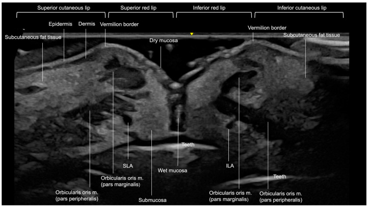
The layers the arteries cross are thin. In Harris’s 2026 study of 126 participants scanned at 18 or 20 MHz, the whole upper lip measured 8.36 mm and the lower lip 9.01 mm; the five layers in the table run 1.4 to 2.3 mm each, with intrarater agreement of 0.71 to 0.89 across the layers.
| Layer | Labio superior | Labio inferior |
|---|---|---|
| Subcutaneous layer | 1.47 | 1.65 |
| Pars marginalis of the orbicularis | 1.37 | 1.43 |
| Connective tissue between the muscle parts | 1.61 | 1.87 |
| Pars peripheralis of the orbicularis | 1.53 | 1.61 |
| Wet mucosa | 2.26 | 2.29 |
Lip filler injection points and the arteries beneath them
Lip filler injection points take their names from surface landmarks: the vermilion border and the Cupid’s bow, the philtral columns, the tubercles, the wet–dry junction, the commissures and the labiomental crease. Each one sits over a known stretch of the labial arteries, their branches or the lip’s veins.
| Injection point | What runs beneath it | Fuente |
|---|---|---|
| Upper vermilion border, lateral to the Cupid’s bow | The SLA, mostly under the orbicularis near the wet mucosa: under the muscle in 78%, inside it in 44%, above it in 8% at Kim’s entry point 1 (percentages overlap where several vessels share a point) | Kim 2024 (9-point study), 50 patients |
| Cupid’s bow apex | The SLA rises: inside the orbicularis in 82%, over it in 28%, under it in 48% | Kim 2024 (9-point study) |
| Upper central tubercle and the upper midline | Dry-mucosa branches and perforators in 80%; an arteriovenous shunt at the midline; the SLA 2.54 mm from the vermilion border | Kim 2024 (9-point study); Kim 2026 |
| Philtral columns and the philtrum | Columellar and septal branches of the SLA in a fibromuscular compartment above the philtrum; columellar arteries in the subcutaneous layer | Lupu 2022; Alizadeh 2024 |
| Lower central lip and between the lower tubercles | Superficial ILA branches and arteriole–venous shunts; the ILA 1.91 mm from the vermilion border at the midline; venous pooling between the tubercles | Kim 2026; Moorefield 2023 |
| Wet–dry junction | The junction where most of the lip’s vessels lie, with the labial arteries submucosal at most sites | Adel 2021; Cotofana 2017 |
| Commissures | The SLA origin 12.1 mm lateral to the corner, inside a 1.5 cm square; the SLA entering the lip within 0.5 cm of the commissure; veins 1 to 1.5 cm lateral to the commissures | Lee 2015; Bravo 2025; Moorefield 2023 |
| Labiomental crease | The horizontal labiomental pattern of the ILA in 69.61% of facial arteries; the mental artery supplying the lower lip in 96%; venous tributaries along the crease | Nguyen 2023; Swancutt 2025; Moorefield 2023 |
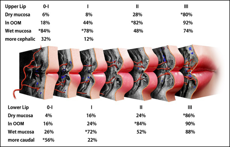
Kim’s 2024 9-point injection technique (9-PIT) ties every point to the artery map. Points 1 and 2 build the upper vermilion border from an entry 1 inch (one needle length) lateral to each Cupid’s bow apex, point 3 shapes the upper central tubercle, points 4 and 5 create new upper lateral tubercles near the wet–dry junction, points 6 and 7 fill the lower tubercles from an entry 1 mm below the vermilion border, and points 8 and 9 thread the lower lateral border from an entry 1 mm lateral to the commissure. In the 50 of 382 patients with sonography records, the SLA ran under the orbicularis at points 4 and 5 in every case; Kim rates points 4, 5, 8 and 9 as very safe and point 3, over the midline shunt, as the point most exposed to bruising. The standard volume was 1 mL of particle HA through a 29G 1-inch needle, and the corners of the mouth rose 1.02 mm.
Kim’s 2026 11-point technique (11-PIT) targets the Cupid’s bow, the vermilion tubercles, the philtral columns and the commissures, and measures each artery at 24 MHz before the first pass. In 25 women, Kim threaded HA slowly in the submucosa with a 29G 1-inch needle, 0.05 to 0.2 mL per point and the most at points 6 and 7 (0.2 mL each), for about 1 mL per patient. At one month, upper-lip thickness had risen from 8.94 to 10.98 mm and lower-lip thickness from 10.30 to 12.25 mm, with no major complications.
The lip’s veins follow their own map. Moorefield’s transillumination study of 26 participants found venous pooling about 2 mm above the Cupid’s bow, along the upper middle tubercle, along the lower wet–dry line and between the lower tubercles, with tributaries 1 to 1.5 cm lateral to the commissures and along the labiomental crease. On the scan, an artery and a vein both fill with color; pressing with the probe tells them apart (Lee 2023).
Lip filler techniques and where each one places the product
Each named technique puts product in its own plane: tenting (“Russian lips”) traces sit superficial to the orbicularis (Kichler 2026), the vertical cannula runs in the submucosa behind the artery (Bravo 2025), Kim’s 9-point and 11-point techniques thread in the submucosa, and Hong’s 2026 technique fills the deep labial fat. Across 16 lip studies and 3,692 patients, Miranda Cerna’s 2026 review counted linear retrograde threading in 12 protocols, serial puncture, cross-hatching or fanning in 4 and bolus or microdeposits in 2; most used 1 to 2 mL per session, and 9 of the 14 that named a tool used a needle alone. The plane decides how close the product comes to the artery.
| Technique | Entry and tool | Plane | Volume and series | Fuente |
|---|---|---|---|---|
| Tenting (“Russian lips”) | 30G needle at the contour, aimed vertically toward the wet–dry junction, repeated every 2–3 mm, skipping the philtral columns | Superficial to the orbicularis | 0.01–0.02 mL per trace; 10 patients. Alizadeh: 0.01 mL per pass, perpendicular to the border | Kichler 2026; Alizadeh 2024 |
| Inverted Mercedes Benz | 22G 50 mm microcannula through both Glogau-Klein points of the upper lip and the lower-lip midline | Superficial muscular plane, away from the wet–dry junction | Retrograde threads and aliquots; 10 women | Adel 2021 |
| Vertical submucosal cannula | 22G 40 mm cannula from the cutaneous lip: 0.5 cm above the vermilion border in line with the lateral border of each nasal wing for the upper lip, 1 cm below it at the midline for the lower lip | Submucosa below the wet–dry junction, behind the artery | 0.05–0.15 mL per compartment; 10 women scanned before, during and after | Bravo 2025 |
| 9-point (9-PIT) | 29G 1-inch needle; entries along the upper vermilion border, 1 mm below the lower border and 1 mm lateral to the commissure | Submucosal linear threads and fanning | 1 mL standard volume; 50 of 382 patients with sonography records | Kim 2024 (9-point study) |
| 11-point (11-PIT) | 29G 1-inch needle; points at the Cupid’s bow, the vermilion tubercles, the philtral columns and the commissures | Slow submucosal linear threading | About 1 mL at 0.05–0.2 mL per point; 25 women | Kim 2026 |
| Contour, then volume | Needle started 2–3 mm inside the commissure, or a cannula entered slightly outside the corner | The border in the dermis and subdermis first, then the submucosa at the dry–wet junction | 0.5–1 mL per lip, 1.5 mL at most | Hong 2025 |
| Deep labial fat | Entries placed away from the commissures; staged dosing | Deep labial fat, for eversion | 0.5–1.0 mL per lip per session | Hong 2026 |
| Multi Vector Lip | 30G ½-inch needle, oblique from the vermilion border | Subcutaneous lines; an intramuscular bolus at the tubercles | 0.02–0.025 mL per line, mean 1.06 mL; 253 patients | Menezes 2025 |
| No-touch | 30G needle 5 mm lateral to the commissure and at the base of the philtral columns; 25G cannula from the commissure | The white roll (needle); the submucosal space between the white and red rolls (cannula) | 410 patients | Surek 2016 |
| Bi-Bi | 27G cannula through each hemi-lip; 33G needle along the border and the Cupid’s bow | Intramuscular (cannula), intradermal (needle) | 30 patients | Stéphane 2022 |
“Russian lips” has a peer-reviewed definition: Adel describes it as tenting, vertical struts of filler along the whole lip that cross both the vermilion line and the wet–dry border, and lists vascular occlusion and spread onto the skin side among its risks. “Swedish lips” and “Korean lips” are style names that clinics use for a look. Peer-reviewed Korean lip protocols include Kim’s 9-point (2024) and 11-point (2026) techniques, both built on sonographic mapping, and Hong’s contour-then-volume sequence (2025) and deep labial fat technique (2026). The M-shaped lip has its own grading, by the angle between the lips’ vertical and horizontal midlines: mild at 15° or less, moderate at 16° to 25° and severe above 25° (Villalobos 2025, 9 patients).
Small volumes per site run through most of these protocols. The Φ (Phi) technique places 0.01 to 0.05 mL per site and caps a session at 1 to 1.5 mL, with repeat sessions 15 to 30 days apart (Keramidas 2021, 833 patients). Walker and Cetto (2021) use a 4-mm 30G needle for precise HA placement in the subcutaneous plane of the lip, and the short needle lowers the pressure needed to extrude it.
Cannula or needle changes the shape of the deposit and the force needed to enter an artery. Beyond the lips, the guide to cannula vs needle across the face gives the gauge, entry and plane for each area. In Gonzalez’s 18 MHz series, a 25G × 50 mm cannula through one entry per hemi-lip at the commissure left elongated deposits, and a 30G × 13 mm needle at the vermilion border left rounded ones. In penetration tests, a 27G cannula entered an artery with the same force as a 27G needle (0.78 vs 0.70 N; Pavicic 2019), Siperstein found every cannula gauge, 27G and 30G included, needed more force than the matching needle, and Ugradar’s 18G and 22G cannulas never penetrated the facial artery wall. Tansatit showed a cannula meeting the artery at a right angle perforating it. Both tools reach the same planes: Blandford found 93% (cannula) and 79% (needle) of HA aimed at the upper vermilion border inside the orbicularis, and Ghannam’s cadaver checks of experienced injectors’ lip techniques put product close to a labial artery in 58.3% of injections, 60.0% with a needle and 57.1% with a cannula.
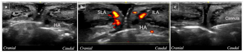
A negative aspirate leaves the artery’s position unknown. The global consensus tells injectors not to rely on it (Signorini 2016), and Goodman’s group found no evidence for aspiration as a safety measure. On ultrasound, Lin’s team watched the needle tip move back 1.1 to 5.3 mm while aspirating and forward 0.6 to 4.1 mm while injecting, distances as large as the 2.54 mm between the upper vermilion border and the SLA at the midline in Kim’s 2026 series, and 30G needles already holding filler gave false-negative aspirates (Moon 2021). Positive aspirates are rare: 35 of 5,106 (0.69%) in Nikolis’s multicenter study, with 88% of blood returns appearing within 10 seconds (Peng 2023). The consensus keeps slow injection, minimal pressure and small volumes per pass at several points; the map tells you where the artery is before the needle goes in.
How to map the labial arteries with ultrasound before lip filler
To map the labial arteries, scan each lip with a linear probe of at least 15 MHz, follow each artery in Color or Power Doppler from its entry near the commissure to the midline, and record its plane and depth at every point you plan to inject. The 2025 Delphi panel on ultrasound in aesthetic injectables recommends vascular mapping before lip filler and a scan for previously injected filler in the area to be treated, with B-mode, Color Doppler, spectral Doppler and image archiving as mandatory functions. The WFUMB 2025 position paper asks for each labial artery to be followed along its complete course and its plane recorded as submucosal, intramuscular or superficial. Lee’s 2026 review of cannulas and needles concludes that ultrasound guidance further enhances plane confirmation and vascular avoidance, and Teixeira, with a 1 to 3 minute map per session on a handheld 20 MHz probe, recorded no vascular complications across 503 patients, 106 of them treated in the lips.
- Scan for old product. Sweep both lips in B-mode before planning, as the Delphi panel recommends for the area to be injected, and record any prior filler and its layer. FaceAtlas AI Filler Scan names the material among seven types and measures its depth and size.
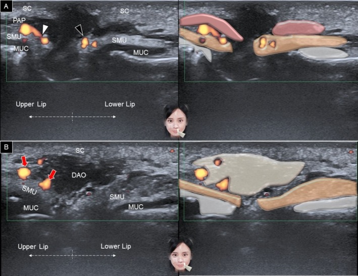
- Take a sagittal scout at the midline. Hold the probe vertically across both lips at the midline, then sweep it out toward each commissure (WFUMB 2025; Wu 2025).

The sagittal scout on Power Doppler with a schematic overlay: the superior and inferior labial arteries in the submucosal layer, and the facial artery medial to the depressor anguli oris at the commissure. Figure 9 from Wu WT, Chang KV, Naňka O, et al. J Cosmet Dermatol 2025;24(4):e70164. doi:10.1111/jocd.70164. CC BY 4.0. - Scan each lip transversely. Place the probe horizontally over the philtrum and slide it down to the upper vermilion; for the lower lip, work along the labiomental crease (Wu 2025). The orbicularis shows as two hypoechoic bands in the red lip and one in the cutaneous lip, and the teeth form the deepest bright line (Vasconcelos-Berg 2025).
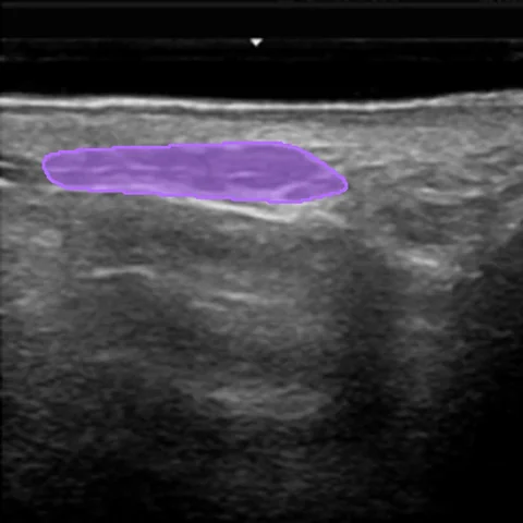
- Follow each artery in Color or Power Doppler. Start where the artery enters the lip, within about 0.5 cm of the commissure (Bravo 2025), and follow it to the midline, scanning across the vessel and steering the color box along it (Sigrist 2024). Record its plane and depth at every point you plan to inject, as Kim’s 2026 study did at six points per lip.
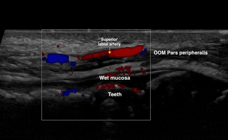
The superior labial artery followed across the upper lip in Color Doppler before the first pass: Suresult L24. - Confirm the waveform with PW Doppler. An arterial spectrum identifies the labial artery; color fills arteries and veins alike, and probe compression separates them (Lee 2023).
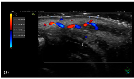
- Mark the skin. Mark the artery on each cross-section and join the marks into a line (Sigrist 2024), with an extra mark wherever it runs most superficially (Vasconcelos-Berg 2025); in Vasconcelos-Berg’s mapped patient, 1.1 mm separated the skin from the artery at its shallowest point.

Mapping before superficial needle work: the labial artery on color Doppler with on-image depth calipers from 0.11 to 0.16 cm, the shallowest 1.1 mm. Figure 18, panel a, from Vasconcelos-Berg R, Desyatnikova S, Bonavia P, et al. Diagnostics (Basel) 2025;15(7):921. doi:10.3390/diagnostics15070921. CC BY 4.0. - Plan the plane and the entry against the map. For needle work at the border, keep the tip shallower than the artery depth measured at that point (Vasconcelos-Berg 2025); Trzeciak’s general ceiling is 3.5 mm, and 4 mm in the superior vermilion. Run cannula volume in a plane the map shows clear of the artery, such as behind it in the submucosa (Bravo 2025), and keep entries away from the corner, where the labial arteries leave the facial artery (Hong 2026). Gonzalez’s team switched to a short needle and a very superficial approach whenever the artery ran in the dry mucosa.
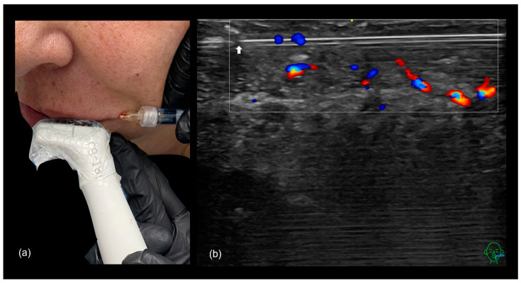
- Inject under the probe where the plan calls for it. For cannula passes through the body of the lip, scan while injecting with the cannula in-plane, placing product above, below or between the two parts of the orbicularis (Vasconcelos-Berg 2025). For border work with a needle, map first and inject against the marks.

Scanning while injecting at 8–18 MHz: a 25G cannula held in-plane under a hockey-stick probe in a sterile cover, and on color Doppler the cannula tip running superficial to and parallel with the superior labial artery. Figure 16 from Vasconcelos-Berg R, Desyatnikova S, Bonavia P, et al. Diagnostics (Basel) 2025;15(7):921. doi:10.3390/diagnostics15070921. CC BY 4.0. - Rescan. Image the deposit in B-mode, confirm flow along both labial arteries in Doppler, and save the cine loop to the patient record.
Doppler settings for a labial artery
To show a labial artery, set the velocity scale to about 3 cm/s with the PRF and wall filter lowered to match (Sigrist 2026), steer the color box along the vessel, and hold a 0° to 60° angle with abundant gel and minimal pressure. Near its origin from the facial artery, a labial artery measures about 1.6 mm across, with a peak systolic velocity near 27 to 28 cm/s (Tucunduva 2016, 20 subjects), and the WFUMB 2025 position paper puts hypodermal arteries at 15 cm/s or below; the settings below make that slow, shallow flow fill the color box.
| Contexto | Value for a labial artery | Fuente |
|---|---|---|
| Modos | B-mode, Color and spectral (PW) Doppler; Power Doppler for the slowest branches | Delphi 2025; WFUMB 2025 |
| Velocity scale | About 3 cm/s, adjusted to the flow | Sigrist 2026 |
| PRF and wall filter | Lowered together with the scale; a published 9-point lip map shows PRF at 1.00 kHz | Sigrist 2026; Tucunduva 2016; Kim 2024 (9-point study) |
| Color box | Steered in the direction of the vessel | Sigrist 2024 |
| Doppler angle | 0° to 60° to the flow, made by tilting the probe | Tucunduva 2016; Velthuis 2021 |
| Ganancia | Adjusted together with the scale | Sigrist 2024 |
| Detection threshold | About 2 cm/s | WFUMB 2025; Sigrist 2026 |
| Acoplamiento | Abundant gel and minimal pressure; heavy pressure compresses the shallow vessels | Sigrist 2026; Wu 2025; Teixeira 2025 |
| Artery or vein | PW waveform and probe compression | Lee 2023 |
| Documento | The plane of each artery at each planned point, saved as stills and a cine loop | WFUMB 2025; Delphi 2025 |
Power Doppler is more sensitive than color Doppler (WFUMB 2025), and the 2025 Delphi panel recommends it alongside the mandatory color and spectral modes. Velthuis’s 2021 Doppler guide scans the upper lip transversely along its edge, with the orbicularis and the teeth as reference points, and describes the superior labial artery changing layers along a tortuous “dancing snake” course. On a Suresult Pro probe, color controls cover steer, color gain, PRF, wall filter, move and zoom, PW controls cover gain, steer, volume and angle, and the lip setting can be saved as one of 3 customizable user sets.
The rescan after the last pass
Rescan the lip after the last pass. In B-mode the deposit shows where the product went: Gonzalez’s 2025 series at 18 MHz found 55% of deposits in the dry submucosa, 23% in the intramuscular layers, 19% in the wet submucosa and 2% in the fat. Color or Power Doppler then confirms flow along both labial arteries, as Gonzalez’s team did after each case. Saved with the map as a cine loop, the rescan becomes the baseline for the next visit and the first comparison image if the patient calls back; the skin signs and the event scan are in the oclusión vascular tras relleno guide.
What frequency you need to see the labial arteries
Mapping the labial arteries takes a linear probe of at least 15 MHz: the 2025 Delphi panel recommends it as the minimum, with 12 MHz serving in some cases, and the WFUMB 2025 position paper sets it as the minimum for aesthetic scanning, at which the axial resolution reaches 100 µm. The recent lip layer and mapping studies read their layers and arteries at 18 to 24 MHz (Harris 2026; Gonzalez 2025; Kim 2026). Jaguś’s team reports that 18 to 22 MHz transducers show vessels about 0.1 mm across, and Velthuis’s 2021 guide notes that the small infraorbital and supraorbital arteries are not always seen at 12 MHz. At 20 MHz the image reaches about 10 mm (Lee 2023), close to the full thickness of a lip, which measured 8.36 to 10.9 mm in Harris’s 2026 and Lee’s 2020 studies; a 10 mm preset therefore fills the screen with the lip.
| Frecuencia | What it resolved in the lip | Estudio |
|---|---|---|
| 24 MHz | SLA and ILA depth at six points per lip, midline to corner, before an 11-point injection | Kim 2026, 25 women |
| 20 MHz | Facial arteries in a 1 to 3 minute map per session on a handheld probe, before cannula or needle | Teixeira 2025, 503 patients |
| 18 or 20 MHz | Five lip layer thicknesses, with intrarater agreement of 0.71 to 0.89 | Harris 2026, 126 participants |
| 18 MHz | The artery’s plane (wet submucosa, dry submucosa or muscle) and the deposit shapes left by cannula and needle | Gonzalez 2025, 27 patients |
| 18 MHz | Two orbicularis bands in the red lip and one in the cutaneous lip, with the labial arteries | Vasconcelos-Berg 2025 |
| 8–18 MHz | The labial arteries on Power Doppler before, during and after a vertical cannula pass | Bravo 2025, 10 women |
| 15 MHz | The consensus minimum for aesthetic scanning | Delphi 2025; WFUMB 2025 |
| 12 MHz | SLA 3.16 mm and ILA 3.44 mm from the dry mucosa in one mapped patient, before a 9-point injection | Kim 2024 (particle HA series) |
| 5–12 MHz | Labial artery diameter, peak velocity and resistance index near the origin | Tucunduva 2016, 20 subjects |
On a Suresult probe the lip fits the shallowest presets of each band: the L24Pro scans at 18/24 MHz from 10 and 15 mm presets, the L20Pro at 16/20 MHz from 10 and 20 mm, and the L14Pro at 10/14 MHz from 20 mm, with deeper presets for the rest of the face and the body.

The 5 best handheld ultrasounds for lip filler, ranked
The Suresult L24Pro ranks first for lip filler at 94.1/100, ahead of the Clarius L20 HD3 (66.8), the GE Vscan Air SL (42.0), the Butterfly iQ3 (35.3) and the Philips Lumify L12-4 (33.1). I ranked the handhelds on what a lip visit asks of a probe: enough frequency to separate the orbicularis, the mucosa and an artery about 1 mm across; Color, Power and PW Doppler to map the labial arteries and confirm the waveform; software that reads prior filler or vessel depth; needle guidance for the passes made under the probe; records for the rescan; and the five-year total for everything above. The weights and formulas are under How I scored.

01 Suresult L24Pro — Best Overall for Lip Filler Mapping
Needle Enhance · Color, Power and PW Doppler · FaceAtlas AI, 3 months included · 18/24 MHz · 10–25 mm · 256 elements · $4,185
Score: 94.1/100
$4,185 · 145 g · up to 4 h · cordless · IP67 · no imaging subscription
Buy the Suresult L24Pro aesthetic ultrasound probe: $4,185
Veredicto: Buy the L24Pro if lips fill much of your filler list and you want the labial arteries, the orbicularis and the mucosa sharp on one screen, three Doppler modes to map and confirm the arteries, and the needle visible as it passes them. It is my pick for injectors who map every lip before they treat it.
- Lip tools: 10 and 15 mm presets that frame the lip from skin to mucosa; Color, Power and PW Doppler (color: steer, color gain, PRF, wall filter, move and zoom; PW: gain, steer, volume and angle); automatic vessel flow measurement with PS, ED, TAMAX and TAMEAN
- Filler software: FaceAtlas AI Filler Scan finds prior filler, names the material among seven (HA, CaHA, PLLA, PCL, PMMA, PVA and collagen) and gives its depth and size; its Color Power Doppler view puts blood flow on the live scan before the needle enters; Dual View sets the raw scan beside the AI layers while you inject; 21 standard mapping points from forehead to jawline, two of them perioral; 3 months included, on 64-bit Android 8.0 or later
- Herramientas para agujas: the In-Plane & Out-Plane Puncture Guide, Needle Enhance, plus Flip and Grid
- Records: a cine buffer set to 100, 200, 500 or 1,000 frames, the longest about 165 s; on-device storage; export as JPEG, PNG, MP4 or DICOM; FaceAtlas AI PDF reports and one-click comparison with the previous visit
- Precio y plan: $4,185, with imaging, Doppler and needle tools free of subscription; FaceAtlas AI from month 4 at $50 a month, $500 a year or $999 for life, the lifetime license sold until 11:59 PM PT, December 31, 2026
- Weight and scan time: 145 g; a 4,400 mAh battery for up to 4 h; connects over its own dual-band Wi-Fi to phones and tablets on iOS, Android or Windows; IP67 and drop-tested
Por qué ocupa este puesto. The lip is thin, and its arteries run close to where the product goes. In Harris’s 2026 study the upper lip measured 8.36 mm and the lower lip 9.01 mm, with each of five layers 1.4 to 2.3 mm thick, and Kim’s 2026 readings at 24 MHz put the SLA 2.54 mm from the vermilion border at the midline. The L24Pro’s 24 MHz top frequency and 10 and 15 mm presets work at that scale, the same frequency Kim used for the six-point artery measurements, and earn the full 25 for superficial detail.
Color, Power and PW Doppler all ship with the probe: color to follow each artery from the commissure to the midline, Power for the slow branches at the midline, and PW to confirm an arterial waveform, covering the modes the 2025 Delphi panel makes mandatory (color and spectral) and recommends (Power), for the full 25. FaceAtlas AI earns the full 15 for software: Filler Scan names prior filler among seven materials and measures its depth, the 21-point map keeps every facial site scanned the same way at each visit, and the layer view maps skin, fat and the SMAS, with 3 months in the price.
For needle guidance, Needle Enhance plus in-plane and out-of-plane guide lines earn the full 10: the tools a cannula pass under the probe depends on. For records, the cine buffer holds up to 1,000 frames, about 165 seconds, each scan saves on the device and exports as DICOM, and FaceAtlas AI produces a PDF report and a one-click comparison with the previous visit: 10 of 10. Owning the L24Pro with the FaceAtlas AI lifetime license for five years totals $5,184, which scores 9.08 of 15 against the field’s lowest five-year total.
Sub-scores: superficial detail 25, Doppler 25, filler and vessel software 15, needle guidance 10, records 10, five-year cost 9.08.
Every Suresult L-series probe is FDA 510(k) cleared under K211321.
L24Pro, L20Pro or L14Pro? All three share 256 elements, three Doppler modes, Needle Enhance, 4 hours of scanning and 3 months of FaceAtlas AI. The L24Pro covers the lip and other shallow sites at 18/24 MHz in presets from 10 to 25 mm. For $465 less, the Suresult L20Pro high-frequency linear probe scans the lip at 16/20 MHz and keeps 30 and 40 mm presets for the deeper chin and nasolabial fold planes, so one probe covers the whole perioral region in the same session. The Sonda lineal Suresult L14Pro at $3,441 runs 10/14 MHz with presets to 80 mm for clinics that scan the body as well as the face. Lips first: the L24Pro. The whole perioral region on one probe: the L20Pro. The L14, L20 and L24 aesthetic ultrasound chooser compares the three bands for aesthetic work.
Ideal para: injectors who map the labial arteries before every lip and want the artery, the deposit and the needle on one 24 MHz screen.Elija un modelo de la familia si: you want the lip, chin and nasolabial folds on one probe (the L20Pro), you scan the body as well as the face (the L14Pro), you want the 24 MHz band at $3,441 (the L24), or you want the 20 MHz band at the lowest Needle Enhance price in the family (the L20).Compare cuidadosamente si: you want a Lips application in the menu with live vessel depth in a membership (the Clarius L20 HD3), or heart, lung and abdominal scans on the same probe (the GE Vscan Air SL).
También en esta familia
Suresult L24 linear ultrasound probe · $3,441 · 88.1/100. Same 18/24 MHz band and 10, 15, 20 and 25 mm presets as the L24Pro, on 192 elements with 2 hours per charge; Color, Power and PW Doppler and Needle Enhance included, and FaceAtlas AI on a monthly, yearly or lifetime license.
Suresult L20Pro linear ultrasound probe · $3,720 · 87.8/100. The lip, chin and nasolabial folds on one probe: 16/20 MHz with 10, 20, 30 and 40 mm presets on 256 elements, three Doppler modes, Needle Enhance and 4 hours per charge, with 3 months of FaceAtlas AI.
Suresult L20 linear ultrasound probe · $3,000 · 82.1/100. The 16/20 MHz band with presets from 10 to 40 mm, 192 elements and 2 hours per charge at 147 g; the same three Doppler modes and Needle Enhance, with FaceAtlas AI by license.
Suresult L14Pro linear ultrasound probe · $3,441 · 77.7/100. Face plus body work: 10/14 MHz with 20, 40, 60 and 80 mm presets on 256 elements, three Doppler modes, Needle Enhance and 4 hours per charge, with 3 months of FaceAtlas AI.
Suresult L14 linear ultrasound probe · $2,139 · 69.6/100. The lowest entry price in the line: 10/14 MHz from 20 to 80 mm on 192 elements, three Doppler modes and in-plane and out-of-plane guide lines, 2 hours per charge at 200 g; FaceAtlas AI by license.
sonda ecográfica para medicina estética Suresult L24Pro · almohadillas de gel para ecografía estética Suresult · Ecógrafo de mano Suresult para medicina estética
02 Clarius L20 HD3 — Best for a Named Lips Application at 20 MHz
8–20 MHz · 4 cm · Lips among the listed applications · Vessel Depth AI, Needle Enhance and PW Doppler with Essentials · $5,395 plus Essentials
Score: 66.8/100
$5,395 plus Essentials · Color and Power Doppler included · 290 g · about 60 min per charge · IP67 · 3-year warranty
Veredicto: Buy the L20 HD3 if a Lips entry in the application list matters to you, you want a cordless 20 MHz scanner with a live readout of the shallowest vessel’s depth, and you will keep the Clarius Essentials membership.
- Lip tools: Vessel Depth AI shows the depth of the shallowest blood vessel on the imaging screen and updates it in real time, with Essentials; Auto Aesthetic Calipers measure skin, fat and tissue thickness; T-Mode AI Aesthetics labels the facial layers, a tool Clarius lists for educational and training purposes
- Imagen: 8–20 MHz linear to 4 cm with a 25 mm field of view; Color and Power Doppler included; PW Doppler with Essentials; applications include lips, aesthetics and vascular work
- Herramientas para agujas: Needle Enhance with Essentials; a center guide line
- Records: 30-second cine; local and cloud storage, DICOM and PDF reports with Essentials
- Precio y plan: $5,395 at the Clarius store, checked October 29, 2026; Essentials yearly at $695, three years at $1,785, or one-time at $2,500
- Weight and scan time: 290 g; roughly an hour of scanning per charge and about 90 min to refill the battery; IP67; 1 m drop rating; 3-year warranty; 30-day returns
Por qué ocupa este puesto. At 20 MHz the L20 HD3 scores 17.86 of 25 for superficial detail, and a Clarius L20 at 20 MHz carried Teixeira’s 503 mapped patients, 106 of them lips, with a 1 to 3 minute map per session. The scanner includes Color and Power Doppler and Essentials adds PW, for 21 of 25. Clarius calls Vessel Depth AI vascular mapping, which earns half the map credit as a membership tool, and the Auto Aesthetic Calipers, sold with no tier named, earn the full depth credit: 7.5 of 15 for software.
Needle Enhance at half credit in Essentials plus the center guide make 7.5 of 10 for needle guidance. The 30-second cine falls short of the 60-second mark, and storage, DICOM and PDF reports arrive with Essentials, for 7 of 10 on records. With one-time Essentials, five years total $7,895, or 5.96 of 15 on cost.
Sub-scores: superficial detail 17.86, Doppler 21, filler and vessel software 7.5, needle guidance 7.5, records 7, five-year cost 5.96.
Limitaciones. PW Doppler, Needle Enhance, Vessel Depth AI, storage and DICOM all sit in the membership, cine playback runs 30 seconds, and no Clarius tool names a filler material.
Ideal para: injectors who want 20 MHz, a Lips application and live vessel depth, and who will stay on Essentials.Descártelo si: you need PW Doppler, Needle Enhance and DICOM without a membership, or software that names old filler before you add more.
También en esta familia
Clarius L15 HD3 · $3,795 plus Essentials · 56.9/100. A 5–15 MHz linear to 7 cm with a 50 mm field of view on the Essentials membership; $6,295 over five years with one-time Essentials.
Especificaciones oficiales del Clarius L20 HD3 · Tienda de Clarius · Precios de Clarius y planes Essentials
03 GE Vscan Air SL — Best for Clinics That Also Scan Hearts and Lungs
Linear 3–12 MHz to 8 cm · sector head to 24 cm · Color and PW Doppler included · $4,999
Score: 42.0/100
$4,999 · 218 g · about 50 min per charge · IP67 · 3-year warranty
Veredicto: Buy the Vscan Air SL if your clinic wants one wireless probe for the lips, the heart, the lungs and the abdomen, with Color and PW Doppler in the purchase price.
- Lip tools: Color and PW Doppler on a 192-element linear head at 3–12 MHz, behind a 40 × 7 mm lens
- Imagen: linear 3–12 MHz to 8 cm and sector to 24 cm; GE lists aesthetics as an optional indication, offered in some markets
- Herramientas para agujas: a centerline marker
- Records: 500 exams stored on the device; DICOM; an OB report
- Precio y plan: $4,999 in the GE HealthCare US shop, checked October 29, 2026; the optional Innovations plan, $500 a year or $1,299 for three years, adds Caption AI and the cardiac and bladder tools
- Weight and scan time: 218 g; about 50 min per charge; IP67; 3-year warranty
Por qué ocupa este puesto. The linear head tops out at 12 MHz, which scores 3.57 of 25 for superficial detail; 12 MHz is the frequency of Kim’s 2024 particle-HA series, which measured both labial arteries from the dry mucosa in one mapped patient. Color and PW Doppler come with the probe, and GE lists no Power Doppler, for 17 of 25. The centerline marker earns 5 of 10 for needle guidance, and on-device storage, DICOM and the OB report earn 7 of 10 for records. At $4,999 with every scored tool included, it has the lowest five-year cost of the four rivals, 9.42 of 15. No GE tool reads filler or vessel depth, so software scores 0.
Sub-scores: superficial detail 3.57, Doppler 17, filler and vessel software 0, needle guidance 5, records 7, five-year cost 9.42.
Limitaciones. 12 MHz sits under the 15 MHz minimum that the 2025 Delphi panel recommends and the WFUMB 2025 position paper sets, Power Doppler is not listed, and the needle aid is a centerline marker.
Ideal para: clinics that want lip mapping plus cardiac, lung and abdominal scans from one dual-head probe.Descártelo si: lips are your main work and you want 18 MHz or more, Power Doppler or needle enhancement.
Official GE Vscan Air SL page · Tienda de GE HealthCare en EE. UU.
04 Butterfly iQ3 — Best Whole-Body Probe With Biplane Needle Views
1–12 MHz on one probe · “Vascular: Superficial” preset at 1–4 cm · Color with Core; Power, PW, Needle Viz and Biplane with Advanced · $3,899 plus a membership
Score: 35.3/100
$3,899 plus Core or Advanced · 300 g · up to 2 h · IPX7 and IP6X · 3-year warranty
Veredicto: Buy the iQ3 if a single probe has to cover the whole body, biplane and out-of-plane needle views matter to you, and you will map lips in its superficial vascular preset on the Advanced plan.
- Lip tools: the “Vascular: Superficial” preset, which Butterfly describes for small facial vessels with slow flow, at 1–4 cm; Color Doppler with Core; Power and PW Doppler with Advanced
- Imagen: one 1–12 MHz chip-based probe for every preset, with the nerve preset reaching 8 cm; Auto Bladder Volume and Auto B-Lines
- Herramientas para agujas: Biplane Imaging, Needle Viz and the Needle: Out-of-Plane preset, which shows the tip within 1 mm of the entry point, all with Advanced; a midline marker
- Records: recorded clips; cloud storage with Core; Basic DICOM sold by quote
- Precio y plan: $3,899 with a 3-year warranty at the Butterfly store, checked October 29, 2026; Core $299 a year; Advanced $420 a year or $1,500 for five years
- Weight and scan time: 300 g; up to 2 h; IPX7 and IP6X; 4-ft drop rating; connects to the phone or tablet by cable
Por qué ocupa este puesto. At 12 MHz the iQ3 scores 3.57 of 25 for superficial detail. Color Doppler sits in Core and Power and PW Doppler in Advanced, each at half credit as a paid mode: 12.5 of 25. Needle Viz at half credit in Advanced plus the midline marker earn 7.5 of 10. Cloud storage with DICOM bought by quote scores 3 of 10 for records, and $5,399 over five years with Advanced scores 8.72 of 15. No Butterfly tool reads filler or vessel depth, so software scores 0.
Sub-scores: superficial detail 3.57, Doppler 12.5, filler and vessel software 0, needle guidance 7.5, records 3, five-year cost 8.72.
Limitaciones. The single probe tops out at 12 MHz, under the 15 MHz consensus minimum, and every Doppler mode needs a paid plan: Color with Core, Power and PW with Advanced.
Ideal para: clinicians who scan the whole body and want biplane needle guidance on one probe.Descártelo si: you want 18 MHz or more for the lip, or Doppler without a membership.
Especificaciones oficiales de Butterfly iQ3 · Butterfly prices and memberships
05 Philips Lumify L12-4 — Best for Practices Already on Lumify
12–4 MHz to 12 cm · PW and steerable color Doppler included · battery-free · $10,307.95 outright at PartsSource
Score: 33.1/100
$10,307.95 at PartsSource or by quote through Philips · under 136 g · up to 7.5 h · no subscription · 5-year basic warranty through Philips
Veredicto: Buy the L12-4 if your practice already runs the Lumify app on other Philips transducers and you want a light linear with all of its software in the purchase.
- Lip tools: steerable color Doppler in Fast Flow and Slow Flow modes, and PW Doppler
- Imagen: 12–4 MHz linear to 12 cm; a 34 mm footprint and a 38 mm aperture
- Herramientas para agujas: a center line marker; biopsy capable
- Records: loops of 1 to 10 s; hundreds of exams on the device; DICOM to PACS
- Precio y plan: sold by quote through Philips Healthcare; $10,307.95 outright at PartsSource with a 90-day warranty, checked October 29, 2026; Philips sells Lumify with no subscriptions, all software and features in the purchase
- Weight and scan time: under 136 g; no battery of its own, so it runs on the phone or tablet’s power, for up to 7.5 h; 5-year basic warranty through Philips
Por qué ocupa este puesto. The L12-4 tops out at 12 MHz, 3.57 of 25 for superficial detail. Steerable color and PW Doppler come with it, and Philips lists no Power Doppler: 17 of 25. The center line marker earns 5 of 10, and 1–10 s loops with storage and DICOM score 3 of 10 for records. At $10,307.95 it carries the highest five-year cost in this comparison, 4.57 of 15. No Philips tool reads filler or vessel depth, so software scores 0.
Sub-scores: superficial detail 3.57, Doppler 17, filler and vessel software 0, needle guidance 5, records 3, five-year cost 4.57.
Limitaciones. 12 MHz sits under the 15 MHz consensus minimum, loops stop at 10 s, and Philips sells it by quote.
Ideal para: practices standardized on Lumify that want a linear for occasional lip mapping.Descártelo si: you want 18 MHz or more, Power Doppler, needle enhancement or a published price from the maker.
What each handheld costs to own for five years
Five years of ownership, counting the handheld plus the software each scored tool needs, total $5,184 for the Suresult L24Pro with the FaceAtlas AI lifetime license, $4,999 for the GE Vscan Air SL, $5,399 for the Butterfly iQ3, $7,895 for the Clarius L20 HD3 and $10,307.95 for the Philips Lumify L12-4. Essentials carries the Clarius PW Doppler, Needle Enhance, Vessel Depth AI, storage and DICOM; Butterfly puts Color in Core and Power, PW and the needle tools in Advanced. Every Doppler mode, both needle tools and the imaging app come with the L24Pro; its only add-on is FaceAtlas AI after month 3.
| De mano | Precio | What the scored tools need | Five-year total, software paid once | On a yearly plan |
|---|---|---|---|---|
| Suresult L24Pro | $4,185 | FaceAtlas AI after the 3 included months; imaging, Doppler and needle tools included | $5,184 with the FaceAtlas AI lifetime license, sold until 11:59 PM PT, December 31, 2026 | $6,685 over five years with the FaceAtlas AI yearly license at $500 from month 4 |
| Clarius L20 HD3 | $5,395 | Essentials, $2,500 paid once, for PW Doppler, Needle Enhance, Vessel Depth AI and DICOM | $7,895 | $8,870 over five years with Essentials billed yearly at $695 |
| GE Vscan Air SL | $4,999 | Included: Color and PW Doppler, the centerline marker, storage and DICOM | $4,999 | Innovations, $500 a year, adds cardiac and bladder AI outside the score |
| Butterfly iQ3 | $3,899 | Advanced for Power and PW Doppler and the needle tools, $1,500 for five years | $5,399 | Advanced at $420 a year: $5,999 in five years |
| Philips Lumify L12-4 | $10,307.95 outright at PartsSource | All Lumify software in the purchase | $10,307.95 | Sold without subscriptions |
Rival prices come from each maker’s US store and the PartsSource listing, read on October 29, 2026; the guía de costes de ecografía estética prices the wider field of aesthetic probes.
Suresult prices each probe with No Hidden Fees: the checkout figure is what you pay, with worldwide delivery, customs clearance and duties covered, and orders leave within 24 hours. Every probe carries a 30-day worry-free trial from the day it arrives and a 2-year warranty, extendable to 3 or 5 years. FaceAtlas AI is licensed one device at a time, with the key sent by email and no auto-renewal. The 3 months that come with the L14Pro, L20Pro and L24Pro are claimed on the FaceAtlas AI claim page and begin when you press Start; the L14, L20 and L24 run the same mensual, anual y vitalicia licenses from day one. For mapping during the injection, the almohadillas de gel para ecografía estética Suresult are sterile, individually sealed 35 mm hydrogel pads that conform to the lips, at $140 for 50.
Cómo puntué
I scored each maker’s card out of 100 on six weighted criteria, set on October 29, 2026, before scoring a single rival: superficial detail 25, Doppler 25, filler and vessel software 15, needle guidance 10, records 10 and five-year cost 15. Together they answer the question an injector asks before buying: which handheld shows the labial arteries and the lip layers clearly enough to plan every pass, maps and confirms the arteries with Doppler, reads old filler or vessel depth, guides the needle or cannula under the probe, keeps the rescan on file, and what the whole kit costs across five years.
- Superficial detail (25): 25 × (top linear frequency in MHz − 10) ÷ 14, held between 0 and 25, so 24 MHz scores 25, 20 MHz 17.86 and 12 MHz 3.57.
- Doppler (25): Color 9, Power 8 and PW 8, counted in full when the purchase includes the mode and at half when a paid plan unlocks it.
- Filler and vessel software (15): naming the filler material 5, a facial vessel or injection-point map 5, layer or depth measurement 5, each in full when included and at half when paid. FaceAtlas AI counts as included on the Pro models, which carry 3 months of it, and so does any tool whose tier the maker leaves unnamed.
- Needle guidance (10): an enhancement mode 5, plus 5 for in-plane or out-of-plane guide lines, biplane views or a center line the maker names for needle work; paid tools at half.
- Records (10): cine of at least 60 s in a maker figure for this exact model 3; on-device or app storage plus DICOM export 3; a visit report or a side-by-side comparison 4.
- Five-year cost (15): the lowest five-year total among every scored model divided by this model’s total, times 15. A Suresult total is the October 29, 2026 listed price plus the FaceAtlas AI lifetime license; Clarius adds one-time Essentials and Butterfly Advanced for five years; the Lumify total is its PartsSource price. The Suresult L14 with the lifetime license sets the reference at $3,138.
| Rank and maker card | Superficial /25 | Doppler /25 | Software /15 | Needle /10 | Records /10 | Cost /15 | Total /100 |
|---|---|---|---|---|---|---|---|
| 1 · Suresult L24Pro | 25 | 25 | 15 | 10 | 10 | 9.08 | 94.1 |
| 2 · Clarius L20 HD3 | 17.86 | 21 | 7.5 | 7.5 | 7 | 5.96 | 66.8 |
| 3 · GE Vscan Air SL | 3.57 | 17 | 0 | 5 | 7 | 9.42 | 42.0 |
| 4 · Butterfly iQ3 | 3.57 | 12.5 | 0 | 7.5 | 3 | 8.72 | 35.3 |
| 5 · Philips Lumify L12-4 | 3.57 | 17 | 0 | 5 | 3 | 4.57 | 33.1 |
Family members sit inside their maker’s card: Suresult L24 88.1, L20Pro 87.8, L20 82.1, L14Pro 77.7 and L14 69.6; Clarius L15 HD3 56.9. The Suresult card leads with the L24Pro because lip work happens at 18 to 24 MHz in the first 10 mm, and the L24Pro brings that band with 256 elements, 4 hours of scanning and FaceAtlas AI included.
Five reruns test the order. Dropping the software credit, dropping cost, scoring the Suresult card on the L20Pro or on the L14Pro, and crediting every rival Doppler mode as included all leave Suresult first and the Clarius L20 HD3 second. The harshest combination for the L24Pro, with FaceAtlas AI at half credit, no credit for the 21-point site map and rival Doppler counted as included, still puts it at 84.1 against 70.8. Two runs reorder the lower places: without cost, the Philips Lumify L12-4 (28.6) moves above the Butterfly iQ3 (26.6), and with rival Doppler counted as included, the Butterfly iQ3 (47.8) moves above the GE Vscan Air SL (42.0). Rival prices, plans and tool lists come from the makers’ US pages and the PartsSource listing, and Suresult figures from its product pages, all read on October 29, 2026.
Lip filler technique questions
Where do you inject lip filler?
Lip filler goes into the vermilion border and the Cupid’s bow for shape, the tubercles for projection, the body of the lip at or behind the wet–dry junction for volume, and the philtral columns and commissures for support, and most published protocols thread it in linear retrograde passes (12 of 16 in Miranda Cerna’s 2026 review). Kim’s 9-point technique enters along the upper vermilion border, 1 mm below the lower border and 1 mm lateral to the commissure, and the 11-point version targets the Cupid’s bow, the vermilion tubercles, the philtral columns and the commissures. Map both labial arteries with Color Doppler before choosing the plane: in Kim’s 2026 series the superior labial artery ran 2.54 mm from the vermilion border at the midline and 6.62 mm at the corner.
Where should you not inject lip filler?
Keep product out of the plane the artery occupies at each point: in most lips that is the submucosa deep to the orbicularis near the wet–dry junction, and at the midline the artery and its branches rise toward the surface. Kim’s 2024 9-point study rates the upper central tubercle (point 3) as the point most exposed to bruising, over a midline arteriovenous shunt, and Kim’s 2026 study finds superficial inferior labial branches and shunts in the central lower lip. The labial arteries leave the facial artery near the commissure, so entries go away from the corner (Hong 2026). The map shows where this patient’s artery runs.
Which lip filler technique is safest for the labial arteries?
The technique that places product in a plane the map shows clear of the artery. In cadaver checks of experienced injectors’ own lip techniques, 58.3% of injections put product close to a labial artery, 60.0% with a needle and 57.1% with a cannula (Ghannam 2019), so the plane matters more than the tool. Published protocols built on a scan include Kim’s 9-point and 11-point techniques, which map the arteries first, and Bravo’s vertical submucosal cannula, which places product behind the artery under Power Doppler; Teixeira’s handheld mapping before cannula or needle recorded no vascular complications in 503 patients.
Should you use a cannula or a needle for lip filler?
Both work in the lip, and they differ in deposit shape and in the force needed to enter an artery. A cannula leaves elongated deposits and a needle rounded ones (Gonzalez 2025, 27 patients at 18 MHz). A 27G cannula enters an artery with the same force as a 27G needle (0.78 vs 0.70 N), 22G and 25G cannulas need more force than needles of the same gauge (Pavicic 2019), and 18G and 22G cannulas never penetrated the facial artery in Ugradar’s test. Most published lip protocols use a needle (9 of 14 that named a tool; Miranda Cerna 2026); a short needle suits the border and the Cupid’s bow, and a cannula suits passes through the body of the lip.
How much filler goes into the lips per session?
Most published lip protocols use 1 to 2 mL per session (Miranda Cerna 2026, 16 studies). Hong’s 2025 contour-then-volume protocol places 0.5 to 1 mL per lip, 1.5 mL at most; the Multi Vector Lip series averaged 1.06 mL across 253 patients (Menezes 2025); Kim’s 2026 11-point technique used about 1 mL per patient; and the Φ technique places 0.01 to 0.05 mL per site and caps a session at 1 to 1.5 mL (Keramidas 2021).
How deep is the superior labial artery?
Pooled across studies, the labial arteries lie 4.59 to 4.90 mm beneath the vermilion surface (Trzeciak 2026 meta-analysis), and the superior labial artery comes closer to the vermilion border toward the midline. Ultrasound in 41 volunteers measured it 5.6 mm deep in the upper lip (Cotofana 2020b), and Lee’s 2020 ultrasound study of 60 volunteers put it 5.3 mm from the vermilion border. Measured point by point from the vermilion border at 24 MHz, it lay 2.54 mm away at the midline, 3.39 mm at the Cupid’s bow apex and 6.62 mm at the corner (Kim 2026), so the scan at the point being injected gives the depth that counts.
How deep should lip filler be injected?
Shallower than the labial artery at the point being injected. Trzeciak’s 2026 meta-analysis, which put the labial arteries 4.59 to 4.90 mm beneath the vermilion surface, recommends a depth limit of 3.5 mm for most of the lip and 4 mm for the superior vermilion. The artery comes closer to the surface at the upper midline, where it ran subcutaneous in 4 of 15 dissected heads (26.7%; Milla 2026), so the Color Doppler map at each point sets the limit for that patient.
Which layer does the labial artery run in?
Mostly the submucosa, between the orbicularis oris and the mucosa: 78.1% of sites in 193 dissected heads (Cotofana 2017), and on ultrasound in 41 volunteers, 58.5% submucosal, 36.2% intramuscular and 5.3% subcutaneous (Cotofana 2020b). The plane changed along the course in 29% of upper lips and 32% of lower lips (Cotofana 2017), and at the upper midline Kim’s 2024 9-point study found dry-mucosa branches and perforators in 80% of patients, so the plane is checked at each point you inject.
What is the difference between the superior and inferior labial artery?
The superior labial artery supplies the upper lip and usually arises above the commissure (80.71% in Ostrowski’s pooled data, a mean 11.95 mm away); the inferior labial artery supplies the lower lip, arises below the commissure in 72.44% at 21.99 mm, and varies more. The ILA was absent in 13.45% of cases against 3.55% for the SLA (Ostrowski 2026), and in Nguyen’s 102 facial arteries the ILA most often followed the horizontal labiomental pattern (69.61%) rather than the classic course along the border (8.82%). On ultrasound near their origin, both measured about 1.6 mm across, with peak velocities near 27 to 28 cm/s (Tucunduva 2016).
Can ultrasound map the labial arteries before lip filler?
Yes. A high-frequency linear probe with Color or Power Doppler shows both labial arteries, their plane and their depth, and a handheld map takes 1 to 3 minutes per session (Teixeira 2025). The 2025 Delphi consensus recommends vascular mapping before lip filler, and the WFUMB 2025 position paper sets out the scan: transverse across each lip, sagittal at the midline, each artery followed along its whole course with its plane recorded. Kim’s 9-point and 11-point techniques and Bravo’s vertical cannula technique are built on this mapping.
What MHz probe do you need for lip filler?
At least 15 MHz, the consensus minimum (Delphi 2025; WFUMB 2025), and 18 to 24 MHz for the lip itself: the recent lip layer and mapping studies (Harris 2026; Gonzalez 2025; Kim 2026) read layers and arteries at 18 to 24 MHz, Jaguś’s team reports vessels about 0.1 mm across at 18 to 22 MHz, and at 20 MHz the image reaches about 10 mm, the thickness of a lip. The Suresult L24Pro covers 18/24 MHz with 10 and 15 mm presets, and the L20Pro covers 16/20 MHz.
Which Doppler settings show a labial artery?
A velocity scale of about 3 cm/s with the PRF and wall filter lowered to match (Sigrist 2026), the color box steered along the vessel, a Doppler angle of 0° to 60° made by tilting the probe, abundant gel and minimal pressure. Power Doppler is more sensitive than color Doppler (WFUMB 2025), PW Doppler confirms an arterial waveform, and the detection threshold is about 2 cm/s.
Does the Russian lip technique raise vascular risk?
Yes: its traces run vertically across the border toward the wet–dry line, where most of the lip’s vessels lie, and Adel, who describes “Russian lips” as tenting, lists vascular occlusion and spread onto the skin side among its risks. Published versions keep each trace small and superficial: 0.01 to 0.02 mL per trace superficial to the muscle, every 2–3 mm, skipping the philtral columns (Kichler 2026), or 0.01 mL per pass (Alizadeh 2024). Mapping first shows where this patient’s artery sits relative to those traces; the signs and treatment of an occlusion are in the vascular occlusion guide.
How do you know if you hit an artery during lip filler?
Scanning shows it: the Color Doppler map gives each artery’s depth and plane before the pass, so the entry and plane stay clear of it, and a probe on the lip during the pass shows the cannula or needle beside the artery in real time. Aspiration is a weak check: the global consensus advises against relying on a negative aspirate (Signorini 2016), 30G needles already holding filler give false negatives (Moon 2021), and the tip moves 1.1 to 5.3 mm back during aspiration (Lin 2022). The skin signs of an occlusion and the scan that follows one are in the vascular occlusion guide.
Is there a major artery in the lip?
Yes. Each lip has its own named artery from the facial artery, the superior labial artery in the upper lip and the inferior labial artery in the lower lip, and the superior labial arteries of the two sides join at the midline (Milla 2026). In 8.4% of 284 hemifaces the facial artery ended as the superior labial artery itself (Loukas 2006). The superior labial artery’s lumen narrows from 0.85 mm at the commissure to 0.56 mm at the midline (Money 2020).
What is the best handheld ultrasound for lip filler?
The Suresult L24Pro, at 94.1/100 in my five-maker comparison: 18/24 MHz with 10 and 15 mm presets, Color, Power and PW Doppler and Needle Enhance in the $4,185 price, and FaceAtlas AI with 3 months included to name prior filler. The Clarius L20 HD3 is second at 66.8, with 20 MHz, a Lips application and Vessel Depth AI in its membership. The GE Vscan Air SL (42.0), the Butterfly iQ3 (35.3) and the Philips Lumify L12-4 (33.1) follow, each topping out at 12 MHz.
Should you rescan after lip filler?
Yes. A B-mode rescan shows where the product went: in Gonzalez’s 18 MHz series, 55% of deposits sat in the dry submucosa, 23% in the muscle, 19% in the wet submucosa and 2% in the fat. Color or Power Doppler then confirms flow along both labial arteries, as Gonzalez’s team did after each case. Saved with the map, the cine loop becomes the baseline for the next visit.
Do you need a sterile probe for lip filler?
For scanning during the injection, yes: the probe works under a sterile cover (Sigrist 2024; Vasconcelos-Berg 2025), and the full setup is in the sterile coupling section of the ultrasound-guided filler injections guide. Mapping before the injection uses gel on intact skin. Suresult’s aesthetic ultrasound gel pads are sterile, individually sealed hydrogel pads, 35 mm across and 4 mm thick, that conform to the lips, keep vessel flow visible on color Doppler and let the needle pass through, at $140 for 50 pads.
Fuentes
- Cotofana S, Pretterklieber B, Lucius R, et al. Distribution pattern of the superior and inferior labial arteries: impact for safe upper and lower lip augmentation procedures. Plast Reconstr Surg. 2017;139(5):1075–1082. — doi.org, October 29, 2026
- Kim JS. Enhancing lip augmentation safety and aesthetics through 11-point injection technique and sonographic vascular mapping. Plast Reconstr Surg Glob Open. 2026;14(4):e7520. — doi.org, October 29, 2026
- Ostrowski P, Bonczar M, Elsaftawy A, et al. The blood supply of the lips: a systematic review and meta-analysis. Aesthetic Plast Surg. 2026;50(15):5949–5964. — doi.org, October 29, 2026
- Velthuis PJ, Alfageme F, Cartier H, et al. The use of ultrasound imaging in aesthetic injectables: a modified Delphi consensus. J Plast Reconstr Aesthet Surg. 2025;104:33–37. — doi.org, October 29, 2026
- Chammas MC, Sigrist R, Alfageme F, et al. WFUMB position paper: consensus on best practice in aesthetic dermatologic ultrasound. Ultrasound Med Biol. 2025;51(11):2173–2193. — doi.org, October 29, 2026
- Lee SH, Gil YC, Choi YJ, et al. Topographic anatomy of the superior labial artery for dermal filler injection. Plast Reconstr Surg. 2015;135(2):445–450. — doi.org, October 29, 2026
- Milla GL, Dos Santos HCS, de Sousa MC, et al. Anatomical distribution of the superior and inferior labial arteries: a cadaveric, systematic review, and meta-analysis study. Aesthetic Plast Surg. 2026;50(15):6446–6456. — doi.org, October 29, 2026
- Nguyen VH, Cheng-Kuan L, Nguyen TA, et al. Facial artery: anatomical variations in the perioral region in cadavers. Arch Craniofac Surg. 2023;24(6):266–272. — doi.org, October 29, 2026
- Swancutt MM, Allard AJ, Ho A, et al. Filling the gap: facial anatomy and safe lower lip injection practices. J Clin Med. 2025;14(9):3214. — doi.org, October 29, 2026
- Money SM, Wall WB, Davis LS, Edmondson AC. Lumen diameter and associated anatomy of the superior labial artery with a clinical application to dermal filler injection. Dermatol Surg. 2020;46(5):678–684. — doi.org, October 29, 2026
- Lupu F, Iliuta CP, Enyedi M, et al. The assessment of the anatomical risk in the perioral region. Maedica (Bucur). 2022;17(4):820–825. — doi.org, October 29, 2026
- Loukas M, Hullett J, Louis RG, et al. A detailed observation of variations of the facial artery, with emphasis on the superior labial artery. Surg Radiol Anat. 2006;28(3):316–324. — doi.org, October 29, 2026
- Cotofana S, Alfertshofer M, Schenck TL, et al. Anatomy of the superior and inferior labial arteries revised: an ultrasound investigation and implication for lip volumization. Aesthet Surg J. 2020;40(12):1327–1335. — doi.org, October 29, 2026
- Lee KL, Lee HJ, Youn KH, Kim HJ. Positional relationship of superior and inferior labial artery by ultrasonography image analysis for safe lip augmentation procedures. Clin Anat. 2020;33(2):158–164. — doi.org, October 29, 2026
- Kim JS. Changes in volume of lips in 3-dimensional analysis and projection of lips in sonography after injection of particle-type hyaluronic acid filler utilizing a 9-point injection technique. Aesthet Surg J Open Forum. 2024;6:ojae076. — doi.org, October 29, 2026
- Tansatit T, Phumyoo T, McCabe H, Jitaree B. Translucent and ultrasonographic studies of the inferior labial artery for improvement of filler injection techniques. Plast Reconstr Surg Glob Open. 2019;7(9):e2399. — doi.org, October 29, 2026
- Trzeciak M, Lipski M, Gładysz T, et al. Anatomical depth of labial arteries in dermal filler procedures: a meta-analysis with clinical implications. Aesthet Surg J. 2026;46(5):478–485. — doi.org, October 29, 2026
- Gonzalez C, Callejas E, Nuñez C, et al. Lip volumization with hyaluronic acid: comparative ultrasonographic evaluation of cannula and needle techniques in a multicenter study. Cureus. 2025;17(2):e79325. — doi.org, October 29, 2026
- Vasconcelos-Berg R, Desyatnikova S, Bonavia P, et al. Best practices for the use of high-frequency ultrasound to guide esthetic filler injections—part 3: lower third of the face. Diagnostics (Basel). 2025;15(7):921. — doi.org, October 29, 2026
- Harris S, Schelke L, Orlovska M, et al. Ultrasound evaluation of lip anatomy and filler placement: a cross-sectional study of injection accuracy, migration, and demographic variation. Plast Reconstr Surg. 2026;158(1):65e–75e. — doi.org, October 29, 2026
- Kim JS. 9-point injection technique for lip augmentation and lip corner lifting using sonographic imaging of the labial artery pathway. Aesthet Surg J. 2024;44(10):1080–1090. — doi.org, October 29, 2026
- Alizadeh N. New Aesthetic Unit (NAU) method: a comprehensive method based on accurate anatomical assessment and precise multilayering panfacial treatment for hyaluronic acid fillers. Aesthetic Plast Surg. 2024;48(23):4735–4749. — doi.org, October 29, 2026
- Moorefield AK, Rose-Reneau Z, Wright BW, et al. Venous tributaries of the lip: implications for lip filler injection. Plast Reconstr Surg. 2023;152(2):257e–263e. — doi.org, October 29, 2026
- Adel N. A new approach for lip filler injection using an inverted Mercedes Benz sign. Plast Reconstr Surg Glob Open. 2021;9(12):e3999. — doi.org, October 29, 2026
- Bravo BSF, Bravo LG, Cornachini BG, et al. Navigating the labial artery: a safer approach to submucosal lip filler techniques. Life (Basel). 2025;15(4):509. — doi.org, October 29, 2026
- Lee W. Hyaluronic acid filler injection guided by Doppler ultrasound. Arch Plast Surg. 2023;50(4):348–353. — doi.org, October 29, 2026
- Miranda Cerna VR. Hyaluronic acid injection techniques for lip augmentation—comparison of linear, retrograde, and microdeposit approaches: a systematic review. Aesthet Surg J Open Forum. 2026;8:ojag059. — doi.org, October 29, 2026
- Kichler M, Barros M, de Freitas C, et al. 4D lips technique based on anatomy for naturalization. Plast Reconstr Surg Glob Open. 2026;14(1):e7270. — doi.org, October 29, 2026
- Hong GW, Choi W, Yoon SE, et al. Anatomical-based diagnosis and filler injection techniques: lips and philtrum. Life (Basel). 2025;15(2):315. — doi.org, October 29, 2026
- Hong GW, Corrêa MC, Song JK, et al. Deep labial fat filler technique for effective lip eversion. J Craniofac Surg. 2026;37(6):1688–1692. — doi.org, October 29, 2026
- Menezes MVA, Dezanetti CBG, Penna FWP, et al. Aesthetic treatment of the lips with hyaluronic acid filler: the Multi Vector Lip technique. J Cosmet Dermatol. 2025;24(9):e70445. — doi.org, October 29, 2026
- Surek CC, Guisantes E, Schnarr K, et al. “No-touch” technique for lip enhancement. Plast Reconstr Surg. 2016;138(4):603e–613e. — doi.org, October 29, 2026
- Stéphane S, Candice SM, Florence B. “Bi-Bi” technique for lip augmentation: a retrospective study on 30 cases. J Cosmet Dermatol. 2022;21(10):4339–4344. — doi.org, October 29, 2026
- Villalobos DM. Innovative classification of M-shaped lips for enhanced refill outcomes: a case series. J Cosmet Dermatol. 2025;24(6):e70239. — doi.org, October 29, 2026
- Keramidas E, Rodopoulou S, Gavala MI. A safe and effective lip augmentation method: the step-by-step Φ (Phi) technique. Plast Reconstr Surg Glob Open. 2021;9(2):e3332. — doi.org, October 29, 2026
- Walker L, Cetto R. Lip augmentation using hyaluronic acid filler and a 4-mm needle: a safer, more natural, and predictable approach. J Clin Aesthet Dermatol. 2021;14(1):E61–E63. — pmc.ncbi.nlm.nih.gov, October 29, 2026
- Pavicic T, Webb KL, Frank K, et al. Arterial wall penetration forces in needles versus cannulas. Plast Reconstr Surg. 2019;143(3):504e–512e. — doi.org, October 29, 2026
- Siperstein RD, Broadfield DC, Yerke Hansen P, et al. Arterial wall and tissue penetration force with various cannulas and needles. Aesthet Surg J. 2023;43(12):1521–1529. — doi.org, October 29, 2026
- Ugradar S, Hoenig J. Measurement of the force required by blunt-tipped microcannulas to perforate the facial artery. Ophthalmic Plast Reconstr Surg. 2019;35(5):444–446. — doi.org, October 29, 2026
- Tansatit T, Apinuntrum P, Phetudom T. A dark side of the cannula injections: how arterial wall perforations and emboli occur. Aesthetic Plast Surg. 2017;41(1):221–227. — doi.org, October 29, 2026
- Blandford AD, Hwang CJ, Young J, et al. Microanatomical location of hyaluronic acid gel following injection of the upper lip vermillion border: comparison of needle and microcannula injection technique. Ophthalmic Plast Reconstr Surg. 2018;34(3):296–299. — doi.org, October 29, 2026
- Ghannam S, Sattler S, Frank K, et al. Treating the lips and its anatomical correlate in respect to vascular compromise. Facial Plast Surg. 2019;35(2):193–203. — doi.org, October 29, 2026
- Signorini M, Liew S, Sundaram H, et al. Global aesthetics consensus: avoidance and management of complications from hyaluronic acid fillers—evidence- and opinion-based review and consensus recommendations. Plast Reconstr Surg. 2016;137(6):961e–971e. — doi.org, October 29, 2026
- Goodman GJ, Magnusson MR, Callan P, et al. Neither positive nor negative aspiration before filler injection should be relied upon as a safety maneuver. Aesthet Surg J. 2021;41(4):NP134–NP136. — doi.org, October 29, 2026
- Lin F, Goodman GJ, Magnusson M, et al. Movement of the syringe during filler aspiration: an ultrasound study. Aesthet Surg J. 2022;42(10):1109–1116. — doi.org, October 29, 2026
- Moon HJ, Lee W, Kim JS, et al. Aspiration revisited: prospective evaluation of a physiologically pressurized model with animal correlation and broader applicability to filler complications. Aesthet Surg J. 2021;41(8):NP1073–NP1083. — doi.org, October 29, 2026
- Nikolis A, Metelitsa A, Avelar L, et al. Rates of positive aspiration prior to facial hyaluronic acid filler injections: outcomes of a multicenter, cross-sectional study. Aesthet Surg J. 2025;45(12):1285–1290. — doi.org, October 29, 2026
- Peng YP, Lin YW, Yang YY. Aspiration before tissue filling with hyaluronic acid—safety enhancement by using a lidocaine-primed syringe. J Cosmet Dermatol. 2023;22(9):2457–2463. — doi.org, October 29, 2026
- Lee KWA, Chan LKW, Lee CH, et al. Cannula is safer than needle in filler injection? JPRAS Open. 2026;48:415–424. — doi.org, October 29, 2026
- Teixeira DA. A new time-saving technique for ultrasound-guided filler injection: a 3-year retrospective study. Aesthet Surg J Open Forum. 2025;7:ojaf061. — doi.org, October 29, 2026
- Wu WT, Chang KV, Naňka O, et al. Lip sonoanatomy and relevance to aesthetic filler injections: a pictorial review. J Cosmet Dermatol. 2025;24(4):e70164. — doi.org, October 29, 2026
- Sigrist R, Desyatnikova S, Chammas MC, et al. Best practices for the use of high-frequency ultrasound to guide aesthetic filler injections—part 1: upper third of the face. Diagnostics (Basel). 2024;14(16):1718. — doi.org, October 29, 2026
- Sigrist RMS, Gonzalez C, Schelke L, et al. Doppler ultrasound findings in filler-related facial vascular adverse events: an international multicenter study. Diagnostics (Basel). 2026;16(11):1587. — doi.org, October 29, 2026
- Tucunduva MJ, Tucunduva-Neto R, Saieg M, et al. Vascular mapping of the face: B-mode and Doppler ultrasonography study. Med Oral Patol Oral Cir Bucal. 2016;21(2):e135–e141. — doi.org, October 29, 2026
- Velthuis PJ, Jansen O, Schelke LW, et al. A guide to Doppler ultrasound analysis of the face in cosmetic medicine. Part 2: vascular mapping. Aesthet Surg J. 2021;41(11):NP1633–NP1644. — doi.org, October 29, 2026
- Jaguś D, Skrzypek E, Migda B, et al. Usefulness of Doppler sonography in aesthetic medicine. J Ultrason. 2021;20(83):e268–e272. — doi.org, October 29, 2026
- Clarius L20 HD3: store page with Essentials plans. — store.clarius.com, October 29, 2026
- Clarius L20 HD3 specifications. — clarius.com, October 29, 2026
- Clarius L15 HD3: store page. — store.clarius.com, October 29, 2026
- GE HealthCare Vscan Air SL: US shop. — gehealthcare.com, October 29, 2026
- GE HealthCare Vscan Air SL: product page. — gehealthcare.com, October 29, 2026
- Butterfly Network: pricing and memberships. — butterflynetwork.com, October 29, 2026
- Butterfly iQ3 specifications. — butterflynetwork.com, October 29, 2026
- Philips Lumify L12-4 linear array transducer. — usa.philips.com, October 29, 2026
- Philips Lumify handheld ultrasound: purchase model. — usa.philips.com, October 29, 2026
- PartsSource: Philips Lumify L12-4 listing. — partssource.com, October 29, 2026
- Suresult L24Pro. — suresultmed.com, October 29, 2026
- Suresult L20Pro. — suresultmed.com, October 29, 2026
- Suresult L14Pro. — suresultmed.com, October 29, 2026
- Suresult L24. — suresultmed.com, October 29, 2026
- Suresult L20. — suresultmed.com, October 29, 2026
- Suresult L14. — suresultmed.com, October 29, 2026
- FaceAtlas AI. — suresultmed.com, October 29, 2026
- Licencia mensual de FaceAtlas AI. — suresultmed.com, October 29, 2026
- Licencia anual de FaceAtlas AI. — suresultmed.com, October 29, 2026
- Licencia vitalicia de FaceAtlas AI. — suresultmed.com, October 29, 2026
- Almohadillas de gel Suresult para ecografía estética. — suresultmed.com, October 29, 2026
- Suresult certifications and compliance. — suresultmed.com, October 29, 2026
Choose the probe around your lip list
For lips first, with the labial arteries, the deposit and the needle on one 24 MHz screen, I recommend the L24Pro at $4,185. For the lip, chin and nasolabial folds on one probe, I choose the L20Pro at $3,720. Clinics that also scan the body get the L14Pro at $3,441, with presets to 80 mm. Orders leave within 24 hours, every Pro comes with a 30-day worry-free trial and a 2-year warranty you can extend to 3 or 5 years, and customs and duties are already in the checkout price: No Hidden Fees.

