

For seroma and hematoma checks after liposuction and abdominoplasty I pick the Suresult L14Pro at $3,441, because its 20 to 80 mm presets hold the flap, the collection and the fascia in one view. Examination found a seroma in 35% and ultrasound in 90% of 60 women two weeks after abdominoplasty (Andrades 2007); the one published tap threshold is a collection over 20 cc above the fascia (Nahas 2007). Color, Power and PW Doppler and Needle Enhance come with the probe for the tap. Serial scans put the peak on day 11, at 38.1% (Di Martino 2015). An office series of 310 abdominoplasties averaged 3.5 guided taps per seroma (Swanson 2023). For the encapsulated seroma, capsulectomy cured 6 of 6 in one series and, with negative-pressure therapy, 15 of 15 recurrent chronic seromas in another, 5 of them post-traumatic (di Summa 2013; Marangi 2020). Below are the look of a seroma and its look-alikes, the rescan calendar, the tap threshold, the guided technique step by step and six handhelds ranked for the work.


Alcance de la evaluación: I come to this page as an independent device reviewer with deep ultrasound knowledge. I traced every rate, depth, volume and threshold below to its primary paper, then scored one handheld per maker on seven criteria fixed on November 1, 2026, before any rival had a score: the Suresult L14Pro, Mindray TE Air e5M, Clarius L7 HD3, Butterfly iQ3, GE Vscan Air CL and Philips L12-4, with the Suresult L20Pro, L14 and L20 scored inside the Suresult card. I read maker pages, datasheets, manuals and US stores from October 29 to November 1, 2026, rechecked every rival price on November 1, and searched PubMed, PMC and Europe PMC on November 1.
Quick answer: which handheld should you buy for seroma checks after body contouring?
The Suresult L14Pro ($3,441) is the handheld I recommend for seroma and hematoma checks after liposuction, abdominoplasty and BBL, at 95.0/100 in my six-maker ranking. Its 10/14 MHz linear runs on 256 elements with presets of 20, 40, 60 and 80 mm. Mean skin-to-fascia depth runs 37 to 42 mm across four abdominal regions before large-volume liposuction, inside the 60 mm preset, and 21 to 22 mm a week after it, inside the 40 mm preset (Sudha 2026), so the flap, the fluid and the fascia stay on one screen. All three Doppler modes, Needle Enhance and both puncture guides ship with the probe, the Suresult app turns three diameters into a volume, and cine holds 1,000 frames, about 165 seconds. Five years of ownership cost $3,441, with no subscription. For thin flaps and pockets within 40 mm, the L20Pro ($3,720) in the same card brings 16/20 MHz.
The Mindray TE Air e5M ranks second at 86.1: Color, Power and PW Doppler, a B-mode Volume tool, Extended FOV, iNeedle and the VA Grid in a $4,499 purchase, checked November 1, 2026, with no subscription.
Seroma scanners compared: six handhelds side by side
The Suresult L14Pro leads the six at 95.0/100, with presets to 80 mm, all three Doppler modes and Needle Enhance in its price. Each handheld, one per maker, is compared on what a post-operative fluid check uses: depth for the flap, the collection and the fascia beneath it, Doppler to clear the superficial epigastric vessels before a tap, a needle aid in the scan plane, a volume from three diameters, and the software each scored tool needs over five years. For the wider office picture, from vessel mapping before surgery to follow-up scans, see the Suresult handheld ultrasound overview for plastic surgery.
| Escáner | Top frequency · depth | Doppler | Herramientas para la aguja | Measurement and volume | Cine, storage and export | Coste a cinco años y suscripción | Ideal para |
|---|---|---|---|---|---|---|---|
 Suresult L14Pro · No. 1 · 95.0/100 | 14 MHz on a 10/14 MHz linear with 256 elements · presets 20, 40, 60 and 80 mm | Color, Power and PW, all with the probe | Needle Enhance; in-plane and out-of-plane puncture guides; Grid and Flip | Length, area and circumference, angle and trace; volume from three diameters in the app | 1,000-frame cine, about 165 s; saved on your own phone, tablet or PC; JPEG, PNG, MP4 and DICOM | $3,441 one-time; imaging, Doppler and needle tools run with no subscription | Flap, collection and fascia in one view, with all three Doppler modes for the entry check |
 Mindray TE Air e5M · No. 2 · 86.1/100 | 9.0 MHz top of a 2.0–9.0 MHz bandwidth, linear B-mode bands to 8.2 MHz · to 40 cm | Color, Power and PW, all standard | iNeedle; VA Grid | Distance and area; Smart Calc; B-mode Volume in the EM FAST, EM AAA, GYN and OB packages; Extended FOV | Cine to 125 frames over 2–5 s; DICOM 3.0; patient data on board; reports to Tricefy | $4,499 one-time, with no subscription plan | Full Doppler, needle and measurement tools at the lowest five-year rival cost |
 Clarius L7 HD3 · No. 3 · 76.0/100 | 13 MHz on a 4–13 MHz linear · to 15 cm | Color and Power with the scanner; PW with Essentials | Needle Enhance with Essentials; Center Guide | Measurements of an area of interest or of the depth from the skin line | 30 s cine; local storage and DICOM with the membership; PDF Reports with Essentials | $6,295 including the $2,500 one-time Essentials license; $7,270 on yearly Essentials at $695 | 13 MHz detail to 15 cm on a membership |
 Butterfly iQ3 · No. 4 · 72.7/100 | 12 MHz from a 1–12 MHz probe · to 30 cm; MSK-Soft Tissue preset to 12 cm | Color on Core; Power and PW on Advanced | Needle Viz and Biplane on Advanced; midline marker | Four linear calipers and an ellipse; Manual Volume from three diameters | Unlimited cloud storage on Core or Advanced; Basic DICOM sold separately; onboard worksheets | $5,399 with Advanced as a one-time $1,500 for five years; $420 a year on the yearly plan | Manual Volume and Biplane views on one whole-body probe |
 GE Vscan Air CL · No. 5 · 67.0/100 | 12 MHz on a 3–12 MHz linear to 8 cm; 2–5 MHz curved head to 24 cm | Color and PW in the purchase | Centerline Marker | Distance, circumference and area | Up to 500 exams on the phone or tablet; wireless DICOM export to PACS | $4,999 one-time; the Innovations plan at $500 a year sits outside the score | Linear and curved heads with Color and PW in one price |
 Philips L12-4 · No. 6 · 61.7/100 | 12 MHz on a 12–4 MHz linear · to 12 cm | Color with Fast Flow and Slow Flow; PW | Marcador de línea central; apto para biopsias | Up to four linear distances, plus ellipse area and circumference | Loops of 1–10 s; email, DICOM to PACS, a shared network or a local directory | $10,307.95 bought outright from PartsSource, every software feature included without a required subscription | Steerable color with Fast Flow and Slow Flow, bought outright |
The five-year column adds to each price whatever tier its scored tools require: one-time Essentials for Clarius, and Advanced paid once for five years for Butterfly, whose Basic DICOM is sold separately outside that total. Suresult, Mindray, GE and Philips put every tool they score on in the purchase. The arithmetic is in the five-year cost section and the weights under How I scored.
La mejor opción para cada uso: selección rápida
- Best Overall for Seroma and Hematoma Checks After Body Contouring: Suresult L14Pro, 95.0/100. Flap, fluid and fascia in one 20–80 mm view, three Doppler modes and Needle Enhance with the probe, a volume from three diameters, and the L20Pro in the same card for thin flaps.
- Best for Full Doppler, Needle and Measurement Tools at the Lowest Five-Year Rival Cost: Mindray TE Air e5M, 86.1/100. Color, Power and PW Doppler, B-mode Volume, Extended FOV, iNeedle and the VA Grid for $4,499, checked November 1, 2026, with nothing to renew.
- Best for 13 MHz Detail to 15 cm on a Membership: Clarius L7 HD3, 76.0/100. A 4–13 MHz linear with Color and Power included, and PW Doppler, Needle Enhance and PDF Reports on Essentials.
- Best for Manual Volume and Biplane Views on One Whole-Body Probe: Butterfly iQ3, 72.7/100. An ellipsoid volume from three diameters on a 1–12 MHz probe, with Biplane and Needle Viz on the Advanced plan.
- Best for Linear and Curved Heads With Color and PW in One Price: GE Vscan Air CL, 67.0/100. A 3–12 MHz linear to 8 cm and a 2–5 MHz curved head to 24 cm, both with Color and PW Doppler and the Centerline Marker in the purchase.
- Best for Steerable Color With Fast Flow and Slow Flow, Bought Outright: Philips L12-4, 61.7/100. Two color-flow modes, PW and a center line marker on a 12–4 MHz linear, every software feature in the outright purchase.
Dónde comprar cada ecógrafo
- Suresult L14Pro: $3,441 at the Suresult store, delivered duties paid, with a 30-day trial, a 2-year warranty and no subscription for imaging or Doppler. Order the Suresult L14Pro linear probe at $3,441
- Mindray TE Air e5M: $4,499 with a 3-year warranty included, checked November 1, 2026, on Mindray North America’s own TE Air store, which sells no subscription plan; the Air Capsule costs $750 extra. TE Air e5M at the Mindray store
- Clarius L7 HD3: $4,490 with the first year of Essentials at the Clarius store, checked November 1, 2026. Sold with an Essentials plan: $695/yr, $1,785/3 yr, or $2,500 one-time; the plan carries PW Doppler, Needle Enhance, local storage, DICOM and PDF Reports, and Clarius Care costs $265 for one year, $530 for two or $795 for three. Clarius L7 HD3 at the Clarius store
- Butterfly iQ3: $4,198 with a year of Core from Butterfly Network, checked November 1, 2026, with a 3-year warranty. Color runs on the Core plan at $299 a year; Advanced, at $420 a year or a single $1,500 that covers five years with no recurring fees, unlocks Power and PW Doppler, Needle Viz and Biplane. Butterfly iQ3 plans and pricing
- GE Vscan Air CL: $4,999 at the GE HealthCare US shop, checked November 1, 2026, with a 3-year warranty; Innovations, an optional plan at $500 a year or $1,299 for three years, brings Auto Bladder Volume, Lung Guidance and HD-SRI. Vscan Air CL at the GE HealthCare shop
- Philips L12-4: PartsSource sells it new for $10,307.95 outright, with 90 days of warranty, or for $7,929.19 on exchange, checked November 1, 2026; Philips includes all software and features with the outright purchase. Philips L12-4 at PartsSource
Seroma on ultrasound: what it looks like, and why examination misses it
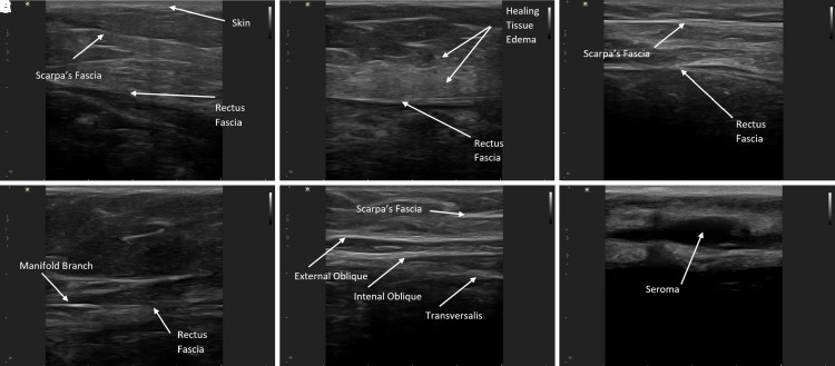
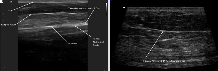
A seroma on ultrasound is an anechoic, compressible pocket with well-defined margins and no flow inside it on color Doppler (Masino 2025). After abdominoplasty it lies under the flap, on the abdominal fascia: Alfonso 2022 scanned every patient at 12 MHz when the closure device’s manifold came out and over any swelling, found seromas in 3 of 71 full abdominoplasties (4.2%) on days 14, 18 and 23, cleared each with 1 to 3 aspirations, and saw no fluid on any scan after day 30. After large-volume liposuction, Sudha 2026 counted any hypoechoic area over 1 cm between the subcutaneous fat and the fascia as a collection and found one in 42 of 50 patients (84%) a week after surgery, most prominently in the flanks.
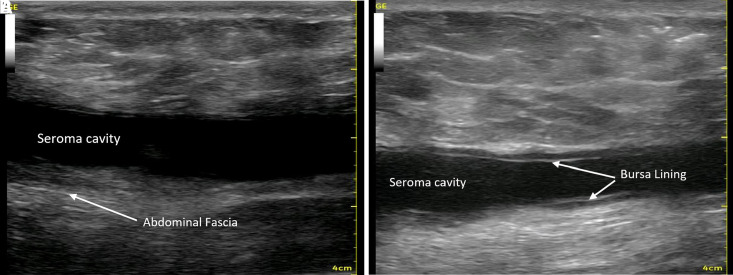
Examination misses most of these collections. In a blinded randomized trial of 60 women, evaluators found a seroma in 35% by examination and in 90% by ultrasound two weeks after abdominoplasty (Andrades 2007), and ultrasound in 56 of 80 abdominoplasty patients found fluid collections in 29 (51.8%), 24 seromas and 5 hematomas (Mohammad 1998). Fluctuance is a clear sign of a collection, while a small one is hard to separate from post-operative edema by hand (Swanson 2018, office ultrasound series). The field has no agreed definition of a clinically significant seroma (Vidal 2017), and among the abdominoplasty studies pooled by Liao 2024, 7 diagnosed seroma clinically, 3 by ultrasound and 6 by both, with 4 unspecified. A scan settles the question at the visit, and the timing section below sets the rescan calendar.
Seroma vs hematoma on ultrasound: echoes, age and Doppler
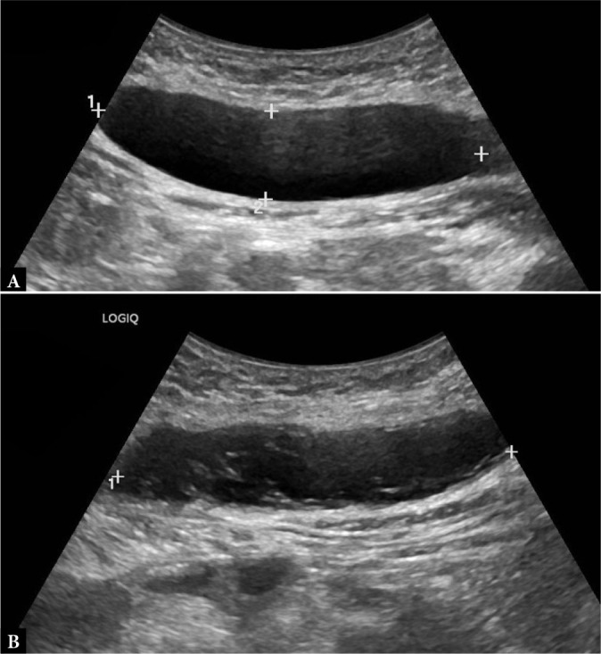
Seroma vs hematoma comes down to the fluid, serous in a seroma and blood in a hematoma, and on ultrasound it shows in the echoes and how they change with time: a seroma stays anechoic and compressible, while a hematoma starts hyperechoic as fresh clot, turns anechoic and can enlarge as the clot liquefies, then fills with sediment, septations and fluid-fluid levels and grows a thin capsule over the following weeks (Pistoia 2025; Masino 2025). Fluid drawn from post-abdominoplasty seromas in 18 women was an exudate, inflammatory in the first week and taking on some features of lymph by days 15 to 20, with protein, LDH and cholesterol levels below those of blood and above those of lymph (Andrades and Prado 2007). Neither collection shows flow inside it on color Doppler, and Masino 2025 uses that absent internal signal to separate a hematoma from a tumor or an abscess, whose wall can be hyperemic. B-mode and color Doppler are limited for active bleeding, which contrast-enhanced ultrasound shows as microbubbles escaping the vessel (Pistoia 2025).
| Collection | Echo pattern | Margins, wall and contents | Doppler color | Timing or setting | Fuente |
|---|---|---|---|---|---|
| Seroma | Anechoic and homogeneous | Well-defined margins; compressible | No flow inside the collection | Under the abdominoplasty flap or between fat and fascia after liposuction; peak on day 11 of serial scans | Masino 2025; Alfonso 2022; Sudha 2026; Di Martino 2015 |
| Hematoma, hyperacute | Hyperechoic, from fresh coagulated blood | Turns anechoic as the clot liquefies, and can enlarge | Active bleeding shows on contrast-enhanced ultrasound as microbubble leakage | The first, hyperacute phase | Pistoia 2025 |
| Hematoma, acute | Anechoic or mildly hypoechoic, with fine echoes | Usually compressible; margins poorly defined early | No internal flow; mild peripheral hyperemia in some | 0–48 hours | Masino 2025 |
| Hematoma, early subacute | Heterogeneous, with echogenic sediment | Fine septations; fluid-fluid levels in some | No internal vascularity; peripheral hyperemia persists in some | 3–7 days | Masino 2025 |
| Hematoma, late subacute | More complex internal echoes | Thin capsule; partial organization | Avascular inside; mild peripheral flow in some | 1–3 weeks | Masino 2025 |
| Hematoma, chronic | Hypoechoic or mixed | Well defined, with a capsule; peripheral calcification or echogenic septa in some | No internal flow; thin reactive rim | Beyond 3–6 weeks | Masino 2025 |
| Post-surgical abscess | Complex, hypoechoic or anechoic | Thick, irregular, ill-defined wall; variably compressible | Hyperemia around the wall in some; no significant flow in the published figure case | Along a scar with fever, 2 weeks after surgery in the published figure case | Masino 2025; Porrello 2022; Soliman 2024 |
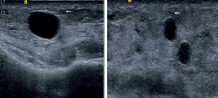
| Oil cyst (cystic fat necrosis) | Anechoic and oval | Sharp, well-defined edge with a capsule; posterior enhancement | No flow in the published case | Knee subcutis after trauma, at 14 MHz | de Wilde 2022 |
One published case shows both collections in the same patient. Two weeks after an abdominoplasty that followed sleeve gastrectomy, a 26-year-old woman had an anechoic, homogeneous seroma on the right and an inhomogeneous hematoma on the left, and neither held internal flow (Masino 2025). The same review advises a high-frequency linear for superficial collections, a curvilinear probe for deep ones, and a panoramic view to document a large superficial lesion in full.
Seroma after abdominoplasty and seroma after liposuction: when it appears and when to rescan
A seroma after abdominoplasty builds through the second week, and a seroma after liposuction is already present at the first: serial ultrasound of 21 women after standard abdominoplasty found seroma in 4.8% on day 4, 38.1% on day 11 and 19% on day 32, most often in the iliac fossae (Di Martino 2015), while routine scans a week after large-volume liposuction found collections in 84% of 50 patients, every one resolved by one month, spontaneously or with serial aspiration (Sudha 2026). Clinically, the post-op seroma mostly presents 2 to 3 weeks after abdominoplasty (Dujmović 2022). Earlier work from the same group scanned five abdominal regions on days 11 to 14 and 18 to 21: abdominoplasty without quilting sutures had the highest seroma rate at both scans, and after lipoabdominoplasty the largest collections sat in the hypogastrium (Di Martino 2010). In a randomized trial of drains, quilting sutures and fibrin sealant, seroma fell between days 15 and 30 in all three arms (Bercial 2012).
| Postoperative day | Women with a seroma on ultrasound |
|---|---|
| Day 4 | 4.8% |
| Day 11 | 38.1% |
| Day 18 | 33.3% |
| Day 25 | 23.8% |
| Day 32 | 19% |
Rescan a hematoma within 7 to 14 days, and send one that persists beyond 4 to 6 weeks, grows or changes its look on to advanced imaging: Masino 2025 sets that calendar for hematomas with indeterminate features or a rebleeding risk, such as anticoagulation. For a seroma, the office tap series set the clock: Swanson 2023 made the first tap at a mean of day 11.6 (8 to 15) and the last at day 21 (8 to 38), and Alfonso 2022 found no fluid on any scan after day 30. A larger hematoma after abdominoplasty arrives sooner, usually in the first 24 hours, as swelling, localized pain and ecchymosis; a small one can be needle-aspirated, and a large active bleed brings hemodynamic instability (Vidal 2017). Scan the same sites in the same planes at every visit, so a change in the numbers is a change in the collection.
Seroma and hematoma rates by procedure, and what raises them
Seroma follows 4.1% of abdominoplasties, the highest rate in the TOPS database of 214,504 aesthetic patients, and 1.8% of liposuctions, with hematoma at 1.0% and 0.4% (Sergesketter 2023). A registry counts the collections that reached a clinician; pooled studies put seroma after abdominoplasty at 10.9% (Salari 2021) and after circumferential lower-trunk contouring at 13%, with hematoma at 3% (Carloni 2016).
| Procedure | Seroma | Hematoma | Cohort | Fuente |
|---|---|---|---|---|
| Abdominoplasty | 4.1% | 1.0% | 24,946 patients, TOPS registry | Sergesketter 2023 |
| Abdominoplasty, pooled | 10.9% | 143 studies, 27,834 patients; seroma prevalence only | Salari 2021 | |
| Liposuction | 1.8% | 0.4% | 56,756 patients, TOPS registry | Sergesketter 2023 |
| Isolated aesthetic liposuction, pooled | 2% | 1% | 60 studies, 21,776 patients | Aljerian 2024 |
| Circumferential lower-trunk contouring (body lift) | 13% | 3% | 28 studies, 1,380 patients | Carloni 2016 |
| All aesthetic procedures | 1.1% | 0.7% | 214,504 patients, TOPS registry | Sergesketter 2023 |
| All aesthetic surgery, major hematoma | 0.91%; men 1.4%, women 0.9% | 129,007 patients, CosmetAssure; major hematomas only, those needing an emergency visit, admission or reoperation within 30 days | Kaoutzanis 2017 |
Five findings shift the odds. Weight: after massive weight loss, a BMI of 30 or more carried seroma in 4.4% against 1.5% below 30 across 2,533 drain-free abdominoplasties (Borriello 2026), and each BMI unit raised the odds 1.1-fold (Najera 2011). Added liposuction: seroma rose from 16.0% to 31.2% when liposuction joined the abdominoplasty, and flank liposuction raised the odds 3.3-fold (Najera 2011). Sex, for hematoma: men had major hematomas at 1.4% against 0.9% in women, and male sex carried a relative risk of 1.98 on multivariate analysis (Kaoutzanis 2017). Progressive tension sutures cut seroma against drains, RR 0.34 across 24 studies (Rao 2024), and tranexamic acid lowered its odds, OR 0.51 across 7 studies (Almahzoum 2026).
What counts as a large seroma: seroma size, volume and the tap threshold
A large seroma, by the one published tap threshold, holds more than 20 cc above the fascia on ultrasound, and seroma size is followed as a volume from three diameters at each visit. Nahas 2007 scanned all 21 women on days 15 to 18 after abdominoplasty, found a mean collection of 8.2 cc, tapped only the two over 20 cc and saw no recurrence in either; Dujmović 2022 cites it as the only research on the question, with the tap set against the risk of a chronic seroma or a pseudobursa. Trials set their own seroma definitions, such as more than 30 mL on a day-20 scan in a randomized trial of post-bariatric anchor-line abdominoplasty (Inforzato 2020).
| Series | Contexto | Volumen | What followed | Fuente |
|---|---|---|---|---|
| 21 women, routine scans | Days 15–18 after abdominoplasty | Mean 8.2 cc; taps only over 20 cc above the fascia | Two women tapped; neither recurred | Nahas 2007 |
| 42 post-bariatric patients, randomized | Day-20 scan after anchor-line abdominoplasty | Over 30 mL counted as a seroma | The trial’s seroma definition | Inforzato 2020 |
| 30 arms after arm liposuction | Seromas found on ultrasound in 5 arms (16.7%) | Mean 15.4 cc (10–20) | Ultrasound-guided aspiration | Benito-Ruiz 2025 |
| 167 abdominoplasties, office series | 9 clinical seromas (5.4%) | Mean aspirate 74 cc (3–240) | Mean 5 office aspirations (1–12), the last on day 28 (15–43) | Swanson 2013 |
| 310 abdominoplasties, office series | 15 seromas (4.8%), tapped under ultrasound guidance | Mean aspirate 112 mL (5–700) | Mean 3.5 taps (1–7), from day 11.6 to day 21; no hematomas | Swanson 2023 |
| Figure case with a standard drain | 3 weeks after abdominoplasty, on the fascia | About 200 cc | Partial drainage, with the cavity lining visible afterward | Alfonso 2022 |
| Chronic encapsulated seroma | Swelling from week 3, still present at 3 years | About 540 mL aspirated | Capsulectomy with revision abdominoplasty | Goldman 2018 |
| Pseudobursa | Evacuated repeatedly under ultrasound | 2,010 mL in one evacuation | Excision at mini-abdominoplasty; no recurrence at 3 months | Roje 2006 |
Measure the collection in two planes at right angles: length and depth in one, width in the other. The Suresult app calculates a volume from those three diameters, so each visit leaves one number to compare, beside the calipers’ length, area and circumference. Trials track seroma size the same way: Martins 2022 measured seroma volume on ultrasound on days 7 and 14 in a randomized trial of abdominal binders and found no difference between the women with and without one. Keep the same landmarks, planes and preset at every visit, so the volume moves only when the collection does.
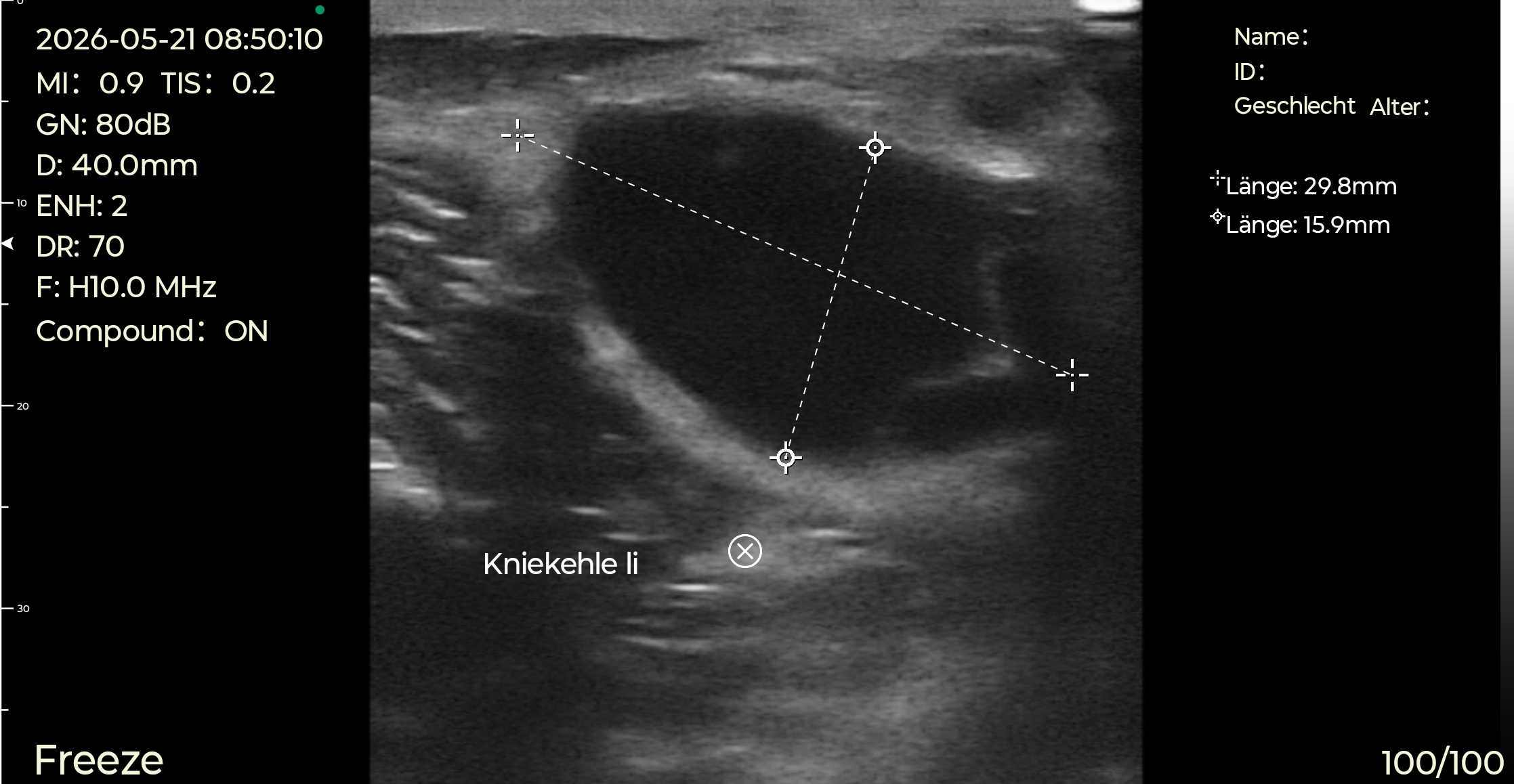
Seroma aspiration under ultrasound: how to aspirate a seroma, step by step
Seroma aspiration under ultrasound puts the needle where the scan shows the fluid: blind aspiration with a needle and syringe is common practice, while ultrasound delineates the margins of the seroma, places the needle and tracks the drainage (Stebbins 2011). One office series ran every tap under ultrasound guidance, 15 seromas in 310 abdominoplasties, with no hematomas (Swanson 2023), and a case report drained a recurring right-lower-quadrant seroma after abdominal liposuction the same way (Glashofer 2005). Here is how to aspirate a seroma in six steps.
- Check the entry on Doppler. Map the superficial epigastric vessels along the planned path before the needle goes in: at the level of the anterior superior iliac spine the superficial inferior epigastric vein lies a mean 3.5 mm deep, deeper as BMI rises, and the artery, found at that level on 22% of sides, a mean 4.9 mm (Kuo 2025, 48 MHz, 50 hemiabdomens). The reason is on record: a 430 mL subcutaneous abdominal-wall hematoma followed a rectus sheath block, with the superficial epigastric artery its most probable source (Shiraishi 2025). Which Doppler mode shows what is set out in the Doppler guide.
- Plan the path in-plane. With the needle along the probe’s long axis, the whole shaft shows as a bright line down to the tip; out-of-plane, the needle crosses the beam and shows only as a dot where it meets it (Saraiva 2026). The L14Pro and L20Pro draw in-plane and out-of-plane puncture guides on the image, add Needle Enhance for the shaft, and carry Grid and Flip.
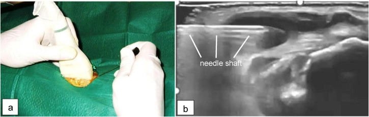
- Cover the probe and the gel. A probe used for guided procedures is a semi-critical device, and non-sterile gel can contaminate the prepared skin; 27.91% of 86 guided-injection studies reported a sterile probe cover (Kweon 2025), and after emergency-department procedures, 15.1% of polyethylene and 2.4% of polyurethane covers were perforated (Rippey 2025); the full set-up is laid out under sterile coupling for guided injections. The Suresult Ø35 × 4 mm sterile gel pads ($140 for 50, shipping and tax included) span the whole 25 mm array of the L14Pro and L20Pro, a needle entering within 5 mm of the array’s end passes through the pad on an in-plane pass, and through its 4 mm the 80 and 40 mm presets image 76 and 36 mm of tissue.
- Choose the needle. An 18 G sheathed needle drew 10 mL of serous fluid from a recurrent post-traumatic collection above the fascia over the sacrum, after three earlier taps of 110, 30 and 8 mL (Leach 2020). The Morel-Lavallée literature, which covers post-traumatic collections in the same plane between the subcutaneous fat and the fascia, recommends large-bore needles for thick fluid and leaves a 12–14 Fr catheter in place when a drain or a sclerosant is needed (Molina 2021).
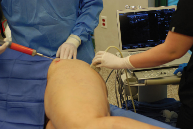
- Tap under view. Keep the tip inside the cavity as it empties and follow the walls in. In the clips from Alfonso 2022, on a 3 cm display, an IV catheter enters a seroma cavity under Scarpa’s fascia, and the cavity then collapses around it as the fluid drains.
- Rescan and log. Rescan the emptied cavity before the dressing goes on, record the three diameters and the volume drawn, and book the next scan. The ultrasound-guided office series averaged 3.5 taps per seroma (1–7), with mean first and last taps on day 11.6 and day 21 (Swanson 2023); Swanson’s earlier 551-case study averaged 5 office aspirations (1–12) for its 9 clinical seromas, the last at a mean of day 28 (Swanson 2013).
Pseudobursa after a tummy tuck: the encapsulated seroma and its seroma capsule
A pseudobursa is an encapsulated seroma: fluid held inside a seroma capsule of collagen sheets with no epithelial lining, which multiple aspirations do not relieve. Ersek and Schade 1990 described it in 9 patients after abdominolipectomy combined with suction, and advised waiting at least 6 weeks between suction and the largest resections. On ultrasound the chronic form shows fluid inside a dense fibrous capsule: in one woman the swelling began in week 3 after a tummy tuck and was still present at 3 years, about 540 mL of blood-tinged serous fluid was aspirated, and capsulectomy with a revision abdominoplasty ended it (Goldman 2018).
Capsulectomy is the cure the series agree on. In 6 patients whose scars turned asymmetric 3 months after abdominoplasty, ultrasound confirmed an encapsulated chronic seroma, and capsulectomy under local anesthesia worked in all 6 with no recurrence (di Summa 2013). Capsulectomy with negative-pressure wound therapy resolved all 15 recurrent chronic seromas in another series, 10 post-operative and 5 Morel-Lavallée lesions, with no recurrence (Marangi 2020). Roje 2006 evacuated a pseudobursa under ultrasound many times, 2,010 mL at one sitting, then excised it at a mini-abdominoplasty with progressive tension sutures, and it had not recurred at 3 months. The chronic seroma management ladder runs the same way: repeated puncture and aspiration resolves most seromas, bleomycin, doxycycline and talc have been used for recurrent ones, and surgery comes last (Vidal 2017).
The volume drawn flags recurrence early. In Morel-Lavallée lesions, aspirates over 50 mL came from 83% of the lesions that recurred against 33% of those that resolved, a well-developed pseudocapsule raised recurrence after aspiration and compression, and doxycycline sclerodesis resolved all 16 lesions that had failed simple aspiration (Molina 2021). One case report treated a recurring collection under an abdominoplasty flap, first drained of 440 cc, with a 50 cc aspiration and 15 mg of bleomycin, and ultrasound confirmed its resolution (Kiper 2026). A late cyst after liposuction can also be lymphatic: a thigh collection first read as a pseudobursa proved a lymphocele on lymphoscintigraphy and was resected after drainage failed (Wang 2017).
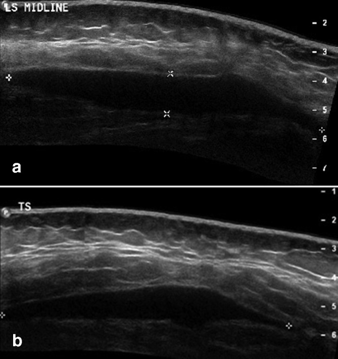
Seroma vs abscess, fat necrosis and oil cysts after body contouring
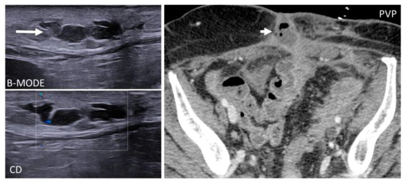
A seroma is told from an abscess, fat necrosis and an oil cyst after body contouring by each one’s own tell: an abscess has a thick, irregular wall that color Doppler can show hyperemic (Masino 2025; Porrello 2022), liquefied fat necrosis sits inside bright fat as multiple oval anechoic areas (Soliman 2024), and an oil cyst is anechoic and oval, with a sharp edge, a capsule and posterior enhancement (de Wilde 2022).
Seroma vs abscess turns on the wall and the patient. An abdominal-wall abscess is a superficial, complex collection with ill-defined margins, predominantly hypoechoic, and color Doppler can show peripheral hyperemia (Porrello 2022); a subcutaneous abscess is variably compressible with a hyperemic wall (Soliman 2024). Infection is the second most common complication of abdominoplasty, at an estimated 1% to 3.8% in Vidal 2017’s review, which also pictures an infected seroma two weeks after a lipoabdominoplasty. At the bedside, point-of-care ultrasound found skin and soft-tissue abscesses with 94.6% sensitivity and 85.4% specificity across 14 studies and 2,656 patients, and with 98.7% and 91.0% in adults (Gottlieb 2020). Mycobacterium abscessus after liposuction runs slower: symptoms appeared about a month after surgery, and identifying the organism took a mean of 33 days from presentation (Tuan 2024).
Rao 2014 proposes defining fat necrosis at the bedside as a palpable, discrete, persistent subcutaneous firmness of at least 1 cm. On ultrasound, liquefying fat lobules show as multiple oval anechoic areas inside hyperechoic fat (Soliman 2024), and in a series of 33 patients with trunk and limb fat necrosis, most lesions showed internal fat lobules, septa and a surrounding pseudocapsule on ultrasound and MRI, with 17 in the buttock or thigh (Robinson 2008). Rescan a tapped fat-necrosis collection within days: after extensive body contouring, about 100 cc of noninfectious fluid drawn under ultrasound from fat necrosis in the rectus abdominis was back, with the same mass, 4 days later, and surgical drainage followed (Boudousq 2025). At the cystic end of the spectrum, the oil cyst is anechoic with a sharp edge, a capsule and posterior enhancement at 14 MHz (de Wilde 2022).
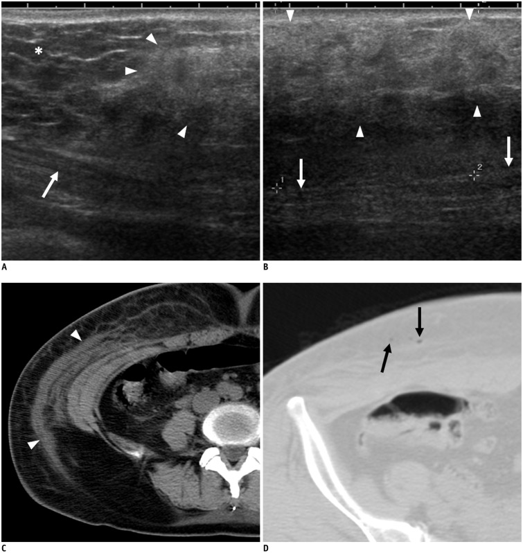
In the first days after liposuction the treated fat changes its own echo: three days out, the abdominal wall shows a heterogeneous hyperechoic area in the subcutaneous layer, set against the normal fat beside it, the edema and infiltration of the treated tissue (You 2015). A seroma stands out against it as a dark, defined pocket.
Fibrosis after lipo on ultrasound: bright fibrous bands in place of fluid
Fibrosis after lipo shows on ultrasound as hyperechoic fibrous strands in the subcutaneous fat, the reverse of a seroma’s dark pocket; a review of five studies pictures them in an abdominoplasty at 6 weeks (Widgerow 2026). At 20 MHz, Claytor 2021 scanned three fixed points after medial thigh liposuction and graded induration, edema and fibrous banding at weeks 1, 2, 4, 7 and 10, reading a brighter image as denser tissue. At secondary liposuction, a year or more after the first, intraoperative ultrasound improved the detection of fibrotic lesions (Tambasco 2025). Reported rates run from 1% for fibrosis or induration after isolated liposuction (Aljerian 2024) through 2.3% over five years of follow-up (Gentile 2026) to transient fibrosis in 2.7% after high-definition liposuction (Willet 2023) and 3.2% after deep trunk liposuction (Valente 2026).
Fibrosis after liposuction responds to physiotherapy: twelve sessions of therapeutic ultrasound followed by manual lymphatic drainage, three a week, reduced fibrosis, swelling and pain in 18 women a mean of 70 days after liposuction or lipoabdominoplasty (Masson 2014), and a review of 7 studies found manual lymphatic drainage reduced swelling after cosmetic surgery such as liposuction and abdominoplasty, with better results when started on days 2 to 5 and given in 6 to 12 sessions (Alsairefi 2026).
BBL donor and recipient sites: seromas, fat necrosis cysts and guided fat placement
After a BBL, seromas collect at the donor site: in 185 buttock augmentations with ultrasonic liposuction and ultrasound-guided fat grafting, seroma was the most common complication, 10 patients (5.41%), mostly in the lower anterior abdomen, and every one was drained successfully (Elsaftawy 2024). The recipient site raises a different collection: a soft-tissue blowout deformity after gluteal fat transfer often holds a fat necrosis cyst, which is evacuated through a liposuction cannula with its capsule fenestrated, or removed through a direct buttock-lift incision (Costa 2026).
Ultrasound already guides the grafting. The Aesthetic Society’s practice advisory calls for ultrasound-guided documentation of cannula placement before and during fat injection, and caps BBL at 3 cases a day (Del Vecchio 2022). BAAPS states that intraoperative ultrasound guides subcutaneous fat placement and avoids inadvertent deep lipofilling (Tillo 2023). Yet in a survey of 178 board-certified plastic surgeons, 48% of those performing BBL, 80 surgeons, did not use ultrasound (Finkelstein 2023). One office’s year of scans shows the same probe working after surgery: of 1,000 scans, 28 checked post-operative abdomens for seromas, 10 drained seromas, 4 evaluated buttock abscesses and 3 assessed buttock fat injection during surgery (Swanson 2018, office ultrasound series).
| Series | Seroma | Otros hallazgos | Fuente |
|---|---|---|---|
| 7,000 consecutive BBL procedures | Seroma (23%) listed among the minor complications | Anemia 6.6%, fat necrosis 1.9%, infection 0.43%, hematoma 0.14% | Güzey 2026 |
| 1,815 patients, ultrasound-guided gluteal fat transfer | 1.2% | Complications 4%; skin ischemia 1.2%; surgical site infection 0.8%; no macroscopic fat embolism and no deaths | Vidal-Laureano 2024 |
| 4 studies, 6,235 women, ultrasound-guided grafting | 2.94 per 100 | Minor complications 6.32 per 100; fat necrosis 0.09 per 100; infection 0.23 per 100; mortality and embolism 0 | Milani Reis 2025 |
| 185 buttock augmentations, ultrasound-guided grafting | 10 patients (5.41%), mostly in the lower anterior abdomen; all drained | One abscess (0.54%); no liponecrosis | Elsaftawy 2024 |
Gluteal fat sits within a linear’s reach. Across 150 adults, the superficial fatty layer was thickest over the buttock, about 2 cm on average and from 6 to 38 mm (Cotofana 2020); a meta-analysis of 734 adults put the ventrogluteal subcutaneous tissue at 19.93 mm (Gulnar 2023); and after fat transfer averaging 287 mL per buttock, the fat layer gained a mean 0.66 cm on the right and 0.86 cm on the left, with no fat necrosis or oily cysts on the scans (Swanson 2016, buttock fat transfer study). Those depths sit inside the L14Pro’s 40 and 60 mm presets, and the superficial layer inside the L20Pro’s 40 mm.
What depth and frequency a seroma scan needs, and which Suresult probe covers it
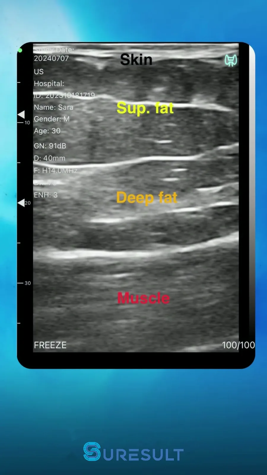
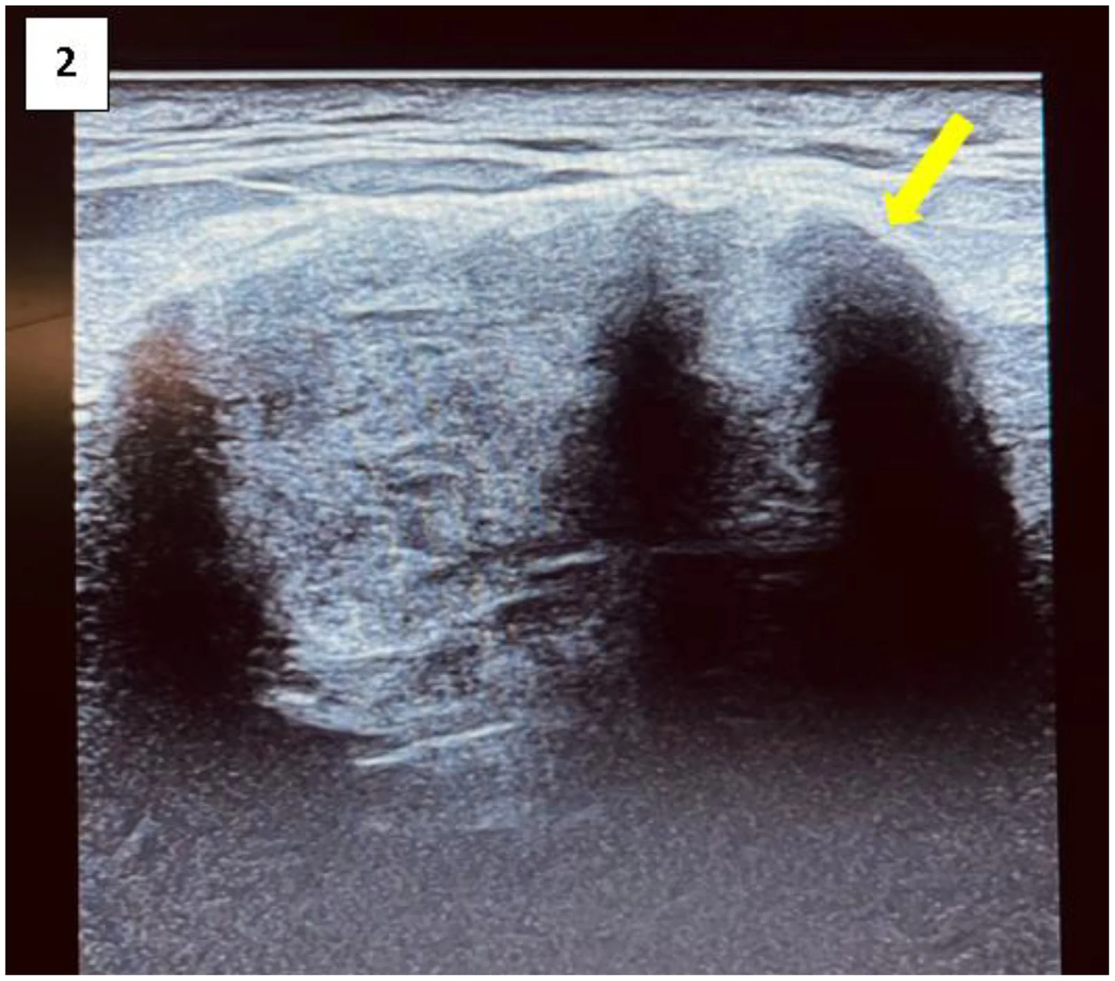
A seroma scan needs a linear of 7 to 15 MHz or higher for the flap and the collection, and a lower-frequency curvilinear probe once a collection runs deep (Pistoia 2025; Masino 2025). The source depths set the presets: mean skin-to-fascia depth measured 37 to 42 mm across four abdominal regions before large-volume liposuction and 21 to 22 mm a week after it at 7 to 12 MHz (Sudha 2026), Alfonso 2022 imaged abdominoplasty seromas at 12 MHz on a 4 cm display, and Masino 2025 advises a panoramic view for a large superficial lesion such as a 13.6 × 4.9 × 5.9 cm post-traumatic subacute gluteal hematoma.
| Site or collection | Depth or size in the source | Ecógrafo de mano Suresult | The spec that decides | Fuente |
|---|---|---|---|---|
| Flap a week after large-volume liposuction | Mean skin to fascia 21–22 mm across four regions | L20Pro | 16/20 MHz on the 30 and 40 mm presets | Sudha 2026 |
| Abdomen before large-volume liposuction | Mean skin to fascia 37–42 mm across four regions | L14Pro | 10/14 MHz on the 60 mm preset | Sudha 2026 |
| Abdominoplasty seroma on the fascia | About 200 cc on a 4 cm display at 12 MHz | L14Pro | The 40 and 60 mm presets hold flap, fluid and fascia | Alfonso 2022 |
| Arm before arm liposuction | 22.25 mm (12.9–37.2) at maximal convexity | L20Pro | The 40 mm preset at 16/20 MHz | Benito-Ruiz 2025 |
| Gluteal superficial fat | About 2 cm, from 6 to 38 mm | L20Pro or L14Pro | The 40 mm preset on either | Cotofana 2020 |
| Ventrogluteal subcutaneous tissue | 19.93 mm | L20Pro | The 30 and 40 mm presets at 20 MHz | Gulnar 2023 |
| Superficial epigastric vein and artery at the anterior superior iliac spine | Mean depths 3.5 mm and 4.9 mm | L20Pro | 20 MHz with Color, Power and PW Doppler | Kuo 2025 |
| Recurrent post-traumatic supra-fascial collection over the sacrum | About 4 to 5.5 cm deep; 10 × 10.9 × 1.4 cm | L14Pro | The 60 and 80 mm presets | Leach 2020 |
| Rectus sheath hematoma | 136 × 120 × 69 mm on a convex probe | D3Ultra or C5SH-Pro | Convex presets from 90 to 300 mm at 3.2/5.0 MHz | Huang 2024 |
| Post-traumatic subacute gluteal hematoma | 13.6 × 4.9 × 5.9 cm, panoramic view advised | L10W for width; D3Ultra or C5SH-Pro for depth | An 80 mm linear field on the L10W; convex depth to 300 mm | Masino 2025 |
That spread is why the L14Pro leads: its 20 to 80 mm presets at 10/14 MHz keep the skin, the flap, the collection and the fascia in one image, and reach the 4 to 5.5 cm depth of a recurrent post-traumatic collection over the sacrum. The L20Pro high-frequency linear probe ($3,720) takes the thin flap after liposuction, the superficial gluteal fat and the superficial epigastric vessels at 16/20 MHz within 40 mm. For collections deeper or wider than the L14Pro and L20Pro frame, the D3Ultra handheld ultrasound ($2,976) is a dual-head 3-in-1 with convex presets from 90 to 300 mm and linear presets to 100 mm; the C5SH-Pro convex probe ($4,185) images to 300 mm on 256 elements; and the L10W wide linear probe ($2,232) spreads an 80 mm field at 7.5/10 MHz, so more of a broad pocket and the needle path show in one view. Frequency by depth is set out in the guía de ecografía de alta frecuencia, and the linears side by side in the portable linear probe ranking.
The 6 best handhelds for seroma and hematoma checks after body contouring, ranked
My ranking for post-operative seroma and hematoma work puts the Suresult L14Pro first at 95.0/100, then the Mindray TE Air e5M at 86.1, the Clarius L7 HD3 at 76.0, the Butterfly iQ3 at 72.7, the GE Vscan Air CL at 67.0 and the Philips L12-4 at 61.7. Each card is one maker’s lead handheld, scored on the seven criteria under How I scored, with every rival price read on November 1, 2026.
01 Suresult L14Pro — Best Overall for Seroma and Hematoma Checks After Body Contouring
10/14 MHz linear on 256 elements · presets of 20, 40, 60 and 80 mm · Color, Power and PW Doppler with the probe · Needle Enhance with in-plane and out-of-plane puncture guides · $3,441
Puntuación: 95,0/100
4 hours of scanning on a 4,400 mAh battery · 145 g · 156 × 60 × 18 mm · IP67 and drop-tested · no subscription · $3,441
See the Suresult L14Pro linear ultrasound probe: $3,441
Veredicto: Buy the L14Pro if you follow liposuction, abdominoplasty and BBL patients and tap your own seromas. I rank it first because the flap, the collection and the fascia fit one 20–80 mm view at 10/14 MHz, all three Doppler modes clear the entry before the needle, Needle Enhance and the guides carry the tap, and a volume from three diameters goes into the record at every visit.
- Profundidad: 40 mm frames a seroma on the fascia under an abdominoplasty flap, 60 mm keeps the fascia in view under mean skin-to-fascia depths of 37 to 42 mm before large-volume liposuction, and 80 mm reaches a post-traumatic collection lying 4 to 5.5 cm down over the sacrum
- Doppler: Color, Power and PW with the probe, to clear the superficial epigastric vein and artery, at mean depths of 3.5 and 4.9 mm at the anterior superior iliac spine, before the needle goes in
- Needle: Needle Enhance plus in-plane and out-of-plane puncture guides, Grid and Flip on a 25 mm array that the Ø35 × 4 mm sterile gel pad covers end to end
- Measurement: length, area and circumference, angle, trace and distance, with Measure Magnify; the Suresult app calculates a volume from three diameters
- Registros: up to 1,000 frames of cine, about 165 seconds; images and video kept on your own phone, tablet or PC; JPEG, PNG, MP4 and DICOM export
Por qué ocupa este puesto. The L14Pro takes the maximum on five of the seven criteria: depth, on its 80 mm preset; detail, at 14 MHz; Doppler, with Color, Power and PW in the purchase; needle guidance, with Needle Enhance and the guides; and five-year cost, where $3,441 is the lowest total among the six card leads and sets the scale. Measurement earns 7 of 10 for calipers plus a volume from three diameters, and records 3 of 5 for 1,000 frames of cine with storage and DICOM.
Sub-scores: depth 20/20, detail 15/15, Doppler 20/20, needle guidance 15/15, measurement 7/10, records 3/5, five-year cost 15/15.
The L14Pro and every Suresult handheld on this page are autorizada mediante 510(k) K211321.
L14Pro or L20Pro? Both Pros carry 256 elements, Color, Power and PW Doppler, Needle Enhance and up to 4 hours of scanning at 145 g, so depth decides. The L14Pro’s 10/14 MHz band runs to 80 mm, deep enough for the fascia under a thick abdominal flap and for the collections beneath it. The sonda lineal de alta frecuencia Suresult L20Pro ($3,720) runs 16/20 MHz on presets of 10 to 40 mm for the thin flap after liposuction, the superficial gluteal fat and the superficial epigastric vessels. Both include 3 months of FaceAtlas AI for facial work.
Ideal para: surgeons and their PAs, NPs and nurses who scan, measure and tap post-operative collections on the abdomen, flanks and buttocks with one probe. Elija un modelo de la familia si: your collections stay within 40 mm (the L20Pro), or you want the L14Pro’s band and presets on 192 elements at $2,139 (the L14). Compare cuidadosamente si: one device also has to image the whole body in a convex mode (the Suresult D3Ultra in the family block, or the Mindray TE Air e5M).
También en esta familia
Suresult L20Pro 16/20 MHz linear ultrasound · $3,720 · 73.9/100. The card’s pick for thin flaps and superficial pockets within 40 mm: 16/20 MHz on 256 elements, presets of 10, 20, 30 and 40 mm, all three Doppler modes and Needle Enhance. The depth criterion rewards reach beyond 40 mm, where the L14Pro’s 60 and 80 mm presets lead the card.

Suresult L14 wireless linear probe · $2,139 · 88.0/100. Scored for the record: the L14Pro’s 10/14 MHz band and 20–80 mm presets on 192 elements, with Color, Power and PW Doppler, the in-plane and out-of-plane puncture guides, 2 hours of scanning and 200 g; the L14Pro adds Needle Enhance and 256 elements.
Suresult L20 high-frequency linear probe · $3,000 · 75.0/100. Scored for the record: the L20Pro’s 16/20 MHz band and 10–40 mm presets on 192 elements, with all three Doppler modes, Needle Enhance, 2 hours of scanning and 147 g.
Ecógrafo de mano de doble cabezal Suresult D3Ultra · $2,976. Unranked, for collections deeper than the L14Pro and L20Pro reach: a dual-head 3-in-1 with convex presets from 90 to 300 mm and linear presets to 100 mm, with all three Doppler modes and Needle Enhance.
Suresult C5SH-Pro convex ultrasound probe · $4,185. Unranked: a 3.2/5.0 MHz convex on 256 elements with presets from 90 to 300 mm, for a deep hematoma such as one inside the rectus sheath.
Suresult L10W wide linear ultrasound probe · $2,232. Unranked: an 80 mm field at 7.5/10 MHz on 256 elements, so more of a broad pocket and the needle path show in one view.
02 Mindray TE Air e5M — Best for Full Doppler, Needle and Measurement Tools at the Lowest Five-Year Rival Cost
Convex and linear modes in one transducer · 2.0–9.0 MHz bandwidth, linear B-mode bands to 8.2 MHz · images to 40 cm · Color, Power and PW Doppler standard · B-mode Volume and Extended FOV · $4,499
Score: 86.1/100
128 elements · 44.2 × 11.5 mm footprint · 229 g · 3-year warranty · no subscription plan · $4,499
Veredicto: Buy the TE Air e5M if you want all three Doppler modes, both needle tools and the full measurement set in one purchase with nothing to renew: Color, Power and PW Doppler, iNeedle and the VA Grid, a B-mode Volume tool and Extended FOV, on a transducer that switches between convex and linear modes for whole-body imaging. Its linear B-mode bands run in three steps from 2.0 to 8.2 MHz, so it reads the flap and the collection at lower frequencies than the 14 MHz pick.
- Doppler: Color, Power and PW, all standard
- Herramientas para agujas: iNeedle needle-visualization enhancement and the VA Grid, both standard on the e5M
- Measurement: distance and area; Smart Calc for automatic diameter and area; B-mode Volume in the EM FAST, EM AAA, GYN and OB packages; Extended FOV of 180° on the linear and 70° on the convex
- Registros: 125 frames of cine, preset from 2 to 5 seconds; DICOM 3.0 storage; patient data managed on board; exam images and reports sent to Tricefy
- Price and clearance: $4,499 from Mindray North America’s TE Air store, checked November 1, 2026, with a 3-year warranty and no subscription plan; cleared by the FDA under 510(k) K240906 on July 23, 2024
Por qué ocupa este puesto. The e5M is the one handheld in this ranking with the full score on Doppler, needle guidance and measurement together: all three Doppler modes in the purchase, iNeedle with the VA Grid, and calipers, Volume and Extended FOV for the full 10. Images to 40 cm earn the full depth credit. Detail scores 6.67 of 15 on the 9.0 MHz top of its bandwidth, records 3 of 5 for DICOM storage and the Tricefy report, and $4,499, the lowest five-year total among the rivals, 11.47 of 15.
Sub-scores: depth 20/20, detail 6.67/15, Doppler 20/20, needle guidance 15/15, measurement 10/10, records 3/5, five-year cost 11.47/15.
Limitaciones. Detail tops out at 9.0 MHz, the edge of its bandwidth, and the linear B-mode bands at 8.2 MHz; B-mode Volume runs in the EM FAST, EM AAA, GYN and OB packages; cine holds 2 to 5 seconds.
Ideal para: practices that scan the whole body as well as the abdominal wall and want every scored tool paid once. Descártelo si: you want 14 MHz detail in the flap and the superficial vessels, or cine long enough to replay a whole tap.
03 Clarius L7 HD3 — Best for 13 MHz Detail to 15 cm on a Membership
4–13 MHz linear · images to 15 cm · 38 mm field of view · Color and Power included, PW with Essentials · Needle Enhance with Essentials; the Center Guide · $3,795 plus Essentials
Score: 76.0/100
288 g · 147 × 76 × 32 mm · B, M, Color and Power modes on the scanner · $3,795 plus Essentials
Veredicto: Buy the L7 HD3 if you want a 13 MHz wireless linear with images to 15 cm and will hold an Essentials membership for PW Doppler, Needle Enhance, local storage, DICOM and PDF Reports. Color and Power Doppler come with the scanner, and the Center Guide draws a line down the middle of the image for injections and line placements.
- Imagen: 4–13 MHz across a 38 mm field of view, to 15 cm; B, M, Color and Power with the scanner
- Membership tools: PW Doppler and Needle Enhance with Essentials, which also adds unlimited cloud storage and PDF Reports
- Needle aid: the Center Guide, a line in the center of the image and on the scanner for injections or line placements
- Measurement and records: measurements of an area of interest or of the depth from the skin line; 30-second cine; local storage and DICOM with the membership
- Precio y plan: $4,490 with the first year of Essentials, checked November 1, 2026. Sold with an Essentials plan: $695/yr, $1,785/3 yr, or $2,500 one-time
Por qué ocupa este puesto. Images to 15 cm take the full depth credit and 13 MHz 13.33 of 15 for detail. The scanner includes Color and Power, and Essentials adds PW, for 17 of 20 on Doppler; Needle Enhance on the membership plus the Center Guide earn 11.5 of 15; the calipers take 3 of the 10 measurement points; storage, DICOM and PDF Reports earn 3 records points; and $6,295 over five years with one-time Essentials scores 8.20 of 15.
Sub-scores: depth 20/20, detail 13.33/15, Doppler 17/20, needle guidance 11.5/15, measurement 3/10, records 3/5, five-year cost 8.20/15.
Limitaciones. PW Doppler, Needle Enhance, local storage and DICOM sit in Essentials; cine runs 30 seconds; measurement runs on calipers.
Ideal para: surgeons who want 13 MHz detail through the abdominal wall and down to 15 cm on one linear, and who keep Essentials running. Descártelo si: PW Doppler, needle enhancement and DICOM have to come with the scanner itself, or you want a volume from three diameters on it.
También en esta familia
Clarius C3 HD3: a 2–6 MHz scanner with images to 40 cm, a 73° field of view and 308 g.
Clarius L15 HD3: a 5–15 MHz linear with images to 7 cm, a 50 mm field of view and 290 g, at $3,795 plus Essentials, checked November 1, 2026.
L7 HD3 at the Clarius store · What Clarius scanners and memberships cost in 2026
04 Butterfly iQ3 — Best for Manual Volume and Biplane Views on One Whole-Body Probe
One 1–12 MHz probe · images to 30 cm, MSK-Soft Tissue preset to 12 cm · Color on Core, Power and PW on Advanced · Needle Viz and Biplane on Advanced · Manual Volume · $3,899 plus a plan
Score: 72.7/100
Trapezoidal imaging · midline marker · unlimited cloud storage on Core or Advanced · 3-year warranty · $3,899 plus a plan
Veredicto: Buy the iQ3 if a single whole-body probe with a three-diameter volume tool suits your office: Manual Volume runs the prolate ellipsoid, 0.52 × D1 × D2 × D3, in the Abdomen, MSK-Soft Tissue and Small Organ presets among others. Its Doppler modes come with the Core and Advanced plans, and its needle tools with Advanced.
- Volume: Manual Volume in the Abdomen, Abdomen Deep, Bladder, MSK-Soft Tissue, Musculoskeletal, Nerve and Small Organ presets; Auto Bladder Volume on Advanced
- Doppler: Color and M-Mode on Core at $299 a year; Power and PW on Advanced
- Herramientas para agujas: Needle Viz, Biplane and the out-of-plane needle view on Advanced; a midline marker
- Measurement and records: four linear calipers and one ellipse; unlimited cloud storage on Core or Advanced; onboard customizable worksheets and a calculations report; a PACS connection, with Basic DICOM sold separately
- Precio y plan: $4,198 with a year of Core and a 3-year warranty, checked November 1, 2026; Advanced at $420 a year or $1,500 for five years with no recurring fees
Por qué ocupa este puesto. Images to 30 cm take the full depth credit and 12 MHz 11.67 of 15. Every Doppler mode sits on a paid plan, for 10 of 20; Needle Viz on Advanced plus the midline marker earn 11.5 of 15; calipers and Manual Volume earn 7 of 10, level with the L14Pro; the worksheets, the calculations report, cloud storage and DICOM earn 3 records points; and $5,399, with Advanced bought once for five years, scores 9.56 of 15.
Sub-scores: depth 20/20, detail 11.67/15, Doppler 10/20, needle guidance 11.5/15, measurement 7/10, records 3/5, five-year cost 9.56/15.
Limitaciones. Color sits on Core, and Power, PW, Needle Viz and Biplane on Advanced; Basic DICOM is a separate purchase; one whole-body probe covers 1 to 12 MHz.
Ideal para: offices that want one probe across the whole body, with an ellipsoid volume on the measured collection. Descártelo si: you want Doppler and the needle tools in the purchase, or a dedicated 14 MHz linear for the flap.
Especificaciones de Butterfly iQ3 · Butterfly plans and pricing
05 GE Vscan Air CL — Best for Linear and Curved Heads With Color and PW in One Price
Dual head: 3–12 MHz linear to 8 cm and 2–5 MHz curved to 24 cm · Color and PW Doppler in the purchase · Centerline Marker · $4,999
Score: 67.0/100
205 g · 40 × 7 mm linear lens and 64 × 16 mm curved lens · 3-year warranty · $4,999
Veredicto: Buy the Vscan Air CL if you want a linear and a curved head on one wireless probe, with Color and PW Doppler and the Centerline Marker in the price and the Innovations plan as an option. GE lists free-hand needle and catheter placement and fluid drainage among the linear head’s interventional indications.
- Imagen: linear 3–12 MHz, centered at 7.7 MHz, to 8 cm; curved 2–5 MHz to 24 cm; B, Color, B+Color, Harmonics, M and PW modes
- Needle aid: the Centerline Marker, which GE says “should not be confused with biopsy guidelines indicating needle path on the image”
- Measurement: distance, circumference and area, ellipse, angle, time and velocity; bladder volume from three distances on Innovations
- Registros: up to 500 exams stored on the phone or tablet; wireless DICOM export to PACS
- Precio y plan: $4,999, checked November 1, 2026, with a 3-year warranty; Auto Bladder Volume, Lung Guidance and HD-SRI sit in the optional Innovations plan, at $500 yearly or $1,299 for three years
Por qué ocupa este puesto. The curved head’s 24 cm takes the full depth credit and the linear’s 12 MHz 11.67 of 15. Color and PW in the purchase earn 13 of 20 on Doppler, the Centerline Marker 8 of 15 on needle guidance, calipers 3 of 10 and storage with DICOM 1 of 5, and $4,999 with every scored tool included scores 10.32 of 15.
Sub-scores: depth 20/20, detail 11.67/15, Doppler 13/20, needle guidance 8/15, measurement 3/10, records 1/5, five-year cost 10.32/15.
Limitaciones. Doppler covers Color and PW; needle work runs on the Centerline Marker; the bladder volume tool sits in Innovations.
Ideal para: practices that want a curved head for deep abdominal views and a linear head for the wall in one purchase. Descártelo si: you want Power Doppler, a needle-enhancement mode or a volume from three diameters on the collection.
También en esta familia
GE Vscan Air SL: a 3–12 MHz linear to 8 cm with a 40 × 7 mm lens, paired with a 1.6–3.7 MHz sector head to 24 cm.
06 Philips Lumify L12-4 — Best for Steerable Color With Fast Flow and Slow Flow, Bought Outright
12–4 MHz linear · images to 12 cm · footprint 34 mm, aperture 38 mm, field of view 34.5 mm · PW and steerable color in Fast Flow and Slow Flow modes · $10,307.95 outright
Score: 61.7/100
Center line marker · biopsy capable · loops of 1–10 s · all software included · $10,307.95
Veredicto: Buy the Lumify L12-4 if you want steerable color with two flow modes, Fast Flow and Slow Flow, on a 12 MHz linear bought outright with every software feature. It images to 12 cm, with PW Doppler and a center line marker.
- Doppler: steerable color with Fast Flow and Slow Flow; PW
- Needle aid: a center line marker; the transducer is biopsy capable
- Measurement: up to four linear distances, plus ellipse area and circumference
- Registros: loops of 1 to 10 seconds; hundreds of exams stored; export to PACS by DICOM, by email, or to a shared network or local directory
- Precio y plan: $10,307.95 new and outright at PartsSource, with 90 days of warranty, or $7,929.19 on exchange, checked November 1, 2026; Philips sells Lumify outright with every software feature and no subscription, and gives a 5-year basic warranty on Lumify transducers bought through Philips
Por qué ocupa este puesto. Images to 12 cm take the full depth credit and 12 MHz 11.67 of 15. Color and PW earn 13 of 20, the center line marker 8 of 15, calipers 3 of 10 and storage with DICOM 1 of 5. At $10,307.95 the L12-4 carries the highest five-year total in the ranking, which scores 5.01 of 15.
Sub-scores: depth 20/20, detail 11.67/15, Doppler 13/20, needle guidance 8/15, measurement 3/10, records 1/5, five-year cost 5.01/15.
Limitaciones. Doppler covers color and PW; the needle aid is a center line marker; loops run 1 to 10 seconds; the published price comes from PartsSource.
Ideal para: clinics that want two color-flow modes and PW on a 12 MHz linear for the abdominal wall, with every software feature paid once. Descártelo si: you want Power Doppler, a needle-enhancement mode or a lower five-year cost.
También en esta familia
Philips Lumify C5-2: a 5–2 MHz transducer with images to 30 cm, a 60 mm aperture and a 67.5° field of view.
What each handheld costs over five years, plan included
Over five years the Suresult L14Pro costs $3,441, the price of the probe itself: its imaging app, all three Doppler modes and Needle Enhance carry no subscription, so nothing else enters the total. On the prices I read on November 1, 2026, the Mindray TE Air e5M comes to $4,499 with nothing to renew, the Clarius L7 HD3 to $6,295 once Essentials is bought outright, the Butterfly iQ3 to $5,399 with Advanced prepaid for five years, the GE Vscan Air CL to $4,999 with every tool it scores on in the price, and the Philips L12-4 to $10,307.95 outright at PartsSource. For every rival I took the cheapest published route to the tools it is scored on.
| De mano | Purchase price | Plan its scored tools need | Five years with one-time software | Five years with yearly software |
|---|---|---|---|---|
| Suresult L14Pro | $3,441 | Every tool it scores on comes with the probe: Color, Power and PW Doppler, Needle Enhance, the puncture guides and the imaging app, with no subscription | $3,441 | $3,441, with no subscription to renew |
| Mindray TE Air e5M | $4,499 | Every tool it scores on is standard: Color, Power and PW Doppler, iNeedle, the VA Grid, B-mode Volume, Extended FOV and DICOM | $4,499 | $4,499; Mindray sells no subscription plan for the probe |
| Clarius L7 HD3 | $3,795, sold with an Essentials plan | Essentials at $2,500 one-time adds PW Doppler, Needle Enhance, local storage, DICOM and PDF Reports | $6,295 | $7,270 at $695 a year for five years; $6,970 with one three-year term at $1,785 and two yearly renewals |
| Butterfly iQ3 | $3,899, sold with a membership plan | Advanced at $1,500 for five years adds Color, Power and PW Doppler and Needle Viz; Basic DICOM, which the records score credits, is a separate purchase outside the total | $5,399 | $5,999 at $420 a year on Advanced |
| GE Vscan Air CL | $4,999 | The price covers Color and PW Doppler, the Centerline Marker, calipers and DICOM export | $4,999 | $4,999; the optional Innovations plan, $1,299 for three years and then $500 a year, makes it $7,298 and sits outside the score |
| Philips L12-4 | $10,307.95 | Every software feature is in the outright purchase | $10,307.95 | $10,307.95; Philips sells the L12-4 outright with no subscriptions |
Suresult prices on this page update live from the Suresult store. The rival figures are the published prices read on November 1, 2026: Mindray’s from its North America TE Air store, Clarius’s, Butterfly’s and GE’s from their own stores and pricing pages, and the Philips L12-4’s from PartsSource.
Suresult prices are final, with No Hidden Fees: delivery is duties-paid, each probe comes with a 30-day trial and a 2-year warranty, and imaging and Doppler run with no subscription for the life of the probe. The handhelds that run without a subscription are ranked against each other in their own guide.
Cómo asigné las puntuaciones
Seven criteria, with weights set on November 1, 2026 before any rival was scored, make up each card’s score out of 100:
- Depth (20): the full 20 at an image depth of 80 mm or more, none below 40 mm, and 20 × (depth in mm − 40) ÷ 40 in between, on the deepest preset or image depth the maker states.
- Detail (15): 15 × (top frequency in MHz − 5) ÷ 9, held between 0 and 15, so 14 MHz earns the full 15, 13 MHz 13.33 and 12 MHz 11.67.
- Doppler (20): Color 7, Power 7 and PW 6; a mode included in the purchase earns its full points, one behind a paid tier half, and a missing mode none.
- Needle guidance (15): 7 for a needle-enhancement mode and 8 for in-plane or out-of-plane guide lines, a needle-path overlay or biplane, on the same full, half and none rule; I read an access grid and a centerline or midline marker the maker names for needle work as guide lines.
- Measurement and volume (10): 3 for distance and area calipers, 4 for a volume from three diameters or an ellipsoid tool, and 3 for an extended field of view or panoramic imaging, on the same full, half and none rule; I credited volume only where a general preset offers it, so a bladder-only volume tool scores nothing here.
- Records (5): 2 for cine of 60 seconds or more, 1 for app or on-device storage plus DICOM export and 2 for a visit comparison or report; I credited a report or worksheet built from a general exam.
- Five-year cost (15): 15 × the lowest five-year total among the six card leads ÷ this card’s total, each total carrying the cheapest published route to the scored tools; the L14Pro’s $3,441 is the lowest and sets the scale.
| Card and rank | Depth (20) | Detail (15) | Doppler (20) | Needle guidance (15) | Measurement (10) | Records (5) | Five-year cost (15) | Total (100) |
|---|---|---|---|---|---|---|---|---|
| 1 · Suresult L14Pro | 20 | 15 | 20 | 15 | 7 | 3 | 15 | 95.0 |
| 2 · Mindray TE Air e5M | 20 | 6.67 | 20 | 15 | 10 | 3 | 11.47 | 86.1 |
| 3 · Clarius L7 HD3 | 20 | 13.33 | 17 | 11.5 | 3 | 3 | 8.20 | 76.0 |
| 4 · Butterfly iQ3 | 20 | 11.67 | 10 | 11.5 | 7 | 3 | 9.56 | 72.7 |
| 5 · GE Vscan Air CL | 20 | 11.67 | 13 | 8 | 3 | 1 | 10.32 | 67.0 |
| 6 · Philips L12-4 | 20 | 11.67 | 13 | 8 | 3 | 1 | 5.01 | 61.7 |
Card 1’s siblings are scored against the same L14Pro cost reference, each criterion held at its maximum: the L14 88.0, the L20 75.0 and the L20Pro 73.9. The L14 matches the L14Pro on every criterion but needle guidance, where its puncture guides earn 8 and the L14Pro’s Needle Enhance adds the other 7. The L20 and L20Pro take full detail, Doppler and needle points at 16/20 MHz and 0 on depth, since their presets run to 40 mm, where the depth criterion starts counting, and the L20Pro’s $3,720 scores 13.88 on cost. That depth is why the L14Pro leads the card.
I reran the order the six declared ways and under fifteen alternative readings of the sources. The L14Pro stays first in the declared run; with the cost, depth or measurement criterion removed; with every rival’s paid needle and Doppler tools counted as included; and in all thirteen single-reading checks. Four runs change the top of the order. S3 moves full depth credit out to 100 mm, past the L14Pro’s 80 mm preset, and puts the Mindray 1.1 points ahead; pockets that deep go to the convex D3Ultra or C5SH-Pro. S5 scores the card on the L20Pro, whose 40 mm ceiling earns 0 on depth, which is why the L14Pro leads the card. RC14 stacks every reading that favors the rivals, S3 and S6 among them, and puts the Butterfly iQ3 first at 89.2 with the L14Pro third at 85.0; RC15 adds three narrowing readings to RC14, and the L14Pro moves up to second. Rival prices were read on November 1, 2026, and the rival plans and tool lists between October 29 and November 1.
| Run | Suresult card | Mindray TE Air e5M | Clarius L7 HD3 | Butterfly iQ3 | GE Vscan Air CL | Philips L12-4 |
|---|---|---|---|---|---|---|
| S0, the declared run | 95.00 | 86.14 | 76.03 | 72.73 | 66.99 | 61.67 |
| S1, cost criterion removed | 80.00 | 74.67 | 67.83 | 63.17 | 56.67 | 56.67 |
| S2, depth criterion removed | 75.00 | 66.14 | 56.03 | 52.73 | 46.99 | 41.67 |
| S3, full depth credit moved to 100 mm, counted from 60 mm | 85.00 | 86.14 | 76.03 | 72.73 | 66.99 | 61.67 |
| S4, measurement criterion removed | 88.00 | 76.14 | 73.03 | 65.73 | 63.99 | 58.67 |
| S5, card scored on the L20Pro | 75.00 | 87.07 | 76.70 | 73.50 | 67.83 | 62.08 |
| S6, rival paid needle tools and Doppler counted as included | 95.00 | 86.14 | 82.53 | 86.23 | 66.99 | 61.67 |
| RC14, S3 and S6 together, with bladder-only volume credited at its tier, GE’s OB report credited and Butterfly’s trapezoidal view counted as extended field of view | 85.00 | 86.14 | 82.53 | 89.23 | 67.74 | 61.67 |
| RC15, RC14 with centerline and midline markers, the VA Grid and Mindray’s Extended FOV left uncredited | 85.00 | 75.14 | 74.53 | 89.23 | 59.74 | 53.67 |
Seroma, hematoma and aspiration questions after body contouring
What does a seroma look like on ultrasound?
A seroma on ultrasound is a black pocket of fluid: anechoic, compressible, sharply marginated and without flow on color Doppler (Masino 2025). After abdominoplasty it lies on the fascia under the flap (Alfonso 2022); after large-volume liposuction, Sudha 2026 counted any hypoechoic area over 1 cm between fat and fascia as a collection.
How do you tell a seroma from a hematoma on ultrasound?
A seroma is serous fluid and a hematoma is blood, and on ultrasound they differ by their echoes and how they age. A seroma stays anechoic and uniform. A hematoma starts bright with fresh clot, darkens as it liquefies, shows sediment, septations and fluid-fluid levels at 3 to 7 days and grows a thin capsule at 1 to 3 weeks (Pistoia 2025; Masino 2025), and neither holds flow on color Doppler.
How is a seroma found after a tummy tuck?
A seroma after a tummy tuck is found with an ultrasound scan, since examination misses most of them: two weeks after abdominoplasty, blinded evaluators found a seroma in 35% of 60 women by examination and in 90% by ultrasound (Andrades 2007). Ultrasound in 56 of 80 abdominoplasty patients found fluid in 29, 51.8% (Mohammad 1998), and the seroma rate peaked at 38.1% on day 11 of serial scans (Di Martino 2015).
When is a seroma worth aspirating after abdominoplasty?
A seroma is worth aspirating after abdominoplasty when ultrasound shows more than 20 cc above the fascia, the one published tap threshold: of 21 women scanned on days 15 to 18, Nahas 2007 tapped the two over that line, and neither recurred. An ultrasound-guided office series made its first tap at a mean of day 11.6, between day 8 and day 15 (Swanson 2023).
What counts as a large seroma?
A large seroma holds more than 20 cc above the fascia on ultrasound, by the one published tap threshold, against a routine mean of 8.2 cc on days 15 to 18 after abdominoplasty (Nahas 2007). Tapped volumes ran to 700 mL: the 15 seromas tapped after 310 abdominoplasties gave a mean aspirate of 112 mL, from 5 to 700 (Swanson 2023), and one evacuation of a pseudobursa drew 2,010 mL (Roje 2006).
How many times can a seroma be drained?
Seromas were drained a mean of 3.5 times, from 1 to 7, in an ultrasound-guided office series of 15 seromas after 310 abdominoplasties (Swanson 2023); the same surgeon’s earlier series averaged 5, from 1 to 12 (Swanson 2013). A collection that keeps refilling points to a capsule, which repeated aspiration does not relieve; capsulectomy cured 6 of 6 encapsulated seromas in one series and, with negative-pressure therapy, 15 of 15 recurrent chronic seromas in another, 5 of them post-traumatic (di Summa 2013; Marangi 2020).
How long is too long for a seroma?
A seroma has run too long once it outlasts the weeks in which office taps clear it: the last tap fell at a mean of day 21 (8 to 38) in an office series of 310 abdominoplasties (Swanson 2023) and day 28 (15 to 43) in one of 167 (Swanson 2013), and a collection still refilling after that points to a capsule (Ersek and Schade 1990). On serial scans the ultrasound rate halved from 38.1% on day 11 to 19% on day 32 (Di Martino 2015), and after large-volume liposuction every collection had gone by one month (Sudha 2026). A hematoma of uncertain features that persists beyond 4 to 6 weeks goes on to further imaging (Masino 2025).
How is an ultrasound-guided seroma aspiration done, and what size needle?
An ultrasound-guided seroma aspiration is done in plane, after a Doppler check of the entry, with the needle sized to the fluid: an 18 G sheathed needle drained a recurrent post-traumatic collection of serous fluid (Leach 2020), and large-bore needles suit thick fluid (Molina 2021). Map the superficial epigastric vessels on the path first, bring the needle in along the probe’s long axis so the whole shaft shows (Saraiva 2026), keep the tip in the cavity as it empties, and rescan before the dressing.
What is a pseudobursa after abdominoplasty, and can it be tapped away?
A pseudobursa after abdominoplasty is an encapsulated seroma, and taps alone do not clear it: the fluid sits inside a capsule of collagen with no epithelial lining, which multiple aspirations do not relieve (Ersek and Schade 1990). On ultrasound it shows as fluid inside a dense fibrous capsule (Goldman 2018). Capsulectomy is the cure: 6 of 6 with no recurrence (di Summa 2013), and 15 of 15 recurrent chronic seromas, 5 of them post-traumatic, with negative-pressure therapy (Marangi 2020).
How does an infected seroma or abscess look on the scan?
An abscess is complex and hypoechoic, with a thick, irregular, ill-defined wall that color Doppler can show hyperemic, against a seroma’s sharp margin and absent flow (Masino 2025; Porrello 2022). Vidal 2017 pictures an infected seroma two weeks after a lipoabdominoplasty, and point-of-care ultrasound finds skin and soft-tissue abscesses with 94.6% sensitivity and 85.4% specificity (Gottlieb 2020).
Does fibrosis after liposuction show on ultrasound?
Fibrosis after liposuction shows on ultrasound as bright fibrous strands in the subcutaneous fat, the opposite of a seroma’s dark pocket; a review pictures them 6 weeks after an abdominoplasty (Widgerow 2026). At 20 MHz, a medial-thigh liposuction study graded induration, edema and fibrous banding at fixed points from week 1 to week 10 (Claytor 2021). A course of therapeutic ultrasound and manual lymphatic drainage reduced the fibrosis in 18 women treated a mean of 70 days after surgery (Masson 2014).
What causes a seroma after a tummy tuck, and what raises the rate?
A tummy tuck disrupts the lymphatic system under the flap, and the space fills with fluid that starts as an inflammatory exudate (Alsairefi 2026; Andrades and Prado 2007); a high BMI and liposuction added to the abdominoplasty raise the rate. After massive weight loss, a BMI of 30 or more carried seroma in 4.4% against 1.5% (Borriello 2026); adding liposuction lifted seroma from 16.0% to 31.2%, and flank liposuction raised the odds 3.3-fold (Najera 2011). Progressive tension sutures in place of drains lower it, RR 0.34 (Rao 2024), and so does tranexamic acid, OR 0.51 (Almahzoum 2026).
What depth and frequency does an abdominal seroma scan need?
An abdominal seroma scan needs a linear of 7 to 15 MHz or higher, with a curvilinear probe in reserve for deep collections (Pistoia 2025), on a depth that keeps the fascia in view. Mean skin-to-fascia depth ran 37 to 42 mm across four abdominal regions before large-volume liposuction and 21 to 22 mm a week after it (Sudha 2026), the buttock’s superficial fat averages about 2 cm (Cotofana 2020), and a recurrent post-traumatic collection over the sacrum lay about 4 to 5.5 cm deep (Leach 2020).
Which handheld fits a plastic surgery office for seroma checks?
I pick the Suresult L14Pro at $3,441, 95.0/100 in my ranking: its presets to 80 mm keep the fascia in frame under mean skin-to-fascia depths of 37 to 42 mm before large-volume liposuction, at 10/14 MHz with Color, Power and PW Doppler, Needle Enhance and a volume from three diameters. For thin flaps and pockets within 40 mm, the L20Pro ($3,720) brings 16/20 MHz on the same Pro kit. The Mindray TE Air e5M follows at 86.1, with all three Doppler modes, both needle tools and the full measurement set for $4,499, checked November 1, 2026.
Fuentes
- Andrades P, Prado A, Danilla S, et al. Progressive tension sutures in the prevention of postabdominoplasty seroma: a prospective, randomized, double-blind clinical trial. Plast Reconstr Surg. 2007;120(4):935-946. — November 1, 2026
- Nahas FX, Ferreira LM, Ghelfond C. Does quilting suture prevent seroma in abdominoplasty? Plast Reconstr Surg. 2007;119(3):1060-1064. — November 1, 2026
- Di Martino M, Nahas FX, Kimura AK, et al. Natural evolution of seroma in abdominoplasty. Plast Reconstr Surg. 2015;135(4):691e-698e. — November 1, 2026
- Swanson E. Clinical evaluation of 310 abdominoplasties and measurement of scar level. Ann Plast Surg. 2023;91(1):14-27. — November 1, 2026
- di Summa PG, Wettstein R, Erba P, et al. Scar asymmetry after abdominoplasty: the unexpected role of seroma. Ann Plast Surg. 2013;71(5):461-463. — November 1, 2026
- Marangi GF, Segreto F, Morelli Coppola M, et al. Management of chronic seromas: a novel surgical approach with the use of vacuum assisted closure therapy. Int Wound J. 2020;17(5):1153-1158. — November 1, 2026
- Sudha S, Biswal BS, Kumar V, et al. Ultrasonographic quantification of reduction in subcutaneous fat thickness following large-volume liposuction. Cureus. 2026;18(2):e103904. — November 1, 2026
- Masino F, Fanigliulo G, Locorotondo C, et al. Soft tissue hematomas on ultrasound: a case-based review and practical guide to diagnosis. J Ultrason. 2025;25(103):20250038. — November 1, 2026
- Alfonso D, Bengtson B, McGuire P. Defining internal tissue closure: high-resolution ultrasound evaluation of Interi, a novel internal tissue closure system. Aesthet Surg J Open Forum. 2022;4:ojac073. — November 1, 2026
- Mohammad JA, Warnke PH, Stavraky W. Ultrasound in the diagnosis and management of fluid collection complications following abdominoplasty. Ann Plast Surg. 1998;41(5):498-502. — November 1, 2026
- Swanson E. The expanding role of diagnostic ultrasound in plastic surgery. Plast Reconstr Surg Glob Open. 2018;6(9):e1911. — November 1, 2026
- Vidal P, Berner JE, Will PA. Managing complications in abdominoplasty: a literature review. Arch Plast Surg. 2017;44(5):457-468. — November 1, 2026
- Liao CD, Zhao K, Nikkhahmanesh N, et al. Decreasing seroma incidence following abdominoplasty: a systematic review and meta-analysis of high-quality evidence. Aesthet Surg J Open Forum. 2024;6:ojae016. — November 1, 2026
- Pistoia F, Macciò M, Picasso R, et al. Musculoskeletal ultrasound in the emergency department: a narrative review for general radiologists. Emerg Radiol. 2025;33(2):423-438. — November 1, 2026
- Andrades P, Prado A. Composition of postabdominoplasty seroma. Aesthetic Plast Surg. 2007;31(5):514-518. — November 1, 2026
- Porrello G, Vernuccio F, Alvarez-Hornia Pérez E, et al. The benign side of the abdominal wall: a pictorial review of non-neoplastic diseases. Diagnostics (Basel). 2022;12(12):3211. — November 1, 2026
- Soliman SB. A 31-year-old female patient with a thigh skin depression. J Med Ultrasound. 2024;32(2):190-191. — November 1, 2026
- de Wilde R, Hautekiet A, Geers S, et al. Ultrasonographic presentation of nodular cystic fat necrosis after a low-velocity trauma: a case report. J Rehabil Med Clin Commun. 2022;5:2237. — November 1, 2026
- Dujmović A, Jurišić N, Orehovec SS, et al. Pseudocyst formation after abdominoplasty: a case report. Acta Clin Croat. 2022;60(3):548-551. — November 1, 2026
- Di Martino M, Nahas FX, Barbosa MVJ, et al. Seroma in lipoabdominoplasty and abdominoplasty: a comparative study using ultrasound. Plast Reconstr Surg. 2010;126(5):1742-1751. — November 1, 2026
- Bercial ME, Sabino Neto M, Calil JA, et al. Suction drains, quilting sutures, and fibrin sealant in the prevention of seroma formation in abdominoplasty: which is the best strategy? Aesthetic Plast Surg. 2012;36(2):370-373. — November 1, 2026
- Sergesketter AR, Shammas RL, Geng Y, et al. Tracking complications and unplanned healthcare utilization in aesthetic surgery: an analysis of 214,504 patients using the TOPS database. Plast Reconstr Surg. 2023;151(6):1169-1178. — November 1, 2026
- Salari N, Fatahi B, Bartina Y, et al. The global prevalence of seroma after abdominoplasty: a systematic review and meta-analysis. Aesthetic Plast Surg. 2021;45(6):2821-2836. — November 1, 2026
- Carloni R, Naudet F, Chaput B, et al. Are there factors predictive of postoperative complications in circumferential contouring of the lower trunk? A meta-analysis. Aesthet Surg J. 2016;36(10):1143-1154. — November 1, 2026
- Aljerian A, Abi-Rafeh J, Hemmerling T, et al. Complications of aesthetic liposuction performed in isolation: a systematic literature review and meta-analysis. Plast Surg (Oakv). 2024;32(1):19-32. — November 1, 2026
- Kaoutzanis C, Winocour J, Gupta V, et al. Incidence and risk factors for major hematomas in aesthetic surgery: analysis of 129,007 patients. Aesthet Surg J. 2017;37(10):1175-1185. — November 1, 2026
- Borriello C, Barone S, Borriello G, et al. Outcomes of standardized drain-free abdominoplasty after massive weight loss: a retrospective analysis of 2533 post-bariatric patients. JPRAS Open. 2026;51:134-142. — November 1, 2026
- Najera RM, Asheld W, Sayeed SM, et al. Comparison of seroma formation following abdominoplasty with or without liposuction. Plast Reconstr Surg. 2011;127(1):417-422. — November 1, 2026
- Rao G, Daneshi K, Ceccaroni A, et al. A systematic review and meta-analysis evaluating the surgical outcomes of progressive tension suturing compared to drains in abdominoplasty surgery. Aesthet Surg J. 2024;45(1):71-83. — November 1, 2026
- Almahzoum K, Alenezi A, Alajmi RF, et al. Efficacy and safety of tranexamic acid in abdominoplasty: a systematic review and meta-analysis. J Clin Med. 2026;15(17):6886. — November 1, 2026
- Inforzato HCB, Garcia EB, Montano-Pedroso JC, et al. Anchor-line abdominoplasty with Scarpa fascia preservation in postbariatric patients: a comparative randomized study. Aesthetic Plast Surg. 2020;44(2):445-452. — November 1, 2026
- Benito-Ruiz J. Argon plasma-assisted liposuction for arm contouring: safety, efficacy, and skin tightening outcomes. Aesthet Surg J Open Forum. 2025;7:ojaf058. — November 1, 2026
- Swanson E. Prospective clinical study of 551 cases of liposuction and abdominoplasty performed individually and in combination. Plast Reconstr Surg Glob Open. 2013;1(5):e32. — November 1, 2026
- Goldman A, Wollina U, França K, et al. Chronic encapsulated seroma persisting for three years after abdominoplasty and a successful surgical solution. Open Access Maced J Med Sci. 2018;6(1):82-84. doi:10.3889/oamjms.2018.051 — pmc.ncbi.nlm.nih.gov, November 1, 2026
- Roje Z, Roje Z, Karanović N, et al. Abdominoplasty complications: a comprehensive approach for the treatment of chronic seroma with pseudobursa. Aesthetic Plast Surg. 2006;30(5):611-615. — November 1, 2026
- Martins MRC, Moraes BZF, Fabri DC, et al. Do abdominal binders prevent seroma formation and recurrent diastasis following abdominoplasty? Aesthet Surg J. 2022;42(11):1294-1302. — November 1, 2026
- Stebbins WG, Hanke CW, Petersen J. Ultrasound-guided drainage of a seroma following tumescent liposuction. Dermatol Ther. 2011;24(1):121-124. — November 1, 2026
- Glashofer M, Coleman WP, Lewis A, et al. Seroma formation following abdominal liposuction. Dermatol Surg. 2005;31(7 Pt 1):770-771. — November 1, 2026
- Kuo SC, Karakawa R, Imai H, et al. Ultra-high-frequency ultrasound mapping of the superficial circumflex iliac and superficial inferior epigastric vessels: an anatomical study. Diagnostics (Basel). 2025;15(10):1210. — November 1, 2026
- Shiraishi T. Large hematoma following ultrasound-guided rectus sheath block. Local Reg Anesth. 2025;18:39-44. — November 1, 2026
- Saraiva F, Naredo E. The evolving landscape of minimally invasive procedures in musculoskeletal diseases, part I. Front Med (Lausanne). 2026;13:1826391. — November 1, 2026
- Kweon Y, Jeong G, Kim S, et al. A scoping review of clinical studies on procedures of ultrasound-guided injection to ensure hygiene and safety. Healthcare (Basel). 2025;13(10):1165. — November 1, 2026
- Rippey J, Morris F, Moseley C, et al. Post-procedure perforation rates in ultrasound transducer covers used for ultrasound-guided percutaneous procedures in emergency medicine: a comparison between polyurethane covers and polyethylene covers. Australas J Ultrasound Med. 2025;28(4):e70025. — November 1, 2026
- Leach SET, Wotherspoon M, King L. Retrosacral Morel-Lavallée lesion: resolution with ultrasound-guided drainage and sclerotherapy. BJR Case Rep. 2020;6(3):20190120. — November 1, 2026
- Molina BJ, Ghazoul EN, Janis JE. Practical review of the comprehensive management of Morel-Lavallée lesions. Plast Reconstr Surg Glob Open. 2021;9(10):e3850. — November 1, 2026
- Ersek RA, Schade K. Subcutaneous pseudobursa secondary to suction and surgery. Plast Reconstr Surg. 1990;85(3):442-445. — November 1, 2026
- Kiper V, Acu B, Aksüt F, et al. Management of chronic seromas and the role of bleomycin sclerotherapy: a two-case report. Cureus. 2026;18(1):e100746. — November 1, 2026
- Wang Q, Wang J, Li P. Lymphocele following liposuction in the thigh. Aesthetic Plast Surg. 2017;41(6):1408-1412. — November 1, 2026
- Gottlieb M, Avila J, Chottiner M, et al. Point-of-care ultrasonography for the diagnosis of skin and soft tissue abscesses: a systematic review and meta-analysis. Ann Emerg Med. 2020;76(1):67-77. — November 1, 2026
- Tuan HT, Ngoc NA, Ai LD, et al. Complicated surgical site infection with Mycobacterium abscessus after liposuction and affections of corticosteroids in the treatment regimen: three cases report and a systematic review. Aesthetic Plast Surg. 2024;48(7):1365-1385. — November 1, 2026
- Rao A, Saadeh PB. Defining fat necrosis in plastic surgery. Plast Reconstr Surg. 2014;134(6):1202-1212. — November 1, 2026
- Robinson P, Farrant JM, Bourke G, et al. Ultrasound and MRI findings in appendicular and truncal fat necrosis. Skeletal Radiol. 2008;37(3):217-224. — November 1, 2026
- Boudousq E, Moris V. Fat necrosis of the rectus abdominis following extensive body contouring surgery, where is the limit? Case Reports Plast Surg Hand Surg. 2025;12(1):2533190. — November 1, 2026
- You JS, Chung YE, Baek SE, et al. Imaging findings of liposuction with an emphasis on postsurgical complications. Korean J Radiol. 2015;16(6):1197-1206. — November 1, 2026
- Widgerow AD, Casas LA, Claytor B, et al. Topical peptide technology and the scarring spectrum: linking mechanistic pathways to internal scarring (fibrous banding) and external scarring (incisional) and outcomes in postsurgical healing. Aesthet Surg J Open Forum. 2026;8:ojaf169. — November 1, 2026
- Claytor B, Casas L, Ziegler M, et al. Evaluating the efficacy, tolerability, and outcomes of topical tripeptide/hexapeptide formulations before and after liposuction of the medial thighs. Aesthet Surg J Open Forum. 2021;3(1):ojaa055. — November 1, 2026
- Tambasco D, Hoyos A, Perez M, et al. Optimizing safety and precision for secondary liposuctions: integrating intraoperative ultrasound with VASER technology. Aesthet Surg J. 2025;45(2):NP57-NP64. — November 1, 2026
- Gentile P, Sterodimas A. Long-term outcomes and complication profile after modern liposuction: a multicenter prospective cohort study with five-year follow-up. Aesthet Surg J. 2026:sjag193. — November 1, 2026
- Willet JW, Alvaro AI, Ibrahim AK, et al. A systematic review of efficacy and complications of high-definition liposuction. Plast Reconstr Surg. 2023;152(1):57-63. — November 1, 2026
- Valente ASC, Neves LJVA, Lima RC, et al. Deep liposuction: analysis of 962 patients with a natural definition of the abdomen. Plast Reconstr Surg. 2026;158(2):249e-258e. — November 1, 2026
- Masson IFB, de Oliveira BDA, Machado AFP, et al. Manual lymphatic drainage and therapeutic ultrasound in liposuction and lipoabdominoplasty post-operative period. Indian J Plast Surg. 2014;47(1):70-76. — November 1, 2026
- Alsairefi S, Alqallaf N, Ali S, et al. Efficacy of manual lymphatic drainage in reducing postoperative complications after cosmetic plastic surgery: a systematic review. Plast Reconstr Surg Glob Open. 2026;14(10):e8139. — November 1, 2026
- Elsaftawy A, Ostrowski P, Bonczar M, et al. Buttock augmentation with ultrasonic liposuction and ultrasound-guided fat grafting: a retrospective analysis based on 185 patients. J Clin Med. 2024;13(6):1526. — November 1, 2026
- Costa CR, Zimmerman HM. Treatment algorithm for the management of soft tissue blowout deformity after autologous gluteal fat transfer. Plast Reconstr Surg. 2026. — November 1, 2026
- Del Vecchio D, Kenkel JM. Practice advisory on gluteal fat grafting. Aesthet Surg J. 2022;42(9):1019-1029. — November 1, 2026
- Tillo O, Nassab R, Pacifico MD. The British Association of Aesthetic Plastic Surgeons (BAAPS) gluteal fat grafting safety review and recommendations. Aesthet Surg J. 2023;43(6):675-682. — November 1, 2026
- Finkelstein ER, Wo L, Garcia O, et al. The Brazilian butt lift remains the deadliest aesthetic surgery procedure: are plastic surgeons adjusting their surgical practice to promote safety? Aesthet Surg J. 2023;44(1):NP69-NP76. — November 1, 2026
- Güzey S, Ergan Şahin A. Brazilian butt lift: what we learned over 7000 patients? Aesthetic Plast Surg. 2026;50(14):5864-5872. — November 1, 2026
- Vidal-Laureano N, Huerta CT, Perez EA, et al. Augmented safety profile of ultrasound-guided gluteal fat transfer: retrospective study with 1815 patients. Aesthet Surg J. 2024;44(4):NP263-NP270. — November 1, 2026
- Milani Reis A, Roca Mora MM, Bicudo Bregion P, et al. Ultrasound-guided gluteal fat grafting: what is the evidence? A systematic review and meta-analysis. Aesthet Surg J. 2025;46(1):57-62. — November 1, 2026
- Cotofana S, Hexsel D, Avelar LE, et al. Calculating the thickness of the superficial fatty layer of the body using age, gender, and body mass index. J Drugs Dermatol. 2020;19(1):36-44. — November 1, 2026
- Gulnar E, Ordu Y, Biyik Bayram S, et al. Gluteal muscle and subcutaneous tissue thicknesses in adults: a systematic review and meta-analysis. J Res Nurs. 2023;28(3):181-196. — November 1, 2026
- Swanson E. Prospective controlled study of buttock fat transfer using ultrasound and photographic measurements. Plast Reconstr Surg Glob Open. 2016;4(5):e697. — November 1, 2026
- Abourak C, El Harras Y, Imrani K, et al. Giant post-traumatic liponecrotic granuloma of the gluteal region: a case report with multimodality imaging findings. Radiol Case Rep. 2026;21(11):5362-5367. — November 1, 2026
- Huang L, Yi H, Liu W, et al. Ultrasound-guided suction using negative pressure, curettage, and percutaneous drainage to treat rectus sheath hematoma. Heliyon. 2024;10(20):e39328. — November 1, 2026
- Mindray. TE Air e5M handheld ultrasound, product page. — mindray.com, November 1, 2026
- Mindray. Handheld ultrasound, the TE Air family. — mindray.com, November 1, 2026
- Mindray North America TE Air store. TE Air e5M. — mindrayna.my.site.com, November 1, 2026
- Mindray. TE Air e5M performance specifications, P/N 0002-08-40803 Rev 2.0. — mindray.com, November 1, 2026
- U.S. Food and Drug Administration. 510(k) K240906, TE Air Diagnostic Ultrasound System, cleared July 23, 2024. — accessdata.fda.gov, November 1, 2026
- Clarius Mobile Health. L7 HD3 handheld ultrasound scanner, store page. — store.clarius.com, November 1, 2026
- Clarius Mobile Health. C3 HD3 handheld ultrasound scanner, store page. — store.clarius.com, November 1, 2026
- Clarius Mobile Health. L15 HD3 handheld ultrasound scanner, store page. — store.clarius.com, November 1, 2026
- Clarius Mobile Health. Center Guide, Clarius Classroom. — clarius.com, November 1, 2026
- Clarius Mobile Health. A beginner’s video guide to Clarius handheld ultrasound. — clarius.com, November 1, 2026
- Butterfly Network. Butterfly iQ3 specifications. — butterflynetwork.com, November 1, 2026
- Butterfly Network. Pricing and plans. — butterflynetwork.com, November 1, 2026
- Butterfly Network. Butterfly iQ3 user manual, revision I. — manual.butterflynetwork.com, November 1, 2026
- GE HealthCare. Vscan Air CL, US shop. — gehealthcare.com, November 1, 2026
- GE HealthCare. Vscan Air CL data sheet, R3 2025. — gehealthcare.com, November 1, 2026
- GE HealthCare. Vscan Air support: specifications. — vscanair-support.gehealthcare.com, November 1, 2026
- GE HealthCare. Vscan Air support: about Vscan Air. — vscanair-support.gehealthcare.com, November 1, 2026
- GE HealthCare. Vscan Air support: basic measurements. — vscanair-support.gehealthcare.com, November 1, 2026
- GE HealthCare. Vscan Air support: configuration. — vscanair-support.gehealthcare.com, November 1, 2026
- GE HealthCare. Vscan Air support: Auto Bladder Volume overview. — vscanair-support.gehealthcare.com, November 1, 2026
- Philips. L12-4 linear array transducer, product page. — usa.philips.com, November 1, 2026
- Philips. C5-2 transducer, product page. — usa.philips.com, November 1, 2026
- Philips. Handheld ultrasound, product site. — usa.philips.com, November 1, 2026
- Philips. Handheld ultrasound product offering, frequently asked questions. — usa.philips.com, November 1, 2026
- Philips. Handheld ultrasound for anesthesiology. — usa.philips.com, November 1, 2026
- Philips. Handheld ultrasound user resources, English. — philips.com, November 1, 2026
- PartsSource. Philips L12-4 transducer, part 453561734017. — partssource.com, November 1, 2026
- Suresult L14Pro. — suresultmed.com, November 1, 2026
- Suresult L20Pro. — suresultmed.com, November 1, 2026
- Suresult L14. — suresultmed.com, November 1, 2026
- Suresult L20. — suresultmed.com, November 1, 2026
- Suresult D3Ultra. — suresultmed.com, November 1, 2026
- Suresult C5SH-Pro. — suresultmed.com, November 1, 2026
- Suresult L10W. — suresultmed.com, November 1, 2026
- Suresult Ø35 × 4 mm sterile gel pads. — suresultmed.com, November 1, 2026
- Certificaciones y cumplimiento normativo de Suresult. — suresultmed.com, November 1, 2026
Which Suresult probe to order for post-operative seroma checks
Order the L14Pro at $3,441 if you scan and tap collections across the abdomen, flanks and buttocks: it is my pick for a body-contouring office, with presets to 80 mm, all three Doppler modes, Needle Enhance and a volume from three diameters. The L14 at $2,139 carries the same 10/14 MHz on 192 elements. If your follow-ups stay in thin flaps and pockets within 40 mm, the L20Pro at $3,720 brings 16/20 MHz on the same Pro kit, and for collections deeper than 80 mm the D3Ultra at $2,976 reaches 300 mm on a convex head. Every order is delivered duties-paid with a 30-day trial and a 2-year warranty, imaging and Doppler run with no subscription, and the price is final: No Hidden Fees.


